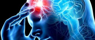
Symptoms of the disorder
Non-straightness of the vertebral arteries in itself is not a pathology. This deviation occurs against the background of diseases of the spine and leads to disorders of the functioning of the brain, which are accompanied by:
- frequent dizziness;
- increased blood pressure;
- pre-fainting states;
- coordination disorders;
- problems with the vestibular apparatus;
- nausea;
- decreased visual function.
The described symptoms are a consequence of deterioration of cerebral circulation, which arose against the background of tortuosity of the aortic vessels. To eliminate these signs, it is necessary to act on the original cause of the deviation, that is, on the disease that caused the non-straightness of the vertebral arteries. By getting rid of an orthopedic problem, you can improve blood circulation and reduce the severity of symptoms. Manual therapy and special massage will help with this. That is, a deep impact is needed on the vertebrae and ligaments of the spine affected by the disease. Only such therapy can restore normal blood flow and improve the patient’s condition.
In addition to manual therapy, drug treatment can also be prescribed - taking drugs that improve venous outflow. Such products include “Antistak”, “Anavenol”, “Eskuzan”. The use of vascular creams, such as “Capillar” and others, is also considered effective.
Before taking any therapeutic measures, it is necessary to consult a doctor who will conduct an examination, assess the condition of the blood vessels and prescribe adequate treatment.
Vertebral artery syndrome (VAS) is a complex of symptoms resulting from disruption of blood flow in the vertebral (or vertebral) arteries. In recent decades, this pathology has become quite widespread, which is probably due to the increase in the number of office workers and people leading a sedentary lifestyle who spend a lot of time at the computer. If previously the diagnosis of SPA was made mainly to elderly people, today the disease is diagnosed even in twenty-year-old patients. Since any disease is easier to prevent than to treat, it is important for everyone to know for what reasons vertebral artery syndrome occurs, what symptoms are manifested and how this pathology is diagnosed. We will talk about this, as well as the principles of SPA treatment, in our article.
Treatment methods
Basically, one of three methods of combating the disease is used. In some cases, it is possible to use two methods of treating spinal vascular stenosis together.
Drug therapy
The method is based on the prescription and use of vascular drugs that help maintain strength and elasticity. Medicines are also prescribed to control blood pressure, thin the blood and reduce blood clots. Along with medications, courses of physical therapy are prescribed, and manual therapy and hirudotherapy are also indicated.
Surgery
This procedure is aimed at surgical correction to eliminate traumatic complications and disorders in the structure of the spine. Stenosis can also be treated with stenting. A reinforced metal frame is placed in the artery, which prevents further rupture and narrowing of the vascular bed. Stents are designed for an average of 15 years of successful functioning. To reduce the risk of rejection, the steel frame is coated with special plastic.
Traditional methods of treatment
Such methods have not yet found recognition among representatives of official medicine. But, according to available data, medicinal decoctions and tinctures help to normalize vascular tone, restore preset blood circulation parameters and regulate blood pressure. As a prophylactic agent used to narrow the vascular systems of the vertebral arteries, folk remedies appear to be quite effective.
To determine the optimal type of therapy, the appropriate specialist issues a referral for several diagnostic procedures. Duplex scanning of certain arteries is recognized as one of the most informative and reliable methods for obtaining a full-scale picture of pathological changes. As an additional option, an MRI of the area of stenosis may be prescribed.
The final decision on treatment methods should be made by the attending physician together with the patient. If the patient complains of regular dizziness, chronic shortness of breath, general weakness and irritability, and vasoconstriction is determined to be at least 70%, then these signs are a serious reason for surgery.
Vessels
Arteries of the Head and Neck: Anatomy, Diagram, Atherosclerosis
Feb 06, 2020 Kokh V. A.
16582
Vessels
Brachiocephalic Arteries: What They Are, Anatomy, Cerebral Oximetry
Feb 03, 2020 Kokh V. A.
6837
Vessels
Fundamentals of Anatomy and Physiology
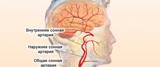
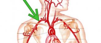
Blood enters the brain through four large arteries: the left and right common carotid and the left and right vertebral. It is worth noting that 70-85% of the blood passes through the carotid arteries, so disruption of blood flow in them often leads to acute cerebrovascular accidents, that is, ischemic strokes.
The vertebral arteries supply the brain with only 15-30% of blood. Violation of blood flow in them, as a rule, does not cause acute, life-threatening problems - chronic disorders occur, which, however, significantly reduce the patient’s quality of life and even lead to disability.
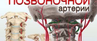
The vertebral artery is a paired formation originating from the subclavian artery, which in turn arises on the left - from the aorta, and on the right - from the brachiocephalic trunk. The vertebral artery goes upward and slightly backward, passing behind the common carotid artery, enters the opening of the transverse process of the sixth cervical vertebra, rises vertically through similar openings of all overlying vertebrae, through the foramen magnum enters the cranial cavity and follows to the brain, supplying blood to the posterior parts of the brain : cerebellum, hypothalamus, corpus callosum, midbrain, partially temporal, parietal, occipital lobes, as well as the dura mater of the posterior cranial fossa. Before entering the cranial cavity, branches depart from the vertebral artery, carrying blood to the spinal cord and its membranes. Consequently, when blood flow in the vertebral artery is disrupted, symptoms arise that indicate hypoxia (oxygen starvation) of the areas of the brain that it supplies.
Causes and mechanisms of development of vertebral artery syndrome
Along its length, the vertebral artery comes into contact with both the hard structures of the spinal column and the soft tissues surrounding it. Pathological changes that occur in these tissues are the prerequisites for the development of SPA. In addition, the cause may be congenital characteristics and acquired diseases of the arteries themselves.
So, there are 3 groups of causative factors of vertebral artery syndrome:
- Congenital structural features of the artery: pathological tortuosity, course anomalies, kinks.
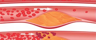
- Diseases that result in a decrease in the lumen of the artery: atherosclerosis, all kinds of arteritis (inflammation of the artery walls), thrombosis and embolism.
- Compression of the artery from the outside: osteochondrosis of the cervical spine, abnormalities of bone structure, trauma, scoliosis (these are vertebrogenic, that is, associated with the spine, causes), as well as tumors of the neck tissue, their scar changes, spasm of the neck muscles (these are non-vertebrogenic causes).
Often, SPA occurs under the influence of several causative factors.
It is worth noting that RAS develops more often on the left, which is explained by the anatomical features of the left vertebral artery: it arises from the aortic arch, which often has atherosclerotic changes. The second leading cause, along with atherosclerosis, is degenerative diseases, that is, osteochondrosis. The bone canal in which the artery passes is quite narrow, and at the same time mobile. If there are osteophytes in the area of the transverse vertebrae, they compress the vessel, disrupting blood flow to the brain.
In the presence of one or more of the above reasons, factors predisposing to the deterioration of the patient’s well-being and the appearance of complaints are sudden turns or tilts of the head.
Causes
Modern medicine divides the causes of non-straightness of the arteries in the cervical spine into 2 categories - vertebrogenic and non-vertebrogenic.
Vertebrogenic causes are associated with pathologies of the development of the spine or changes in its structure, both in children and adults. In children, this pathology can occur for the following reasons:
- Cervical spine injuries. They can occur at any age, including the birth process, which is quite dangerous for a child;
- Anomalies in the development of the spine;
- Pathological spasms of the neck muscles. Often droops as a result of hypothermia. Changes in the tone and location of the muscle affect the position of the artery, as a result of which the capacity of the latter decreases.
Vertebrogenic causes of pathology in adults arise for other reasons, among which the main ones are the following diseases:
- Swellings of the cervical spine or occipital part of the brain;
- Osteochondrosis;
- Ankylosing spondylitis;
- Trauma, as in children, can also affect the condition and location of the artery.
- Interesting read: Exercise therapy for ankylosing spondylitis
Among non-vertebral causes, it is worth noting the following groups of diseases:
- Inflammation, arteriosclerosis, embolism and other diseases that cause loss of artery elasticity and tone;
- Diseases affecting the shape of arteries, kinks, etc.;
- Pathologies associated with changes in the shape of arteries as a result of compression. Causes include abnormal positioning of the ribs, scarring of tissue as a result of surgery, and so on.
The danger of the non-straight course of both arteries is that it increases the likelihood of ischemic attacks in various parts of the brain. If you ignore the symptoms of the disease, there is a risk of cerebral stroke. Rehabilitation after a stroke is not always possible, and complete correction of its consequences is impossible.
Symptoms of vertebral artery syndrome
Patients with vertebral artery syndrome often experience dizziness and headaches.
The pathological process in SPA goes through 2 stages: functional disorders, or dystonic, and organic (ischemic).
Stage of functional impairment (dystonic)
The main symptom at this stage is headache: constant, aggravated by head movements or prolonged forced positions, burning, aching or throbbing in nature, covering the back of the head, temples and moving forward to the forehead.
Also at the dystonic stage, patients complain of dizziness of varying intensity: from a feeling of slight instability to a feeling of rapid rotation, tilting, and falling of one’s own body. In addition to dizziness, patients are often bothered by tinnitus and hearing impairment.
Various visual disturbances may also occur: sand, sparks, flashes, darkening of the eyes, and when examining the fundus, a decrease in the tone of its blood vessels.
If at the dystonic stage the causative factor is not eliminated for a long time, the disease progresses and the next, ischemic stage occurs.
Ischemic or organic stage
At this stage, the patient is diagnosed with transient disorders of cerebral circulation: transient ischemic attacks. They are sudden attacks of severe dizziness, impaired coordination of movements, nausea and vomiting, and speech disorders. As mentioned above, these symptoms are often provoked by a sharp turn or tilt of the head. If, with such symptoms, the patient takes a horizontal position, there is a high probability of their regression (disappearance). After an attack, the patient feels tired, weak, tinnitus, sparks or flashes before the eyes, and headache.
Symptoms and signs
Symptoms of stenosis - neck pain, headaches, memory impairment
Spinal artery stenosis leads to deterioration of blood flow mainly in the posterior parts of the spinal cord. The cells of the nervous system are very sensitive to oxygen and energy deficiency, therefore there is a violation of the functional state, which is accompanied by the following clinic:
- Pain in the neck, which initially occurs infrequently, usually after physical exertion or prolonged stay in one position. Then the discomfort becomes constant. Painful sensations usually precede the development of functional disorders of the spinal cord, as they become a manifestation of degenerative-dystrophic pathology.
- Dizziness and decreased hearing acuity, tinnitus, which reflect a violation of the functional state of the inner ear against the background of insufficient nutrition of the structures of the central nervous system.
- Headaches are a common sign of poor circulation in the brain; discomfort is usually localized in the back of the head and temples.
- Weakness in the arms, numbness of the skin of the upper extremities, which is associated with simultaneous damage to the spinal roots in the cervical region against the background of degenerative-dystrophic changes in the spine.
- Increased muscle tone in the muscles of the upper extremities with a subsequent decrease in strength in the arms.
- Visual impairment – decreased visual acuity, “blurred” objects, “fog,” double vision.
The appearance of one or more signs that indicate a violation of the functional state of the spinal cord, with simultaneous discomfort in the neck, requires examination to exclude stenosis of the vertebral arteries.
Clinical variants of vertebral artery syndrome
These are:
- drop attacks (the patient suddenly falls, his head is thrown back, he cannot move or stand up during the attack; consciousness is not impaired; within a few minutes, motor function is restored; this condition occurs due to insufficient blood supply to the cerebellum and caudal parts of the brain stem);
- syncopal vertebral syndrome, or Unterharnsteidt syndrome (with a sharp turn or tilt of the head, as well as in the case of a prolonged stay in a forced position, the patient loses consciousness for a short time; the cause of this condition is ischemia of the region of the reticular formation of the brain);
- posterior cervical sympathetic syndrome, or Bare-Lieu syndrome (its main symptom is constant intense headaches of the “removing the helmet” type - localized in the occipital region and spreading to the front of the head; pain intensifies after sleeping on an uncomfortable pillow, when turning or tilting head; the nature of the pain is pulsating or shooting; may be accompanied by other symptoms characteristic of SPA);
- vestibulo-atactic syndrome (the main symptoms in this case are dizziness, a feeling of instability, imbalance, darkening of the eyes, nausea, vomiting, as well as disorders of the cardiovascular system (shortness of breath, pain in the heart and others);
- basilar migraine (the attack is preceded by visual disturbances in both eyes, dizziness, unsteadiness of gait, tinnitus and blurred speech, after which intense headache occurs in the back of the head, vomiting, and then the patient loses consciousness);
- ophthalmic syndrome (complaints from the organ of vision come to the fore: pain, a feeling of sand in the eyes, lacrimation, redness of the conjunctiva; the patient sees flashes and sparks before the eyes; visual acuity decreases, which is especially noticeable when there is strain on the eyes; fields partially or completely fall out vision);
- cochleo-vestibular syndrome (the patient complains of decreased hearing acuity (perception of whispered speech is especially difficult), tinnitus, a feeling of swaying, body instability or rotation of objects around the patient; the nature of the complaints changes - they directly depend on the position of the patient’s body);
- syndrome of autonomic disorders (the patient is concerned about the following symptoms: chills or a feeling of heat, sweating, constantly wet cold palms and feet, stabbing pain in the heart, headaches, and so on; often this syndrome does not occur on its own, but is combined with one or more others );
- transient ischemic attacks, or TIA (the patient notes periodically occurring transient sensory or motor disturbances, disturbances in the organ of vision and/or speech, unsteadiness and dizziness, nausea, vomiting, double vision, difficulty swallowing).
Diagnosis of vertebral artery syndrome
Based on the patient’s complaints, the doctor will determine the presence of one or more of the above syndromes and, depending on this, will prescribe additional research methods:
- X-ray of the cervical spine;
- magnetic resonance or computed tomography of the cervical spine;
- duplex scanning of the vertebral arteries;
- vertebral Dopplerography with functional loads (flexion/extension/rotation of the head).
If the diagnosis of SPA is confirmed during further examination, the specialist will prescribe appropriate treatment.
Diagnostics
When making a diagnosis, the specialist interviews the patient about the presence of any chronic diseases or symptoms.
In addition, to exclude errors, the doctor prescribes a number of examinations:
- Doppler ultrasound, with the help of which a specialist obtains data on the speed and direction of blood flow, as well as the patency of the vertebral arteries;
- Compression-functional tests help to find a way to avoid brain hypoxia during the period of vessel compression;
- Duplex scanning allows you to view the condition of the vessel walls, as well as the structure and nature of the narrowing of the lumen;
- MRI and Angiography allow you to study the condition of the great vessels of the head;
- Radiography.
Treatment of vertebral artery syndrome
The effectiveness of treatment for this condition directly depends on the timeliness of its diagnosis: the earlier the diagnosis is made, the less thorny the path to recovery will be. Complex spa treatment should be carried out simultaneously in three directions:
- therapy for pathology of the cervical spine;
- restoration of the lumen of the vertebral artery;
- additional treatment methods.
First of all, the patient will be prescribed anti-inflammatory and decongestant drugs, namely non-steroidal anti-inflammatory drugs (meloxicam, nimesulide, celecoxib), angioprotectors (diosmin) and venotonics (troxerutin).
In order to improve blood flow through the vertebral artery, agapurine, vinpocetine, cinnarizine, nicergoline, instenon and other similar drugs are used.
To improve the metabolism (metabolism) of neurons, use citicoline, gliatilin, cerebrolysin, actovegin, mexidol and piracetam.
To improve metabolism not only in nerves, but also in other organs and tissues (vessels, muscles), the patient takes mildronate, trimetazidine or thiotriazoline.
In order to relax the spasmodic striated muscles, mydocalm or toldel will be used, vascular smooth muscles - drotaverine, better known to patients as No-shpa.
For migraine attacks, antimigraine drugs, such as sumatriptan, are used.
To improve the nutrition of nerve cells - B vitamins (Milgamma, Neurobion, Neurovitan and others).
To eliminate mechanical factors compressing the vertebral artery, the patient may be prescribed physical treatment (manual therapy, post-isometric muscle relaxation) or surgical intervention.
During the recovery period, neck massage, physical therapy, acupuncture, and sanatorium-resort treatment are widely used.
Treatment
Treatment can be outpatient or inpatient. The treatment regimen is determined by the underlying disease (the cause of vasoconstriction). If there is a narrowing of the vertebral artery on the right or left, then complex therapy is required. It includes:
- Use of systemic drugs.
- Massage of the collar area.
- Gymnastics (physical therapy).
- Proper nutrition.
- Manual therapy.
- Wearing a Shants collar.
- Physiotherapy.
- The use of folk remedies (herbal infusions, decoctions).
- Surgical intervention.
Medication
The following medications can cure this pathology:
- Statins (Aterostat, Symvor). Indispensable for arterial stenosis due to atherosclerosis.
- Selective calcium channel blockers (Nimodipine, Nimopin, Nimotop). They are indicated for neurological symptoms secondary to cerebral ischemia.
- Antiplatelet agents, anticoagulants and thrombolytics (Clopidogrel, Aspirin, Thrombo Ass, Heparin, Streptokinase). Prescribed for thrombosis.
- NSAIDs.
- Muscle relaxants.
- Chondroprotectors (prescribed for osteochondrosis). Arthra, Dona, Chondroguard and Teraflex are used.
- Eufillin.
- Metabolic agents (Meldonium, Mildronate, Mildroxin).
- Neuroprotectors (Cinnarizine, Pentoxifylline, Nootropil, Lucetam, Piracetam, Trimetazidine, Cavinton, Vinpocetine, Mexidol, Picamilon, Pikogam, Cerebrolysin, Glycine).
Non-drug
If the right or left artery is narrowed, then physiotherapy (reflexotherapy, ultraphonophoresis, magnetic therapy) is required. If vasoconstriction occurs due to atherosclerosis, you should adhere to a diet (avoid fatty foods, sweets and baked goods).
Surgical
In severe cases, surgical correction is required. Can be carried out:
- Decompression (reduction of intracranial pressure). Required for the development of ischemic stroke.
- Endarterectomy (resection of the affected artery).
- Reconstructive operations.
- Removal of bone growths (osteophytes).
- Vertebral stabilization.
- Removal of the tumor.
- Stenting.
- Installation of implants in the spine.
Important information: How to treat stenosis of the right carotid artery and what are the symptoms of narrowing
Prevention
Measures to prevent stenosis of the vessels supplying the brain are: quitting smoking, treating atherosclerosis, proper nutrition, exercise, preventing spinal diseases and thrombosis.
Prevention of vertebral artery syndrome
The main preventive measures in this case are an active lifestyle and healthy sleep on comfortable bedding (it is highly desirable that they be classified as orthopedic). If your work involves keeping your head and neck in one position for a long time (for example, working at a computer or activities associated with continuous writing), it is strongly recommended to take breaks from it, during which you perform gymnastics for the cervical spine. If the complaints mentioned above appear, you should not wait for their progression: the right decision would be to consult a doctor as soon as possible. Do not be ill!
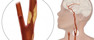
COLOR DUPLEX SCAN OF EXTRACRANIAL PARTS OF BRACHIOCEPHAL ARTERIES.
1. The intima-media complex of the common carotid arteries is not thickened (up to 1.0 mm), the intima is compacted and fragmented.
ON THE LEFT, in the area of the carotid bifurcation on the left, a concentric atherosclerotic plaque with the inclusion of calcium salts is located, stenosing the lumen of the vessel by 20%.
ON THE RIGHT, at the mouth of the right internal carotid artery, along the posterior wall, a heterogeneous atherosclerotic plaque with a predominance of the dense component and ulceration of the surface, 1.7 cm long, is located, stenosing the lumen of the vessel by 30%.
Linear blood flow velocity (LBV) in the common carotid arteries:
left – 112 cm/s; on the right – 80 cm/s.
Linear blood flow velocity (LBV) in the internal carotid arteries:
left – 52 cm/s; on the right – 40 cm/s.
2. Non-straightness of the course of the vertebral arteries between the transverse processes of the cervical vertebrae.
Option for the entry of the right vertebral artery into the bone canal at the level of the transverse process of the 5th cervical vertebra.
C-shaped tortuosity of the extravertebral section of the right vertebral artery.
Blood flow through the left vertebral artery with signs of high peripheral vascular resistance.
Linear blood flow velocity (LBV) in the vertebral arteries:
LSC on the left – 19 cm/s; LSC on the right – 30 cm/s.
Diameter of the vertebral arteries between the transverse processes of the cervical vertebrae:
right – 4.0 mm.
3. In the left supraclavicular region, the internal jugular vein, dilated to 34 mm, is located. The right internal jugular vein is not dilated. The veins are not dilated, are passable, and phasic blood flow is detected.
CONCLUSION:
1. Atherosclerosis of the extracranial parts of the brachiocephalic arteries with stenosis of the area of the carotid bifurcation on the left by 20%, the mouth of the right internal carotid artery by 30%.
2. Non-straightness of the course of the vertebral arteries between the transverse processes of the cervical vertebrae, which is obviously due to the presence of osteochondrosis of the cervical spine, C-shaped tortuosity of the extravertebral section of the right vertebral artery.
Option for the entry of the right vertebral artery into the bone canal at the level of the transverse process of the 5th cervical vertebra.
3. Ectasia of the internal jugular vein on the left.
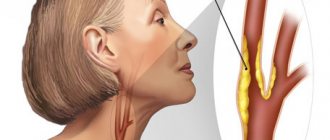
Extravasal compression of the vertebral arteries is one of the causes of a whole complex of manifestations of serious disturbances in the blood supply to the brain. The fact is that blood enters the brain through two main channels: the carotid (two carotid arteries) and the vertebrobasilar (two vertebral arteries). In cases where the functions of one or more of these blood vessels are disrupted, normal nutrition of the brain stops: it receives an insufficient amount of useful substances, including oxygen, and the patient in this case suffers from a whole complex of neurocirculatory disorders known as “Vertebral Artery Syndrome.” " This complex medical concept requires an integrated approach and careful identification of the root cause of the disease, since the process of making a correct diagnosis is quite complex and requires a comprehensive examination of the patient.
Arteries originating from the subclavian arteries are responsible for supplying blood to the brain, cerebellum and inner ear. They are located in a special canal, in the cervical vertebrae, close to the elements of the spinal column. Therefore, if the structure of the spine is disturbed, there is a danger of reducing the lumen of the channel through which this blood vessel passes - there is a possibility of extravasal influence of nearby tissues on it, its compression and disruption of blood flow.
Signs and main causes of PA syndrome
With extravasal compression of the vertebral arteries (left, right or both), they are compressed by intervertebral hernia, tumors of various etiologies, and other anomalies in the structure of the cervical vertebrae. As a rule, compression of the artery develops at the level of 4-5 cervical vertebrae. Whatever the reason, stenosis (narrowing of the lumen of the vessel) can cause disruption in the normal flow of blood to the brain.
photo: possible causes of extravasal compression of the VA
The main symptoms of this complex disease are severe, incessant headaches, dizziness, vomiting, and nausea. Headaches are usually localized in the cervical-occipital part and radiate to the frontal lobes of the head. This symptom is called “helmet removal pain.” It intensifies after sleeping on an uncomfortable pillow, with sudden movements of the neck, it can cause an attack and a shaking ride. There are different types of headaches - throbbing, shooting, aching. Pain is often felt when simply touching the scalp.
This disease is quite often accompanied by disturbances in the cochleo-vestibular apparatus: attacks of dizziness, a feeling of instability and staggering, a feeling of noise and ringing in the ears, decreased hearing, and nausea are possible. In complicated cases, loss of consciousness often occurs with a sudden movement of the neck, or a sudden fall while maintaining consciousness (drop attacks). Visual disturbances are also possible, which begin with increased eye fatigue, decreased vision with any visual load; sensations of pain, redness and lacrimation, a feeling of the presence of a foreign body in the eyes, flashing spots. There are frequent attacks when part of the panorama temporarily falls out of the field of vision when moving the head to the side, as a consequence of a transient ischemic attack.
The causes of these disorders are different: congenital, acquired as a result of heavy loads, during the progression of cervical osteochondrosis , or due to other factors. Depending on the location of pain and symptoms, patients are diagnosed with “Right VA Syndrome” or “Left VA Syndrome”.
Diagnosis of extravasal compression of the vertebral arteries
To clarify the diagnosis, the attending neurologist prescribes additional examinations, which should include the following procedures:
- MRI – magnetic resonance imaging, which allows to identify abnormalities of the bone bed of the vertebral artery (VA);
- SCT – reveals features and areas of compression of the blood vessel;
- X-ray of the cervical spine - allows you to identify herniated intervertebral discs or other formations that impede normal blood flow;
- Duplex scanning of the vertebral arteries - determines the nature of the lesions on the internal walls of the VA and allows you to determine the exact location of compression;
- Vertebral Dopplerography - with this examination you can determine the strength of blood flow and its deviation from normal values;
- According to indications, angiography is prescribed - a contrast agent is injected into the artery and three-dimensional images of the damaged vessels are taken using an X-ray machine.
The use of these diagnostic techniques in combination will allow us to determine the exact cause and localization of VA compression and prescribe appropriate treatment.
Methods used in the treatment of extravasal compression of the VA
The prescribed treatment should be aimed at reducing swelling and inflammation that develop when a blood vessel is mechanically compressed. This is accompanied by a violation of venous blood flow - as a result, a mutual stagnation process develops. Thus, this factor must be taken into account in treatment. For this purpose, a neurologist must prescribe complex drugs that can restore impaired hemodynamics. This stage is very important and responsible in the treatment of the disease, since incorrectly selected medications can intensify its manifestations and even cause various kinds of complications, including hemorrhagic stroke. Doctors usually prescribe decongestants and non-steroidal anti-inflammatory drugs.
For severe headaches, the doctor may prescribe a novocaine blockade of the PA and sympathetic plexus. In some cases, if conservative treatment is ineffective, the doctor may decide to perform a surgical intervention, during which decompression of the vertebral column is carried out , removal of osteophytes and growths that contribute to compression of blood vessels in the vertebral body. This complex operation is performed in specialized medical centers by neurosurgeons experienced in performing such interventions.
After the removal of acute inflammatory phenomena, it is necessary to carry out special therapy aimed at regenerating damaged neurons and restoring brain functions affected by prolonged oxygen starvation. The use of these medications will help improve tissue microcirculation and blood supply to the entire brain stem. As a result, all important functional processes occurring in the brain should return to normal.
This stage of treatment is especially important for patients who have an increased risk of developing transient ischemic attacks in the presence of vertebral syndrome - to prevent persistent neurological deficit and the development of secondary complications.
In addition to basic medications, patients should take antispasmodics, antiallergic drugs and multivitamins. You should definitely remember that all prescriptions must be made by a doctor - otherwise self-medication can lead to disastrous consequences.
Treatment options
Depending on the cause that caused the disease, treatment is prescribed. As the main treatment, drug therapy is prescribed, which may consist, for example, of non-steroidal anti-inflammatory drugs, as well as antibiotics, which help restore cartilage tissue and reduce muscle tone. After this, orthopedic massage is usually prescribed, which improves blood flow and prevents relapse of the disease.
As for drug therapy, this is only an analgesic process. Various tablets are prescribed to dull the pain and improve venous outflow. The person’s well-being will become better, but this is temporary, because the deformation of the vessel cannot be corrected by taking medications.
If there is a threat of ischemic stroke, surgical intervention is prescribed. The surgeon performs stenting, which allows the vessel to expand, or prosthetics (replacing a natural vessel with an artificial one). The process of lengthening and shortening of the artery is also possible.
Physiotherapeutic procedures for PA syndrome
We cannot ignore another important stage of rehabilitation therapy – physiotherapeutic procedures. Massages of the collar zone, acupuncture procedures, and other physiotherapeutic measures: UHF, electrophoresis with medications and physical therapy activities aimed at relieving pain and other clinical manifestations of this disease have a good effect.
Upon completion of the course of treatment, patients should undergo additional rehabilitation treatment in specialized sanatoriums or resorts, where they have the opportunity to take hydrogen sulfide and radon baths, therapeutic mud, and professional massage of the affected area to consolidate the results of therapy.
Prevention of the development of PA syndrome
Early and correct diagnosis of the disease is of great importance for effective treatment. Given that this vascular pathology is a fairly common phenomenon, it would not be superfluous to carry out a preventive examination of children at risk of vertebral pathology, starting from the neonatal period. Ultrasound diagnostic methods are very effective at the general stage, and when deviations from the norm are detected, various additional diagnostic procedures are performed.
Patients who have suffered acute manifestations of PA syndrome must be constantly monitored by a neurologist and regularly undergo courses of preventive vascular treatment. very useful. It is necessary to avoid hypothermia and various injuries that can trigger a relapse of the disease. Work in transport, at heights, heavy loads, sports and work with moving mechanisms are not allowed.