Etiology of the disease
The retina is one of the most important structures of the eye. Thanks to its unique structure, it instantly responds to minor disturbances in the functioning of the nervous and vascular system. Retinal angiopathy of both eyes cannot be an independent disease. Failure in the functioning and innervation of the vessels of the retina, which signals the presence of a pathological cause that can affect vision.
These include the following:
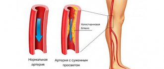
Arterial hypertension. An increase in pressure leads to a decrease in the elasticity of blood vessels, making them more vulnerable. This process represents the destruction of the inner layer of the shell and fibrotization. Then microcirculation disturbances occur.
With hypertension, the vessels increase in size, the lumen decreases, the blood flow slows down, the blood thickens and blood clots form, which prevent the passage of blood through the vessels. Blockage of blood vessels leads to rupture and massive hemorrhage into the surrounding tissue. To compensate for the processes that are caused by blood thickening, sodas acquire a more branched structure.
At the first stage of hypertension, vascular pathology affects a third of patients, at the second stage, about 50% of the total number of patients suffer from this pathology, and at the last stage, this pathological process affects absolutely all people who suffer from arterial hypertension.
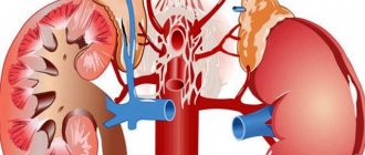
- Diabetes. Endocrine pathology leads to diabetic angiopathy, which can affect absolutely all vessels. But small-caliber vessels suffer the most. High glucose levels lead to the development of vascular pathology. Diabetic angiopathy of the eye is characterized by: occlusions, thickening and thinning of the structure of blood vessels, proliferation of the capillary network, reduction of the lumen of blood vessels and, as a consequence, deterioration of blood patency. Endocrine disease in mild cases leads to loss of vision, and in severe cases to blindness.
- Injuries to the cervical vertebrae and visual analyzer, as well as traumatic brain injuries and prolonged chest compression. All of the above factors contribute to an increase in intracranial pressure. These conditions can lead to hemorrhages. This process is accompanied by hemorrhage in the retina of both eyes.
- Hypotension. Constantly reduced blood pressure leads to the expansion of the lumen of blood vessels, the formation of a branched structure, and a decrease in the speed of blood flow, which leads to a change in the rheological properties of the blood. Its thickness increases, which increases the risk of blood clots. The above reasons provoke an increase in the permeability of the vascular wall.
Predisposing factors for angiopathy:
- Arterial hypertension.
- Bad habits that negatively affect the state of the nervous and vascular system. These include smoking and alcohol abuse.
- Acute and chronic poisonings that are associated with harmful working conditions or living in an area with unfavorable sanitary and epidemic conditions.
- Senile age.
- Congenital anomalies of the vascular system (aneurysms, malformations).
- Osteochondrosis of the cervical spine.
- Systemic pathology of the vascular system or vasculitis.
- Traumatic damage to the organ of vision.
- Some diseases of the hematopoietic system.
- Exposure to radiation.
Conditions in which angiopathy develops in rare cases:
- Angiopathy in pregnant women. The early stages of the disease do not lead to serious consequences. If no measures are taken to treat the disease, it can transform into a more advanced form. In the final stages, the disease can lead to detachment of the retina. Retinal angiopathy of both eyes usually begins to appear in the second half of pregnancy. Its appearance is promoted by reasons associated with increased blood pressure or other extragenital diseases, which are accompanied by disturbances in the tone and morphology of the vascular wall.
- Angiopathy of newborns. The onset of the disease is preceded by postpartum complications and injuries. Retinal damage in newborns is characterized by proliferative changes, narrowing of the lumen of blood vessels and impaired blood patency.
- Juvenile angiopathy. The cause of the disease has not been established. This type of vascular damage is characterized by the formation of small hemorrhages in the structures of the eye. In severe cases, the disease can provoke cataracts and glaucoma. Cases of partial and complete loss of vision are common.
Recommendations for patients with angiopathy
You can significantly increase the effectiveness of treatment, reduce the risks of complications and the likelihood of further progression of angioneurosis by following the following recommendations:
- The diet must include a sufficient amount of foods that are natural sources of vitamins and substances necessary for the proper functioning of the visual apparatus and blood vessels of the fundus (chicken and quail eggs, vegetables, herbs, lettuce, offal, butter, sour cream, nuts, etc. .).
- Self-massage of the cervical-collar area should be performed regularly, unless there are contraindications (such massage has a positive effect on blood circulation in this area and prevents compression of nerve endings due to myofascial syndrome).
- When there is increased stress on the eyes (working at a computer, long reading, activity in poorly lit rooms), it is necessary to perform therapeutic exercises for the eyes.
- When engaging in traumatic sports, it is necessary to protect your eyes from damage.
Natural sources of vitamins for the eyes
It is necessary to visit an ophthalmologist for early detection of pathological changes in the fundus every year (for patients with diabetes mellitus - at least 2-3 times a year).
Retinal angiopathy is a vascular pathology that can provoke more severe ophthalmological disorders and, with a progressive course, lead to low vision and complete blindness. The maximum risk of angiopathy is present in patients with arterial hypertension, diabetes mellitus, endocrine disorders and heart disease. Treatment in the initial stages is medicinal and can be supplemented with physiotherapeutic methods. In severe cases and in the presence of complications, microsurgical or vitreoretinal intervention is indicated.
Traditional methods of treatment
Folk remedies for the treatment of angiopia cannot be the basis of the entire course: such a disease requires not only an integrated approach, but also the use of specially developed and clinically tested medications. However, such methods can help with treatment and speed up the healing process.
For 20 grams of horsetail grass, take 30 grams of the knotweed plant and 50 grams of hawthorn flowers. This mixture of herbs is poured with 200 grams of boiling water and left to infuse for half an hour. The broth is then filtered through cheesecloth to remove any remaining herbs.
Take the product half an hour before meals (three times a day), one tablespoon.
Crushed valerian root (15 grams) is mixed with the same amount of lemon balm, and then 50 grams of yarrow are added to this mixture.
This mixture should be poured with a glass of water and placed in the refrigerator for three hours (but not in the freezer). After this, the product needs to be heated for 15 minutes in a water bath, and the cooled product is diluted with boiled water so that the result is 250 grams.
This glass of infusion must be drunk throughout the day, but not in one gulp, but in portions of several sips.
20 minutes is enough for the product to infuse, after which it is filtered and ready for use. One part of the product is drunk in the morning on an empty stomach, the second after dinner. These drugs should not be abused and you should not continue treatment with them for more than one or two weeks.
Diagnostics
Diagnosing angiopathy in a patient is not difficult at all. A much more labor-intensive process is searching for the cause of such a pathology.
The main means for studying the situation is ophthalmoscopy, which is carried out in two ways:
- Direct ophthalmoscopy is the usual magnification of the fundus image by 15 times thanks to lenses and a light source.
- Indirect ophthalmoscopy is an improved analogue of direct ophthalmoscopy, allowing for a more adequate image and using a remote light source and special lenses.
Ophthalmoscopy is performed without preparation. Only in rare cases, before it is performed, drops are instilled into the eyes to dilate the pupil and help to better diagnose the type of angiopathy.
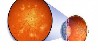
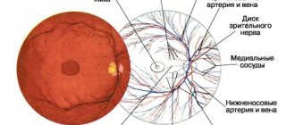
The normal picture is as follows:
- retinal color - red;
- the optic disc has pronounced boundaries, has a clear rounded shape, its color is definitely pink);
- no side formations (infiltrates, hemorrhages) are observed in the fundus;
- there is no tortuosity of the veins, the proportion between the sizes of veins and arteries is maintained;
- The macula (blind spot) is visible with maximum clarity.
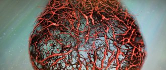
The very first change caused by angiopathy is a change in the appearance of blood vessels. They become tortuous, and the entire process of blood circulation in the human body is destabilized.
Causes
There are several factors that determine the formation of pathology. The main ones are listed in the table, a number of others are listed below.
Table. Causes of constriction of blood vessels in the fundus:
| What causes | A comment |
| With general high blood pressure, the inner layer of the fundus capillaries is destroyed, and the vascular network becomes denser (fibrotization). This impedes the movement of blood, blood clots appear, which causes hemorrhages. In people with stage 1 hypertension, 30% of the blood vessels in the eye narrow, by stage 2. - at 50% and at 3 tbsp. All patients have pathological changes. |
| We are talking about injuries to the eyes, head and cervical spine. Prolonged and powerful compression of the chest can also cause changes in the vascular tone of the eyes. Depending on the nature of the impact, capillaries can either narrow or expand. |
| The disease negatively affects the entire circulatory system due to increased sugar content. Occlusions are formed, blood enters the retina, the endothelium grows, and the vascular lumen narrows. This leads to deterioration of circulation in the fundus and a slow decline in visual function. |
| As a result of blockage of veins and arteries by cholesterol plaques, many pathologies develop, for example, hypertension, thrombosis and others. In this case, the blood flow to the retina is poor, and the muscles of the ophthalmic artery and its collaterals begin to contract spasmodically. |
Retinal detachment is one of the possible consequences of narrowing of the fundus vessels
In addition to the main reasons for the narrowing of the fundus vessels listed in the table, there is a list of factors that can directly or indirectly negatively affect the capillaries and small arteries of the eyes:
- arterial hypertension, regardless of origin;
- high intracranial pressure;
- brain tumors;
- systemic vasculitis;
- blood diseases;
- hemolytic anemia;
- poisoning of the body with certain toxic substances, for example, when working in hazardous industries;
- radiation exposure;
- osteochondrosis of the spine in the cervical region;
- congenital vascular pathologies;
- thrombosis;
- aging changes (presbyopia);
- smoking and alcoholism;
- physical inactivity.
Note. The genetic line of inheritance of pathogenesis can be traced, for example, narrowing of blood vessels in the fundus is more common in people belonging to the African race, or in children if their parents have this problem.
All of the above leads to retinal ischemia, which causes myopia and vision problems. If the situation is sufficiently advanced, then the destructive processes become irreversible, dystrophy and necrosis of individual areas develop, which cause blindness.
The main culprits in this process are diabetes mellitus and arterial hypertension. When treatment is started late, the cost of neglecting one's own health usually becomes quite high.
The pathology must be examined by specialists, the cause must be found and immediate treatment initiated. Otherwise you may lose your vision
Classification
Idiopathic angiopathy is a consequence of dysregulation of the muscle tone of the arterial walls, usually developing due to malfunctions of the autonomic nervous system. In newborns, the primary form can occur as a result of intrauterine development anomalies, birth injuries, oxygen starvation caused by asphyxia or other teratogenic factors.
Juvenile angiopathy, also known as Izla disease, is an example of an idiopathic form. More often detected in male patients, in 90% of cases it is bilateral in nature
Juvenile angiopathy of the arteries and arterioles of the retina (OU-bilateral) is a disease that is characterized by a gradual development, which makes early diagnosis important
At the initial (inflammatory) stage, periphlebitis (inflammation of the tissue surrounding the veins) of the peripheral elements of the circulatory system of the retina develops. Immune mechanisms play an important role in pathogenesis. The development of the pathological process is associated with an increase in the level of interleukins (cytokines - cells of the immune system synthesized by leukocytes), which can have anti-inflammatory and pro-inflammatory (mobilization of the inflammatory response) effects.
The next stages of development are disruption of blood microcirculation, infiltration (impregnation) of the vascular endothelium with leukocytes, and increased effects of oxidative stress. As a result, occlusion (impaired patency) of the arteries occurs with the formation of areas deprived of blood supply and a transition to the ischemic stage.
As a result, vascularization occurs (the formation of new elements of the vascular network), which often leads to hemorrhages in the area of the vitreous body (a gel-like, transparent substance that fills the space between the retina and the lens) and provokes the process of retinal detachment. Such violations indicate a transition to the proliferative stage.
Background angiopathy is damage to elements of the circulatory system that develops against the background of a primary disease (cardiovascular pathologies, blood diseases, head injuries, cerebral blood flow disorders, central nervous system infections). Background angiopathy, affecting the vessels that supply the retina of the eye, is a disease that is caused by a primary pathology, which makes it possible to distinguish types taking into account etiological factors:
- Hypertensive. Due to a persistent increase in blood pressure.
- Hypotonic. Due to a steady decrease in blood pressure.
- Diabetic. Due to an increase in blood glucose concentration.
- Traumatic. Due to mechanical damage to tissues.
- Dysoric, also known as Morel's angiopathy. Due to deposits of amyloid (protein fractions) in the tissues of the vascular wall. Often combined with Alzheimer's disease.
Mixed angiopathy, which affects the vessels of the fundus, is diagnosed if the pathogenetic mechanisms are influenced by various factors, for example, cardiovascular pathologies in combination with a history of diabetes mellitus. Retinal neuroangiopathy means simultaneous damage to the elements of the circulatory system and the nerve fibers of the eye. Angiodystonia (decreased tone of the vascular wall due to impaired neurohumoral regulation) of the retinal arteries is a consequence of malfunctions of the autonomic system.
Types and causes of rare angiopathy
There are some types of pathologies that are diagnosed relatively infrequently. For example, juvenile angiopathy.
It is difficult to determine why the blood vessels in a child’s eyes are narrowed and often this question remains unresolved, but the situation is quite dangerous and requires an immediate solution. In premature infants, pathogenesis may be associated with birth trauma or as a result of intrauterine developmental anomalies.
Angiopathy during pregnancy in the early stages of pathogenesis is not something very dangerous, but if the process is neglected, tissue detachment may occur. More often the disease appears at the end of the second and third trimester. As a rule, the cause is hypertension or other pathologies that weaken or narrow small blood vessels.
Hypotonic retinal angiopathy
Hypotension, that is, a strong decrease in blood pressure, is observed in a disease called arterial hypertension. In this case, the pressure drops so much that this process becomes noticeable to the person and leads to a deterioration in well-being.
There are two types of arterial hypertension – acute and chronic. In an acute condition, manifestations of collapse can be observed, in which vascular tone drops sharply. Shock may occur, which is characterized by paralytic vasodilation. All these processes are accompanied by a decrease in oxygen supply to the brain, which reduces the quality of functioning of vital human organs. In some cases, hypoxia occurs, which requires immediate medical attention. And in this case, the determining factor is not the pressure in the vessels, but the rate of its decrease.
Hypotonic retinal angiopathy is a consequence of arterial hypertension and manifests itself in reduced vascular tone in the retina. As a result, the vessels begin to overflow with blood, which reduces the speed of its flow. Subsequently, blood clots begin to form in the vessels due to stagnation of blood. This process is characterized by a sensation of pulsation, which is observed in the vessels of the eyes.
Retinal angiopathy of hypotonic type
Typically, this type of complication disappears with proper treatment of the underlying disease. The tone of blood vessels throughout the body improves, which also affects the condition of the eye vessels. Blood begins to move faster, blood clots stop forming, which affects the improvement of blood supply to the retina, eyeball, and so on.
Retinal angiopathy of the hypotonic type is caused by the main human disease - hypotension. In this case, there is a decrease in vascular tone throughout the body, and also, in particular, in the eyes. Therefore, blood begins to stagnate in the vessels, which leads to the appearance of blood clots in these vessels. Thrombosis of capillaries and venous vessels causes various hemorrhages in the retina and eyeball. Which leads to visual impairment and other eye problems.
[], [], [], []
Causes of red eyes
Redness of the tunica albuginea often appears when some part of the eye is inflamed with the involvement of blood vessels in the pathological process. They fill with blood and expand, causing the sclera to become stained.
The mechanism of local swelling of blood vessels comes down to the effect on them of inflammatory mediators (histamine, serotonin), which can relax the vascular wall. Infectious agents are viruses, bacteria, fungi, protozoa that cause keratitis, blepharitis, stye, purulent lesions of the eyelid. An early symptom of these inflammatory diseases is red eyes.
Allergic eye damage
The anatomical and physiological characteristics of the organ of vision require constant contact with allergens, which are contained in large quantities in the surrounding air.
Allergic inflammation occurs when a foreign substance (antigen) enters the sensitive mucous membrane of the eye - the conjunctiva. The released biologically active substances (histamine, bradykinin) increase vascular permeability and cause their dilation.
Diseases of the eyeball
Sometimes hemorrhages are the first sign of the development of serious eye diseases.
Underestimation of this symptom can lead to serious complications, including loss of vision:
- Retinal disinsertion.
- Retinal vein blockage.
- Eye tumors.
- Inflammation of various structures.
Injections of blood vessels or the appearance of a capillary network on the sclera of the eyes can cause:
- visual fatigue (TV, computer),
- excessive alcohol consumption,
- lack of sleep,
- physical action - when a blow to the area of the eyeball occurs, rupture of blood vessels and hemorrhage under the conjunctiva of the eye occurs,
- eye contusions due to head injuries,
- chemical exposure - a negative effect on the eye of irritating substances at high concentrations in the atmosphere (chlorine, sulfur, asbestos and others),
- foreign body entering the eye,
- Violations of the rules for wearing contact lenses (wrongly chosen, contaminated).
Important! Hemorrhages in the eyes occur quite often in a person’s life and are not a sign of a serious illness if there are no other symptoms (pain, inflammation, blurred vision). But long-term disruption of the blood supply to the eye structures leads to disruption of their functions.
Clinical picture
Retinal angiopathy is a symptom that can be caused by any disease affecting the structure of the vascular wall and associated with circulatory disorders.
Specific manifestations of the disease directly depend on the type of angiopathy:
| Form | Description |
| Diabetic (mixed type angiopathy) | Is the most common. The background development of angiopathy occurs during a long transition of the disease (10-15 years) to the stage of decompensation. Depending on the type of damage to blood vessels, micro- and macroangiopathies are distinguished. With the first, hemorrhage phenomena are observed, with the latter - swelling and multiple hemorrhages in the retina, unevenness of the optic nerve head |
| Hypertensive | The yellow areas on the right are deposits in the fundus of the eye. A sure sign of angioretinopathy The mechanism of formation is associated with a chronic persistent increase in blood pressure. There is a narrowing of the lumen of the vessels of the fundus. Clinically this is manifested by flickering of flies, darkening of the eyes At the stage of angioretinopathy, there is a significant disruption of microcirculation with the formation of hard or soft deposits in the fundus of the eye. At the same time, vision deteriorates noticeably, up to its complete loss |
| Hypotonic type | In contrast to the hypertensive form, dilation of blood vessels and a decrease in blood flow speed are observed. Patients complain of dizziness and a subjective feeling of pulsation of the eyeballs |
| Traumatic | Due to injuries to the head, chest, and abdominal cavity, a sharp increase in intraocular pressure and vascular damage are observed. Possible feeling of darkening in the eyes, loss of vision |
| During pregnancy | If angiopathy occurred before pregnancy (diabetic, hypertensive and other forms), its manifestations can significantly worsen. If a pathological condition occurs during pregnancy, it can be self-limited without any complications. |
In the early stages of the disease, angiopathy can be virtually asymptomatic for a long time.
In this case, patients rarely pay attention to a slight deterioration in vision. However, at the moment of obvious signs of malfunction of the visual analyzer, pathological changes may be irreversible
Common manifestations of retinal angiopathy include:
- decreased visual acuity,
- feeling of “fog” and spots before the eyes,
- narrowing of the field of view,
- sensation of pulsation in the eyeball,
- visible hemorrhages in the conjunctival area.
Symptoms that can help in diagnosing the disease:
- nosebleeds,
- pain in the lower extremities,
- presence of traces of blood in the urine.
In the case of damage to both eyes, ophthalmology uses the abbreviations OU or OI.
Symptoms of vasoconstriction of the fundus
Bursted blood vessels in the eyes are one of the signs of narrowing of the fundus vessels
It is very important to notice the pathogenesis as early as possible and consult a doctor for advice. The problem is that in the early stages the disease is difficult to detect, since the onset is always latent. When processes become apparent, changes are usually irreversible.
Symptoms of vasoconstriction in the eyes:
- blurred vision;
- the appearance of glare, spots or haze;
- narrowing of visual fields;
- sensation of pulse in the eye;
- the appearance of burst vessels or yellow spots on the conjunctiva.
Etiology of pathology
The occurrence of symptoms of angiopathy indicates the presence of a disease associated with impaired blood supply in the body. Pathology of the tone of large vessels and small capillaries in the future can lead to necrosis of the blood supply. If there is a malnutrition of the fundus of the eye, a decrease in acuity, partial or complete loss of vision is observed.
As a rule, retinal angiopathy develops against the background of existing chronic diseases in adult patients over 40, however, over the past few years, this pathology has often occurred in children.
The main reasons that play a role in the development of angiopathy include the following:
| Etiological factor | Description |
| Diabetes | The pathogenesis of the disease is associated with disorders of all types of metabolism. Due to the increased concentration of glucose in the blood and its significant changes during the day, the target organs that are most sensitive to the lack of oxygen are affected. These include the retina of the eye. |
| Injury | As a result of the action of a traumatic factor (traumatic brain injury, etc.), a sharp increase in intracranial pressure occurs, which is accompanied by damage to the vascular wall and hemorrhage in the retina |
| Hypertension | Symptomatic or essential hypertension is characterized by a persistent increase in blood pressure above 139/89 mmHg. rt. Art. and related circulatory disorders. Damage to the vessels of the fundus is manifested by dilation, branching and tortuosity of the veins, the vessels of the fundus are narrowed. Normally, undetectable small vessels become visible during examination. There is a possibility of small isolated hemorrhages |
| Hypotension | A systemic decrease in blood pressure leads to a decrease in blood flow speed, increased permeability of the vascular wall, and thrombus formation in the vessels of the fundus |
In addition to the above reasons, there are “trigger” factors that contribute to the emergence and aggravation of an existing pathological condition:
- 1. Alcohol abuse.
- 2. Tobacco smoking.
- 3. Chronic and acute intoxication.
- 4. The patient's age is over 50 years.
- 5. Congenital anomalies of the structure of the vascular wall.
- 6. Carrying out professional activities in hazardous production conditions.
Rarer forms of retinal angiopathy include:
| Form | Description |
| Juvenile angiopathy | It is considered one of the most severe forms of pathology. Inflammation of the vascular wall occurs due to an unknown etiology and is accompanied by the occurrence of small hemorrhages in the retina and vitreous body. If diagnosed late, it can lead to severe visual impairment, even complete blindness. |
| Angiopathy in pregnant women | It is one of the indications for caesarean section. Most often develops in the second half of pregnancy against the background of chronic diseases (diabetes mellitus, hypertension, etc.) |
| Angiopathy of prematurity | Etiological factors include various complications during pregnancy and childbirth. It is a vasoproliferative lesion of the retina caused by the immaturity of the ocular structures. |
Treatment and prevention of vascular eye diseases
The following types of medications and methods are used to treat diseases of the ocular vessels:
- Eye drops (Floxal, Albucid, Tobradex). The drops have a local effect and are mainly used in the treatment of diseases of the choroid and retina. Using drops you can relieve inflammation or prevent the spread of infection.
- Eye ointments (Acyclovir, Tetracycline and Hydrocortisone ointment, Demazol). The ointment is applied to the inside of the lower eyelid or to the surface of the eyelids. Just like drops, it has a local therapeutic effect and helps eliminate the symptoms of the disease.
- Vitamins (Complivit Blueberry Forte, Okuwait Lutein Forte). In the complex treatment of any vascular diseases of the eyes, vitamins are definitely needed, as they will help improve blood circulation in the eye area, improve the elasticity of the walls of blood vessels, and reduce the amount of cholesterol in the blood. At the same time, vitamins have a positive effect not only on the blood vessels in the eyes, but also on the entire body as a whole.
- Pills. To treat vascular diseases, tablets are used that improve blood composition, reduce coagulation, and also prevent the formation of blood clots. These can be anticoagulants (Warfarin), statins (Atorvastatin), antiplatelet agents (Acetylsalicylic acid).
- Surgery. Eye surgery is only performed if all drugs and medications are ineffective, the symptoms do not go away and the risk of deterioration of the eye condition is too great, or the condition has already worsened significantly. The operations are performed in special ophthalmic-oncology clinics using hardware therapy.
Since vascular diseases of the eyes usually occur deep at the bottom of the eyeball with inflammation of the retina, treatment becomes very difficult compared to the treatment of diseases on the surface of the eye. Local drugs have very low effectiveness and are often combined with other means of complex treatment of eye vessels.
What preventative measures should you take?
It is always better to prevent a disease than to treat it. Moreover, the process of treating vascular eye diseases is quite difficult and requires a lot of time. To prevent these diseases you need to follow a few simple recommendations:
- Monitor your blood cholesterol levels. It is cholesterol that settles on the walls of blood vessels, leading to a narrowing of the lumen and forming plaques that can lead to thrombosis. To prevent this from happening, you need to control the amount of low-density and very low-density cholesterol in the blood. To do this, you can take tests at the clinic at your place of residence and get recommendations from your doctor on your diet and lifestyle.
- Take a multivitamin complex twice a year. They will help maintain the blood vessels of the eyes in working condition, prevent the walls of blood vessels from thinning and improve the composition of the blood.
- Follow the regime during intense visual work. Working at a computer monitor, reading books, watching TV and other activities that require intense eye work should be combined with a rest regime. Every 40 minutes you need to take a break, during which it is advisable to do a little gymnastics for the eyes: rotating the eyeballs in a circle, changing focus from a near object to a distant one, moving the eyes up, down, right, left. If you feel a burning sensation or a spasm of the blood vessels in the eye appears, it is better to stop working and let your eyes rest for several hours.
In this way, the eye vessels can be kept in good shape and prevent the occurrence of diseases associated with them. If you nevertheless discover the manifestation of the listed symptoms, you must contact a specialist as soon as possible to diagnose the disease and prescribe treatment. The selection of treatment methods and medications should be carried out by an ophthalmologist. Self-medication in this case can lead to adverse consequences, including complete loss of vision, since incorrectly selected drugs can be not only ineffective, but also dangerous.
Do you still think that it is completely impossible to RESTORE blood vessels and the BODY!?
Types of disease
In an adult patient, this type will manifest itself due to failures in the vegetative-vascular system.
Secondary, in other words, “background retinal angiopathy.” It is divided into 6 subspecies:
- Diabetic retinal angiopathy. It is directly related to diabetes mellitus and develops gradually. Due to a malfunction in the metabolic environment, mucopolysaccharides appear. This slows down blood flow due to the fact that the lumen of the vessels decreases. As a result, the likelihood of hemorrhage increases, as the walls of the eye become permeable and thin;
- Hypertensive or hypertensive angiopathy. Occurs if blood pressure is at elevated levels for a long time. In this case, minor hemorrhages and dilatation of the veins of the eyeball may occur.
- Dysoric. Another name is Morel's angiopathy. Occurs if protein metabolism is disrupted in small arteries. This type of disease is typical for older people, especially against the background of Alzheimer's disease.
- Youthful. It is still little studied and is not easily treatable. It manifests itself in the form of blood entering the retinal tissue and inflammation of the blood vessels of the eye may occur;
- Traumatic. It is explained by the narrowing of the capillaries if the vessels are compressed. The cause may be damage to the cervical spine or brain. In this case, if blood flow is disrupted, oxygen starvation will appear, and this will lead to problems;
- Mixed. Formed in systemic pathologies that relate to the vessels of the entire body as a whole. The manifestation of this type is possible in the presence of the following factors: smoking, old age, scoliosis, congenital vascular anomalies, intoxication.
Detachment, ruptures and dystrophy of the retina
These fundus diseases are always secondary. Retinal detachments and ruptures occur as a consequence of vascular retinopathy or vein thrombosis. Hemorrhage enters the retina and contributes to its edema, swelling and detachment from the choroid; with swelling, ruptures, both single and multiple, are possible.
The occurrence of retinal tears can be facilitated by its thinning, caused by genetic (congenital) factors or general diseases.
In most cases, the causes of retinal dystrophy in childhood and young age are hereditary factors. Signs will be scotomas, loss of fields, disturbance (to the complete absence) of color vision.
In more mature (after 50 years) and elderly people, retinal dystrophy is caused by systemic diseases (high blood pressure, diabetes mellitus, etc.), as well as age-related changes in the body. In these cases it is secondary.
Treatment of dystrophy is symptomatic, aimed at localizing the lesion. Drug treatment is indicated (strengthening, absorbable and corticosteroid injections, complex eye drops).
In case of retinal detachment and ruptures, hospitalization is urgent. Symptoms appear quickly - this is a deterioration in visual acuity, lightning before the eyes, a veil, loss of fields. These pathologies can cause complete blindness. Vitreoretinal surgery (laser), cryotherapy, followed by conservative treatment are widely used here.
The prognosis for vision restoration is disappointing.
Moderate retinal angiopathy
Moderate retinal angiopathy is the second stage of the disease, which occurs after the first stage.
With retinal angiopathy of the second degree, the appearance of organic changes in the eyes is characteristic:
- vessels begin to vary more and more in width and size,
- vascular tortuosity also continues to increase,
- in color and structure, the vessels begin to resemble light copper wire, because the central light strips located along the vessels become so narrow,
- with further progression of the narrowing of the light strip, the vessels resemble a kind of silver wire,
- the appearance of thrombosis in the retinal vessels is observed,
- hemorrhages appear,
- characterized by the occurrence of microaneurysms and newly formed vessels, which are located in the area of the optic nerve head,
- when examined, the fundus is pale, in some cases even a waxy tint is observed,
- it is possible to change the field of view,
- in some cases there are disturbances in light sensitivity,
- blurred vision occurs
- Visual acuity begins to be lost, myopia appears.
The first two have already been discussed in previous sections. Now let's touch on the third and most severe stage of the disease.
Causes of retinal angiopathy
In fact, retinal vascular angiopathy does not occur on its own without underlying diseases. This problem develops against the background of a complex change in the functioning of the body’s blood vessels.
Often changes in blood vessels occur not even against diseases, but against conditions of the body, for example, retinal angiopathy during pregnancy.
Since retinal angiopathy is not an independent pathological condition, there are many reasons for its occurrence:
- Osteochondrosis;
- Intoxication;
- Blood diseases;
- Arterial hypertension;
- Brain and spinal cord injuries;
- Features in the structure of blood vessels;
- Elderly age;
- Increased ICP.
The problem is that the blood supply to the fundus of the eye is deteriorating. Against this background, the vessels become too fragile, their walls become thinner, and the vessels can easily collapse.
The situation is complicated by the fact that such changes are irreversible. If a section of the retina has already detached or necrotic changes in blood vessels have begun, then it is no longer possible to establish normal blood supply there.
If we consider in more detail the causes of most angiopathy, then we should first of all immediately determine the nuance: this pathology is usually considered not an independent disease, but a symptom
Therefore, it is important to determine which diseases caused such manifestations
In addition, you need to understand that there is a certain risk group - people who, due to some characteristics of the body, are predisposed to such manifestations.
Based on this, we must understand that if a person has some kind of disease that can provoke such a diagnosis, then it is necessary to at least minimize the influence of factors that only aggravate the situation and further provoke this disease.
It can also occur with various diseases of the blood or immune system and with various age-related changes (for example, at a young age, when the body and all its systems undergo restructuring).
At-risk groups
If we consider the potential risk group, we can identify the following categories of people who are initially most predisposed to the development of such diseases:
- Aged people. According to statistics, in people under 30 years of age, diagnosis practically does not show the presence of this disease.
- Smokers.
- Pregnant women.
- Overweight people.
- Specialists whose retinas are constantly exposed to significant stress at work (for example, welders, workers at metallurgical enterprises).
- People whose body is systematically exposed to intoxication. This means not only workers in such industries, but also patients who are forced to take harmful drugs consistently for a long time.
- Those who have congenital disorders of vascular development.
This does not mean guaranteed development of such a disease if a person has some underlying pathologies or provoking factors. This can be completely avoided. By the way, it is clear that this does not mean the need to urgently quit your job, even if there are no symptoms.
This just means that if a person is initially at risk, then he must not forget about periodic diagnosis. This is what will prevent the serious development of the problem until a late stage. With timely treatment, it is quite possible to soon forget about this disease.
In addition, in the presence of provoking factors, it is important to follow basic safety rules, and also not to forget about eye exercises, adequate rest, sunglasses, limited time watching TV and working at the computer
What does narrowing of the blood vessels in the eyes indicate?
The vessels of the fundus of the eye are a reflection of vascular pathologies occurring in the human body. These are the only blood vessels of the internal organs of the human body that can be seen using non-invasive research methods - various types of ophthalmoscopy.
Vasoconstriction (angiopathy) of the retina is not an independent disease, but almost always indicates the presence of a general somatic pathology in the patient.
I recently read an article that talks about the drug Choledol for cleaning blood vessels and getting rid of CHOLESTEROL. This drug improves the general condition of the body, normalizes the tone of the veins, prevents the deposition of cholesterol plaques, cleanses the blood and lymph, and also protects against hypertension, strokes and heart attacks.
I’m not used to trusting any information, but I decided to check and ordered a package. I noticed changes within a week: the constant pain in my heart, heaviness, and pressure surges that had tormented me before receded, and after 2 weeks they disappeared completely. Try it too, and if anyone is interested, below is the link to the article.
- What causes vasoconstriction in the fundus?
- Clinical signs of pathology
- How to detect pathology?
- Ophthalmoscopy
- Clarifying research methods
Narrowing of the retinal arteries can also be reversible. The causes of reversible (reflex) constriction of the fundus vessels include:
Hypotonic angiopathy
Weakening of vascular tone and a decrease in the rate of blood flow during hypotension form the prerequisites for the appearance of blood clots.
This type of illness is accompanied by a noticeable expansion and branching of the arteries, a sensation of pulsation in the veins, which a person can feel. Many people also experience headaches and dizziness.
Patients often experience weather dependence.
With this disease, hemorrhages occur. They can be localized in the area of the retina or vitreous body. There is also a risk of connective tissue overgrowth. This leads to dangerous consequences such as retinal detachment or the development of cataracts.
Senile macular degeneration
It affects mainly older people, can be one-sided and tends to progress fairly quickly. Sometimes patients can even note the time of onset of blindness. For some time, partial vision correction with glasses is possible. Fundus damage begins from the periphery and spreads to the central fovea of the macula.
At the same time, the veins lying under the arteries are constantly in a state of compression and gradually undergo thinning.
Diagnosis and treatment
Diagnosis of the underlying disease is carried out by a qualified ophthalmologist based on the basic and additional examination methods:
| Method | Description |
| Basic examination methods | They include a thorough collection of the patient’s life history and illness. The attending physician is interested in the patient’s complaints, clarifies when the first signs of visual impairment appeared, and asks what the patient associates with their appearance. An external examination of the patient suggests the cause of the pathology. |
| Ultrasound examination of blood vessels | Allows you to evaluate the speed of blood circulation |
| Perimetry | A method for assessing human peripheral vision. Using special equipment, the sensitivity of the eye to light signals is determined. |
| Ophthalmoscopy | It is carried out using an ophthalmoscope or a special lens. The technique allows the ophthalmologist to examine the fundus of the eye |
| Tonography | Method for assessing intraocular pressure |
| Fluorescein angiography | Allows you to assess the state of blood circulation in the retinal vessels. It is carried out by introducing a radiopaque substance into the vessels and monitoring the process using an X-ray machine |
| Fundus examination with Goldmann lens | Used to evaluate areas of the retina that are not visible during simple fundus examination |
| Magnetic resonance imaging (MRI) | One of the most accurate methods for assessing soft tissues. Based on obtaining ultra-thin sections of the area under study and their evaluation using specialized computer programs |
Therapy for the disease is individual and aimed at eliminating the etiological factor: for decompensation of diabetes mellitus, hypoglycemic agents are prescribed, for hypertension - drug therapy aimed at achieving a normal blood pressure value.
Physiotherapeutic treatment involves the use of physical factors (light, heat, etc.) for therapeutic purposes.
Conservative treatment includes taking the following medications:
| Group | Description | Drugs | Image of one of the medicines |
| Angioprotectors | Strengthens the wall of blood vessels | Calcium dobesilate, Emoxipin (eye drops) | |
| Microcirculation correctors | Medicines that improve microcirculation | Pentoxifylline (Trental, Vazonit, etc.), Actovegin | |
| Vitamin preparations | To reduce the increased permeability of the vascular wall, especially in the presence of hemorrhages, the following are prescribed:
| Ascorbic acid, Rutin, Ascorutin, vitamin P, Venoruton, Troxevasin | |
| Antiplatelet agents | Drugs that have an inhibitory effect on platelet adhesion | Dipyridamole, Abciximab, acetylsalicylic acid, Indobufen | |
| Etiotropic conservative treatment | Aimed at treating a disease whose symptom is retinal vascular angiopathy | Short- and long-acting insulin, oral hypoglycemic drugs (for the treatment of diabetes), beta-blockers (Carvedilol, Metoprolol, Bisoprolol), ACE inhibitors (Captopril, Enalapril, Perindopril), etc. |
Eye drops used to treat retinal angiopathy
It is important to understand that the key role in the treatment of angiopathy is the treatment of the underlying disease. In case of hypertensive form, it is necessary to normalize high blood pressure
If diabetes mellitus has led to pathological changes, treatment aims to reduce blood glucose levels. For this, in addition to drug therapy, a special diet low in easily digestible carbohydrates and teaching the patient how to control their diet and lifestyle are indicated. Moderate physical activity can improve the functioning of the cardiovascular system.
Treatment with folk remedies should be carried out only after consultation with a specialist. In folk medicine, various decoctions and infusions of herbs such as chamomile, hawthorn, St. John's wort, lemon balm and yarrow are used.
If conservative treatment does not lead to the expected therapeutic effect, surgical operations are prescribed to eliminate the pathology: laser coagulation of the retina, vitrectomy (partial or complete removal of the vitreous body of the eye),
Methods for strengthening eye blood vessels
If it turns out that the main cause of hemorrhages is weak blood vessels, first of all care should be taken to ensure that the body receives sufficient amounts of vitamins C, K, P, A, and E along with food.
Drug treatment
Systemic administration of vasodilator drugs is indicated for concomitant diseases. You can strengthen damaged blood vessels with the help of vitamin complexes, vascular preparations, and eye drops.
Vitamins:
- C – helps strengthen blood vessels, takes part in hematopoiesis.
- P - reduces the fragility and fragility of capillaries, reduces the permeability of the vascular wall.
- K – regulates the biosynthesis of proteins involved in blood clotting.
- A – is necessary for the normal functioning of the retina.
- E – accelerates metabolic processes.
Lutein is a combined vitamin complex. Contains blueberry extract, vitamins C, E, A, trace elements zinc, copper, iron.
Optix - tablets contain lutein, beta-carotene, vitamins C, E.
Complivit Oftalmo - contains carotenoids, lutein, folic acid, a complex of vitamins: A, E, C, B1, B6, B12, minerals - selenium, zinc, copper.
Vitamins for the eyes are prescribed when there is increased stress on the organ of vision, to improve metabolic processes, stabilize cell membranes and strengthen blood vessels.
Eye drops
Taurine – reduces intraocular pressure, normalizes blood circulation. Used in the form of eye drops, 1-2 drops 4 times a day.
Oftan-Kataprom - multi-component eye drops dilate blood vessels and improve ocular blood circulation. Emoprox, Emoksipin, Emoksi-Optic (analogs) – have an angioprotective effect and can remove a blood clot.
Increases tissue resistance to oxygen deficiency. They strengthen the vascular wall. Therapy is aimed at resolving intraocular hemorrhages.
Hemorrhages vary in severity and size of the affected segment. It is possible to limit yourself to instilling eye drops only in mild cases.
Traditional methods
The use of folk remedies for eye hemorrhages is limited. Inept intervention can lead to irreparable consequences.
You can make contrasting eye baths: prepare two deep bowls - one with warm, the other with cool water. After immersing your face in a bowl of warm water, open and close your eyes several times, and do the same manipulation in a bowl of cold water.
Hardening with ice cubes. Infusion of chamomile inflorescences - a glass of boiling water per tablespoon - leave for 30 minutes. Pour into an ice tray. After freezing, apply to closed eyelids for 10 minutes.
Brew tea bags in boiling water. After cooling, place on eyes for 15 minutes.
Important! When hemorrhages in the eye occur in the posterior chamber, they are not visible, but symptoms such as deterioration in general condition, blurred vision, and pain in the affected eye are present.
Symptoms of retinal angiopathy
As for symptoms, regardless of the form of the retinal disease, the following signs develop:
- Visual acuity decreases, clouding or complete loss may occur. Poor eyesight. Myopia begins to progress.
- Flashes or sparks appear before your eyes.
- Bleeding occurs, and blood is present in the stool and urine.
- The boundaries of vision are narrowed.
- Pain sensations appear that affect the eyeballs.
- Broken blood vessels are observed in the eyes.
Angiopathy in the initial stage does not have pronounced symptoms, therefore it is a very dangerous disease. It is usually diagnosed when vision begins to deteriorate and the disease is irreversible. General symptoms of angiopathy:
- blurred vision;
- emerging presbyopia;
- the field of view is significantly narrowed;
- pulsates in the eyes;
- fog or spots appear before the eyes;
- the eye vessels burst, yellowness appears on the white of the eye.
Additional symptoms of the disease may be observed: periodic nosebleeds, bloody urination, painful sensations in the legs.
Symptoms
In the early stages of the disease there are no symptoms.
As stenosis develops, the following signs appear:
- headaches of a paroxysmal nature;
- the appearance of dark spots, ripples or a red veil before the eyes;
- periodically occurring throbbing pain in the eyes;
- dizziness;
- photophobia;
- burning, dryness and feeling of fatigue in the eyes;
- decreased visual acuity;
- the appearance of hemorrhages on the conjunctiva.