Definition
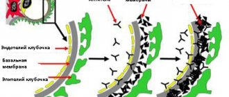
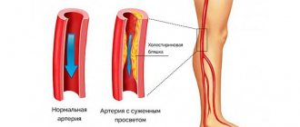
Vascular stenosis is a narrowing of blood vessels due to the deposition of cholesterol formations on their walls - plaques. This is a significant loss for the body, since such deposits prevent the free flow of blood through the vessels, as a result of which many components of the human body do not receive enough oxygen and various nutrients. Often this condition of the blood vessels is associated with impaired vasodilation functions and is dangerous because atherosclerotic plaques over time can completely block the blood supply channel to the body. In addition, the very process of accumulation of plaques is dangerous, which can at some point break off and become a blood clot, threatening blockage of a heart vessel with the possibility of an ischemic stroke.
Many people have little understanding of what vascular stenosis is and the risk of serious consequences from this disease. In the presence of this disease, timely diagnosis becomes very important, because the initial stage for the most part occurs without any noticeable symptoms, therefore it is extremely difficult to identify it at the initial stage. When symptoms begin to appear and become obvious, the risk of ischemic stroke increases many times over.
Thus, speaking about what vascular stenosis is, it should be summarized: this is a pathological condition of the arteries when the blood flow paths are narrowed due to the accumulation of cholesterol plaques in a certain place, narrowing this passage and threatening to block it altogether in the future. Small fragments of plaque can break off and, carried by the blood flow, close one of the smaller vessels.
Causes
Stenosis can be caused by various factors that a person inherits from his parents or acquires during life. The main reasons that lead to narrowing of organs and blood vessels are:
- Congenital anomalies. Caused by pathologies of intrauterine development of the fetus and genetic defects of the parents.
- Thickening of the walls. Infections, toxins and other factors lead to metabolic disorders in humans. As a result, inflammatory processes occur, fibrous fibers grow, cholesterol is deposited, which negatively affects the width of the internal lumen of organs.
- Spasms. In normal condition, the organs and tissues of the human body consist of elastic fibers. Stress, chronic tension, and some diseases lead to a sharp reduction in them, often accompanied by pain. Over time, this causes hypertrophy of the walls of blood vessels and smooth muscles of the organs.
- Pathologies of neighboring organs. Inflammation, trauma, swelling, neoplasms (benign or malignant) in a particular area of the human body can have a compressive effect on adjacent tissues.
- Natural aging of the body. There is a gradual wear and tear of systems and tissues, and destructive changes in their functions.
Causes of the disease
This disease is very insidious, because the absence of symptoms does not allow a timely response to this dangerous prospect. In addition, the symptoms of vascular stenosis are different for different types of disease, and they will be discussed below, with the characteristics of individual types of disease. Most often, stenosis occurs due to the following reasons:
- diabetes;
- atherosclerosis;
- excessive obesity;
- arterial hypertension;
- smoking;
- eating fatty foods, etc.
Types of stenosis
The types of stenosis depend on the location of cholesterol deposits. Most often, poor vascular patency is found in the aorta, veins, arteries - carotid, mesenteric (celiac) and femoral. To understand what vascular stenosis is, it is necessary to characterize each individual type of this disease. Treatment of vascular stenosis is carried out in most cases with medication, as well as by alternating exercise and rest. an anti-cholesterol diet is also prescribed. In particularly difficult cases, surgical treatment is performed.
Classification
Aortic stenosis is classified according to several criteria. Previously, we have already considered one of them - due to its occurrence. According to this classification, aortic stenosis can be congenital or acquired.
At the location of the narrowing
Aortic stenosis is divided according to the localization of the pathological process. It happens:
- Supravalvular.
- Valve.
- Subvalvular.
Valvular localization of AC stenosis is most common. The supravalvular or subvalvular type of this defect occurs quite rarely and is usually congenital.
According to the degree of circulatory disturbance
This classification distinguishes between compensated and decompensated (critical) aortic stenosis.
There are also four degrees of aortic stenosis according to the degree of its severity: from moderate to critical stenosis. The evaluation criteria are: the systolic pressure gradient between the left ventricle and the aorta and the area of the aortic passage.
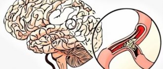
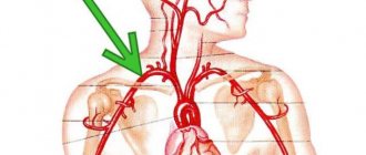
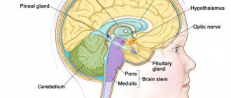
Stenosis of head and neck vessels
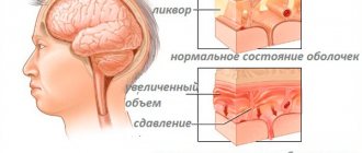
Since the blood supply to the brain occurs through vessels located in the neck, stenosis of the neck vessels should be considered in conjunction with the vessels of the brain. The vessels that are located in the head and neck provide access to the brain of oxygen and nutrients, without which it cannot exist. Without access to oxygen, this organ can survive without pathological changes for no longer than 3-4 minutes, after which the death of nerve endings begins.
Stenosis of cerebral vessels manifests itself in this way: blood circulation in any part of the brain is disrupted. If stenosis develops slowly, cerebrovascular insufficiency is observed, and the severity of the disease is characterized by the degree of damage to the vessel. The main signs indicating stenosis of cerebral vessels are motor dysfunction, memory impairment and negative emotional manifestations. Another form of the disease is possible - cerebral infarction.
The first symptoms of cerebral vascular stenosis appear in the following reactions of the body:
- periodic severe headaches;
- fatigue;
- noticeable distraction;
- extreme irritability;
- frequent changes of mood.
At the first stage, the elasticity of blood vessels decreases, signs of accumulation of cholesterol cells appear and blood flow slightly deteriorates. At the second stage, symptoms intensify, and more serious changes in the patient’s behavior begin to appear:
- noise in ears;
- noticeable memory problems;
- debilitating migraines;
- short fainting spells.
At this time, your gait may change, and there will be a constant urge to urinate frequently. Possible problems with vision. If intensive treatment of cerebral vascular stenosis is not started at this stage, then the onset of a severe third stage will not be far off, when motor functions are already seriously impaired, and it is difficult for a person to maintain balance, and each action needs to be thought over for a long time. During this period, rudimentary signs of dementia begin to appear, and uncontrolled bowel movements and urination become possible. In addition, the emotional state deteriorates significantly, to the point of losing control over one’s actions.
In this case, stenosis of the neck vessels is inextricably linked with brain disease, since the cervical arteries and veins completely provide the brain with blood supply. The neck veins are very rarely affected by plaques; for the most part, the carotid artery and cerebral vessels are affected. The symptoms of the disease are similar to those that appear with brain disease. After all, damage to the vessels of the neck is fraught with impaired blood circulation in the blood vessels of the brain. Sometimes a person does not even suspect that the vessels of the carotid artery are clogged and become blocked until a stroke occurs. The primary signs of narrowing of the walls of blood vessels include flickering of flies and darkening in the eyes, tinnitus, dizziness and weakness in the limbs.
Symptoms and signs of cerebral vascular stenosis
Cerebral artery stenosis often develops in stages, that is, the disease occurs in a chronic form. The pathological process is characterized by a diffuse form of damage to brain cells, therefore clinical manifestations are only general in the brain.
In total, there are 3 stages of progression of stenosis, but in some sources there is a zero stage, in which the disease proceeds latently. Since symptoms are completely absent at the onset of the pathology, this presents difficulties for timely diagnosis.
Clinical manifestations depending on the stage:
- The symptoms already clearly indicate pathological changes in the brain, but the suffering person, as a rule, does not pay attention to them. Headaches are present, but the patient cannot indicate the exact location; more often it is diffuse and paroxysmal. The pain is similar to a migraine and goes away on its own within 30–60 minutes (not relieved by analgesics). There are attacks of dizziness (spontaneously) during which it is difficult to maintain coordination; the symptom goes away after forced rest. The patient's general condition is similar to severe fatigue, but relatives note that the emotional background has changed dramatically - mood swings, irritability. The patient explains this by insomnia - a sign of stage 1, a person cannot sleep for a long time or often wakes up, after which he feels exhausted in the morning;
- Clinical manifestations of stenosis are increasing, and obvious impairments in the intellectual sphere (thinking, logic) are noted. The patient rapidly loses the skill of retaining information (short-term memory), and the behavior resembles a deviant form (inappropriate emotions arise in a certain situation, for example, laughing at grief). Headaches increase in intensity and are accompanied by vomiting, which does not bring a feeling of relief. Spontaneous fainting and convulsive attacks occur (can be combined with epileptic seizures). Coordination of movements is severely impaired, the person cannot control his body (tremor, “shaky” gait, spontaneous inappropriate movements). Dysfunction of the urinary system is noted (innervation is impaired);
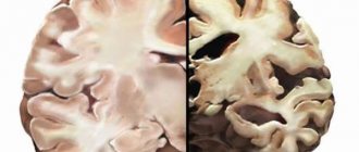
- The brain contains extensive multifocal areas of necrosis. There is no chance of restoring full functionality of the central nervous system; rehabilitation helps partially and is mainly aimed at improving the quality of life. A sick person has no emotions, the world around him is not interesting. The brain does not control many physiological processes, which is why enuresis and involuntary bowel movements occur. The patient does not recognize relatives; there is total amnesia, which is irreversible.
Each patient is individual and the symptoms may manifest themselves in a completely different chronology, but at the same time all of them have signs of vascular dementia.
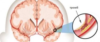
The progression of stenosis inevitably leads to an acute condition - cerebral stroke. An abrupt cessation of blood supply to the brain is manifested by sudden symptoms that rapidly increase - headache, dizziness, vomiting, fatigue, tachycardia.
In addition to general cerebral manifestations, stroke is also characterized by additional symptoms - focal. Manifestations will depend on the location of the stenosis, that is, depending on which artery the spasm occurred in, a certain area of the brain will suffer (paralysis or paresthesia of one part of the body, blindness in one eye).
Treatment and diagnosis
Before starting treatment for cerebral vascular stenosis, it is necessary to conduct a high-quality diagnosis. This can be provided by a neurologist after checking the reflexes and assessing the degree of adequacy of the patient. The most informative tools for studying the condition of the vessels of the neck and brain are the following methods:
- ECG;
- CT scan;
- dopplerography;
- computed angiography.
For treatment, conservative therapy is used, in which the doctor prescribes drugs such as statins, lecithin, fibrates, papaverine or aminophylline, as well as vitamins and minerals. In this case, you should follow a special diet that excludes both fatty foods and salty, smoked, and caffeine foods. The diet should be dominated by seafood, vegetables, fresh herbs, and fruits. You can additionally engage in special exercises, but this can only be done after consulting a doctor, because the risk of a blood clot breaking off is too high if there are contraindications. When treating the most advanced forms of stenosis, it is recommended to perform bypass surgery or even completely replace a vessel blocked by cholesterol plaques.
Complications
If timely treatment of lumbar, cervical or thoracic spinal canal stenosis is not started, pathological manifestations of this disease are possible. Complications are also observed if there have been other spinal injuries (sports injury, fall from a height, etc.). In this case, increased compression of the spinal cord occurs. The resulting hematomas, scars, fragments of the spine, and displaced vertebrae can put pressure on it.
The most severe complications are:
- paresis;
- paralysis of limbs;
- pelvic disorders that manifest themselves due to damage to the nerve roots of the spinal cord with a surgical instrument;
- a slowly and gradually progressing adhesive process, which additionally compresses the roots and the spinal cord itself.
The most rare inflammatory processes occur in nerve elements, membranes and vertebrae. This is due to the fact that after surgery for lumbar spinal stenosis, strong antibiotics are used. But it is still not uncommon that the consequences after surgery of spinal stenosis of the lumbar, cervical and thoracic region give more severe consequences than the disease itself.
Cardiac stenosis
There is a large group of coronary vessels responsible for supplying blood to the heart. They are also at great risk of narrowing the passage with the help of plaques and can be affected by atherosclerosis. Such blocking of the arteries develops coronary artery disease, and with the gradual accumulation of plaques, chronic heart failure appears. Its symptoms include:
- the appearance of shortness of breath at the slightest physical movement;
- heartache;
- severe swelling in the legs;
- abnormal heart rhythm.
In the future, stenosis of the heart vessels leads to blockage of the heart artery by a fragment torn off from the accumulation of cholesterol plaques. In this case, the patient will complain of sharp pain in the heart, radiating to the left shoulder blade, as well as to the arm and jaw. Severe shortness of breath and great weakness appear, the patient is seized with a panicky fear of death. With such a development of the disease, diagnosis should be carried out immediately. To identify the pathology of the heart vessels, ECG, ultrasound of the heart, computed tomography, Doppler ultrasound and vascular angiography, as well as magnetic resonance angiography are performed. When issuing a referral for a study, the doctor himself decides which of these types of diagnostics will be carried out in each individual case in order to facilitate the diagnosis.
Treatment for this type of stenosis
Treatment of cardiac stenosis is prescribed by one of the medical specialists - a neurologist, cardiologist, or surgeon. When treating heart vessels, the following types of surgery are used:
- endarterectomy - removal of a blood clot that causes a narrowing of the vessel;
- stenting - installation of a special stent inside a vessel that expands the walls, as a result of which blood supply improves and the heart can function normally;
- bypass - laying a new channel to supply blood to the heart, bypassing the problem area.
In addition to basic treatment, it is necessary to reconsider the lifestyle that led to problems with the heart vessels.
Treatment
Self-medication for stenosis leads to complications that can include irreversible processes in the patient’s body, which can lead to death. It is necessary to identify the disease in time, because in the first stages it can be cured with medication. If the narrowing of organs and blood vessels is determined in the initial stages, the following methods are applicable for the treatment of children and adults:
- prescription of medications;
- physiotherapy;
- physical therapy complex (physical therapy complex)
- traditional methods of treatment;
- diet therapy.
At stages 2-3 of this disease, surgical intervention may be necessary to correct the pathology. Degenerative stenosis (developing against the background of other diseases) requires special attention. There are highly targeted methods for treating strictures:
- Spinal traction and compression. It is performed under the supervision of a doctor using a special device. If the procedure is performed incorrectly, there is a risk of additional complications.
- Massage. The muscles relax, pain decreases due to stricture of the spinal canals and blood vessels. Particularly effective for eliminating stenosis in children.
- Removal of a blood clot (endarterectomy). A surgical method for releasing the lumen of blood vessels.
- Restoration of electrolyte-water balance. Used to expand the walls of the stomach.
Conservative therapy
Methods of conservative therapy depend on the location and degree of stenosis. In drug treatment, common to all types of strictures is the prescription of drugs with the following properties:
- Anti-inflammatory. Any congestion that causes a narrowing of organs and blood vessels inevitably leads to inflammatory processes. Relieving them until the root cause is cured is the main task of such medicines.
- Painkillers. These drugs are prescribed to patients who suffer from stricture pain.
- Decongestants. The doctor prescribes this type of medicine to the patient to support normal functioning. Such drugs are especially relevant for spinal stenosis.
- Drugs to enhance neuromuscular conduction. All strictures, due to their negative impact on the blood supply to organ systems, lead to atrophy of muscles and nerve endings. Medicines in this group interfere with this process.
- Multivitamin complexes. They are prescribed to patients weakened by the disease.
Conservative therapy for the treatment of vascular stenosis is based on drugs that thin the blood and prevent further increase in plaques:
- Cardiomagnyl. An antiplatelet drug that combines a combination of acetylsalicylic acid and magnesium hydroxide to prevent thrombosis, has an anti-inflammatory and analgesic effect. Reduces the risk of myocardial infarction in patients with complications of cardiovascular diseases. It is a prophylactic against re-formation of blood clots for patients who have suffered a stroke.
Among its advantages is the optimal amount of acetylsalicylic acid recommended by the European Heart Association. The magnesium hydroxide included in the composition protects the gastric lining from irritation by aspirin acid. Among the disadvantages is the ability to block only one mechanism of platelet formation, associated with the synthesis of cyclooxygenase.
- Warfarin. Indirect anticoagulant. Suppresses the effect of vitamin K, which promotes the production of blood clotting factors. As a result, the blood thins out. Has a long half-life and is taken once daily. Prescribed to persons prone to thrombosis in the coronary arteries. The maximum effect from taking the drug is observed 3-5 days from the start of administration and lasts about 5 days after the end of the course.
Among the advantages, the drug can be used in complex therapy with antiplatelet drugs, because it has a different mechanism of action. Among the disadvantages is that it interacts with many substances and even products, which worsens its anticoagulant properties. When interacting with certain medications (Cimetidine, Chloramphenicol, etc.), it increases the risk of bleeding.
Surgery
Surgical intervention is resorted to when non-surgical methods of treating strictures are ineffective. In medicine, the following methods of surgical correction of the lumens of blood vessels and organs are used:
- Stenting – compression of the plaque (neoplasm). First, an invasive examination is carried out - the introduction of a special catheter with a stent (cylindrical frame) to the site of pathology. The vessel or organ is expanded by pressing the plaque/neoplasm against its wall. The artificial structure remains in the vessel (organ), promoting free blood flow.
- Bypass is an operation that involves opening the skull. Thanks to complex surgical manipulation, intracranial vessels are redirected to bypass the abnormal formation.
- Vascular plastic surgery. If the subclavian artery is occluded, it is connected to the carotid artery. Sometimes part of the affected vessel is removed and prosthetics are performed (a synthetic implant is used). A similar method is also used to increase the lumen of hollow organs.
- Bypass surgery is the creation of a new path (shunt) for blood flow bypassing the affected area (blockage).
- Endarterectomy – removal of a blood clot. Correction of the affected area by resection.
Disease of the lower extremities. Causes
Such a serious disease as vascular stenosis of the lower extremities is one of the most common diseases of the cardiovascular system. It is accompanied by pathological narrowing of the arteries located in the area of the lower extremities. With this pathology, the arteries begin to narrow and pass blood worse, which significantly depletes the oxygen saturation of tissues and leads to ischemia. In men, vascular stenosis of the extremities occurs approximately twice as often as in women, and with age the risk of blockage of blood vessels increases significantly. Why such discrimination based on gender? Because men are much more likely to abuse alcohol and smoking, which are included in the list of provocateurs of limb stenosis.
Dangerous factors that can lead to the formation of plaques on the walls of blood vessels include:
- previous atherosclerosis;
- tobacco smoking, both active and passive;
- alcohol abuse;
- presence of diabetes mellitus;
- received injuries to the limbs;
- high degree of obesity;
- high blood pressure;
- hereditary predisposition;
- complications after infectious diseases.
Degrees of the disease
Stenosis is assessed according to the level of manifestation and complexity of symptoms. There are many types of strictures of hollow organs and blood vessels of the body. There are several degrees of the disease with varying severity of symptoms:
- The first stage may be asymptomatic. There are plaques on the walls of tubular structures, but they do not yet have any pathological effects on the organ systems:
- Gastrointestinal tract - belching, sour taste, less often vomiting;
- spinal canals – heaviness in the legs, fatigue;
- upper respiratory tract – shortness of breath.
- In the second stage, the symptoms are clearly distinguishable and the disease progresses. The patient's condition worsens, he turns to the doctor for help. Vasoconstriction can cause complex clinical deviations from the norm in the form of attacks:
- from the gastrointestinal tract - frequent vomiting, dehydration;
- cardiovascular system – coronary heart disease;
- brain – transient ischemic attack.
- Last stage. The lumen of tubular organs and vessels is minimal or completely blocked. Pathological changes affect entire organ systems. If serious treatment is not carried out, the patient may die.
Stenosis of the lower extremities. Stages of development of the disease and its symptoms
Stenosis of the lower extremities is asymptomatic at first. Over time, pain appears in the calf muscles, causing lameness, and a kind of compression is felt at the site of the arteries. With further development of the disease, the skin color changes, it becomes dry and rough, leg hair falls out and the nail plates become thicker. In advanced stages, ulcers and blackening of the skin may appear on the fingers. Further, in the absence of treatment, the subcutaneous fat layer begins to atrophy, and necrosis of muscles and tissues also appears, up to gangrene and amputation of the limbs. If the case is very advanced, then blood poisoning may result, followed by death.
For diagnosis, patients are prescribed:
- Doppler ultrasound;
- duplex ultrasound scanning;
- computed axial tomography;
- magnetic resonance angiography with contrast agent;
- advanced biochemical blood test.
Diagnostics
Examination of patients with suspected aortic valve stenosis is carried out under the supervision of a cardiologist.
Approximate list of events:
- Oral questioning of the patient regarding complaints and their duration.
- Collection of anamnestic data. The main role is given to previously suffered pathologies of a cardiac, nephrogenic and endocrine nature. Lifestyle is also taken into account. The more bad habits, the greater the likelihood of deviation.
- Blood pressure measurement. Indicators may be elevated or normal. Also heart rate. At the time of an angina attack - acceleration of activity.
- Daily monitoring. As needed.
- ECG. To assess functional activity. Shows arrhythmias.
- ECHO-KG. Used to determine organic defects. The pressure in the chambers and the aorta itself is measured using the same method.
- Ultrasound of the kidneys. To identify nephrological conditions.
- MRI diagnostics.
As part of an extended examination, a blood test (general, biochemical, hormonal) may be required.
Activities are carried out in both inpatient and outpatient settings.
Stenosis of the lower extremities. Treatment Options
Treatment in the early stages is carried out with medications - vasodilators and thrombus-dissolving drugs. In more complex cases, surgical intervention is used - dilation of the vessel with a special stent, as well as angioplasty or stenting.
Angioplasty is an advanced innovative technology that is performed without tissue incision or anesthesia. A catheter with a balloon is inserted into the artery, which, when it enters a damaged vessel, expands by inflating and can expand it to a passable size. A stent with a drug is inserted into the resulting lumen, which acts as a frame for the vessel. During cryoplasty, instead of air, the balloon is inflated with liquid nitrous oxide, which at ultra-low temperatures helps destroy atherosclerotic plaques.
Bypass surgery is done if there are contraindications to balloon angioplasty. During this operation, blood is directed through a shunt into a new direction that bypasses the site of vessel damage.