© Author: Z. Nelly Vladimirovna, doctor of the first qualification category, especially for SosudInfo.ru (about the authors)
A choroid plexus cyst is detected in the fetus, usually before 6-7 months of its development, since then it, as a rule, disappears safely and never reminds of itself again. But pregnant women, having received an ultrasound report, are worried and consider this a diagnosis, although this condition is not one. A cyst formed during intrauterine development in the choroid plexus does not pose any danger to the health and development of the baby. In addition, it should be distinguished from a cyst of vascular origin , which occurs in the brain as a consequence of certain pathological processes (stroke, aneurysm, infection).
What is a choroid plexus cyst and the mechanism of its formation
Vascular (choroid, choroid, villous) plexus cysts in the fetus occur with a frequency of approximately 1-3% of cases among all observed normal pregnancies. The cysts, half of which are bilateral, disappear around 28 weeks . Even if this does not happen, and the cyst continues to be visualized at a later stage until delivery, its significance will not increase from this.
It poses no danger to either the fetus, a newborn child, or an adult if it remains for life (an extremely rare occurrence). There may be more than one choroid plexus cyst; their number often varies, which also does not change the prognosis.
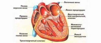
choroid plexus in the structure of the brain
A vascular cyst is an accumulation of cerebrospinal fluid (choroidal, cerebrospinal fluid) inside a plexus produced by it and intended to nourish the brain and spinal cord of a future person. The choroid plexuses themselves are considered one of the early signs of the formation of the central nervous system in the embryo, and the fact that there are two of them indicates the formation of the right and left hemispheres. Why liquid accumulates in certain places and what it all means is unknown to anyone, and there is no particular point in understanding this, since the accumulations do not play any role anyway. On ultrasound they very much resemble a cyst, therefore in conclusion they are designated as a cyst, which will not manifest itself in any way in the future.
Relationship with other pathologies of intrauterine development. Does she really exist?
In medical literary sources, you can find information that there is some connection between the presence of choroid plexus cysts and some congenital pathology caused by a genetic mutation. Moreover, the localization of the cystic cavity on the right, left or both sides at the same time has absolutely no significance. But here it is very important to remember that it is NOT a cyst that provokes developmental anomalies, but on the contrary - a violation of intrauterine development contributes to the formation of vascular cysts , therefore the connection between them is limited only by the presence or increase in the number of choroid plexus cysts and that’s all. Nothing more. Such genetic defects, when a cystic cavity is diagnosed more often than usual, include trisomy 18, known as Edwards syndrome (non-disjunction of the 18th pair and the addition of another chromosome 18 to it, so instead of the normal two, there are 3 of them, and the entire genotype of such an embryo is represented by 47 chromosomes). By the way, it is believed that trisomy 21 (Down's disease) has a much smaller effect on the increase in the incidence of choroid plexus cysts than trisomy 18.
In addition, it is important to note that in the case of other pathology, the primary role does not belong to the cyst, but to the deviations that it accompanies, for example, the same Edwards syndrome, so its significance here is reduced to zero.
Thus, a choroid plexus cyst, no matter right or left, is single or represented by several small formations:
- Equally safe;
- Doesn't play any role;
- Does not participate in any important processes;
- Incapable of growing and being reborn.
Pregnant women should know about it so as not to be afraid and not to be confused with other cystic formations that have similar names, but a completely different genesis and location.
Bilateral choroid plexus cysts on MRI images
Prevention
To prevent the development of choroid plexus cysts, doctors recommend that pregnancy planning be approached with particular seriousness. Regardless of the fact that the pathology is diagnosed in only 3 percent of cases, it is necessary not to expose the body to stress during the period of bearing a child, as well as to eat properly.
In addition, several months before conception, as well as during, doctors advise both parents to undergo a special course of vitamin therapy.
The actions of vitamin complexes are aimed at:
- reducing the likelihood of developing pathologies;
- strengthening the immune system;
- normalization of cerebral circulation.
Similar names and different origins
Vascular cysts detected at a later date than the choroid plexus cyst detected around the 20th week, or diagnosed by ultrasound of the newborn baby’s brain, may deserve attention during pregnancy. The appearance of cystic formations in this case may indicate a previous or existing infection in the mother, in particular, this most applies to the herpes virus and cytomegalovirus.
The later formation of vascular and ramolitic (localized in the substance of the brain) cysts is due to the fact that against the background of a viral infection, cystic cavities are formed in the presence of the brain itself. In addition, there is a high probability of infection of the child when passing through the birth canal of a mother infected with the virus. This explains the appearance of such cystic formations, quite often multiple and located mainly in the frontal and temporal regions, in a newborn baby. A brain cyst that originates from foci of necrosis is called ramolitic. In this case, necrosis of the nervous tissue occurs due to damage by the herpes virus or CMV.
Diagnostic methods
The most effective method for diagnosing formations in the head is computed tomography and magnetic resonance imaging. With their help, it is possible to see the clear outlines of the tumor, assess its size and the degree of impact on nearby tissues. During an MRI, the patient is injected with a special contrast that helps determine the nature of the formation (benign or malignant). To track the dynamics of tumor development, MRI is performed several times.
To determine the cause of cysts in the brain, additional examinations are prescribed:
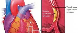
- dopplerometry. Helps determine if there is a narrowing of the vessels delivering arterial blood to the head. Impaired blood circulation is one of the reasons for the occurrence of foci of death of brain cells, which provokes the formation of cysts;
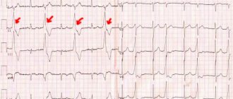
- ECG. Carried out to exclude heart failure;
- blood test for cholesterol and coagulability. It is the increased level of cholesterol and excessive coagulation that can cause blockage of blood vessels and cause the formation of cystic capsules in the head;
- measurement of blood pressure indicators. This is done with a special tonometer; the values are recorded throughout the day and processed by a computer. If a patient experiences periodic pressure surges, this can cause strokes and the subsequent formation of post-stroke cysts;
- a blood test that helps determine the presence of infections and autoimmune diseases. It is carried out if arachnoiditis, multiple sclerosis or the presence of neuroinfections is suspected.
Brain cyst, true form, causes and prognosis
The prognosis of cystic formations in a child depends on the cause, location and size of the cyst, therefore such children undergo PCR diagnostics to determine the virus, and if it is present, the necessary treatment and further observation, which includes mandatory ultrasound examination of the brain (neurosonography) at 3 months, six months and a year of the baby’s life. In most cases, a choroid plexus cyst detected in a newborn, even in the presence of a virus, has a favorable prognosis , disappears by the age of one year and does not resume its development in the future.
A ramolitic cyst can also end its existence in infancy, or it can behave like a formation that arises for other reasons and is called a vascular cyst with a stretch, since its genesis is a violation of the vascular wall, but it itself is localized in the brain tissue (after the fact) .
Thus, the reasons for the formation of a pathological brain cyst may be as follows:
- Infections;
- Birth and other injuries;
- Microstroke;
- Hemorrhagic stroke (a cyst occurs at the site of a hematoma formed as a result of vascular damage);
- Ischemic stroke (tissue necrosis will give rise to a remolition cyst of vascular origin)
- Aneurysm.
It should be noted that if we are talking about damage to the vascular wall, then in this case we mean the arterial wall, since veins, as a rule, do not participate in such processes.
What to remember
- The symptoms of a cyst directly depend on its location and size.
- The main diagnostic methods are tomography, MRI and ultrasound of the brain.
- A brain cyst is a serious diagnosis that requires medical treatment and regular medical supervision.
- Consequences of cysts include hydrocephalus, brain herniation, and sudden death.
- Drug treatment eliminates the causes of the appearance and growth of the cyst.
- Surgical treatment involves removal of the cyst and is carried out only as prescribed by the attending physician.
See you in the next article!
Site headings “Headache”
Eye headache Hypertension Hypotension How to lower blood pressure Diagnosis of headache Stroke Causes, signs and symptoms Pills, medications Ear headache
Possible symptoms and treatment
A brain cyst resulting from a hematoma, stroke, or aneurysm is one of the options for the outcome, which is generally favorable and is sometimes discovered only posthumously, however, along with a cyst formed as a result of a viral infection, it can sometimes have undesirable consequences, manifested by:
- Signs of hypertension in newborns;
- Feeling of compression of the brain;
- Some visual and/or hearing impairments;
- Minor motor coordination disorders;
- Epileptic seizures, which, of course, can be considered the most serious complication.
Clinical manifestations indicating the presence of a cystic formation occur in cases where the cyst compresses neighboring tissues and interferes with their normal functioning, that is, if it is of significant size or “settled” in unacceptable proximity to important centers of higher nervous activity.
In most cases, a brain cyst, like a choroid plexus cyst, does not require special treatment, however, if immunological studies have proven the presence of herpetic, cytomegalovirus or other infection, then treatment directed at the virus is indicated. In the presence of epileptic seizures, the patient is prescribed anticonvulsants, and, if necessary, surgical intervention is performed to eliminate the source.
If the symptoms are mild, but the patient occasionally complains of vascular manifestations of the cyst (dizziness, compression headaches, etc.), he is prescribed medications such as cinnarizine or Cavinton , which are well tolerated by the patient, improve blood supply to the brain and help normalize well-being.
Features of the cyst
An epiphysis cyst is a neoplasm of a non-tumor nature, located on one of the lobes of the organ.
As a rule, its size is small, and there is no tendency to rapidly increase. Morphologically, such a formation is a small cavity that is filled with a specific liquid.
This formation of a benign nature usually does not affect the functioning of the pineal gland and extremely rarely affects the functioning of neighboring organs of the brain.
Important! In the initial stages, the disease is asymptomatic and most often can only manifest itself as a feeling of pressure in the affected area, which is accompanied by mild pain. Timely diagnosis allows you to avoid an increase in formation and further complications and manifestations
Timely diagnosis allows you to avoid an increase in formation and further complications and manifestations.
Cyst sizes
Small neoplasms, which are more common than others, do not exceed 5 mm in size.
They are not accompanied by special symptoms and do not provoke complications.
These cysts are mostly detected incidentally on MRI and respond well to treatment with medications.
As a rule, cystic neoplasms do not require special treatment. With this diagnosis, in most cases, only systematic monitoring is necessary.
However, some people with such a cyst may experience rapid growth and, as a result, various somatic and neurological disorders.