A pseudocyst is a small, non-malignant formation in the form of a capsule, inside of which there is cerebrospinal fluid (CSF), which forms in the baby’s brain structures in utero or during the prenatal and birth periods. Doctors diagnose similar benign structures in the head not only in newborn infants, but also in adult men and women. Another medical term for this abnormality is subependymal cyst.
Peculiarities:
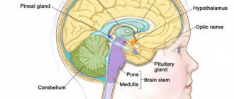
- The pseudocyst is localized only in the lateral ventricles of the brain. The subependymal cyst is separated from the ventricles only by the ependyma (membrane) and a thin layer of medulla containing matrix tissue (germ layer).
- A pseudocyst can form on the left or right (in the right or left ventricle of the brain). Bilateral lesions also occur.
- The diameter of the formations does not exceed the limits from 1 – 2 to 7 – 9 mm.
- They can be either single or multiple.
- The disease is detected in approximately 5 newborn babies out of 100.
- A pseudocyst in the head of a newborn child does not pose a threat to life, physical or mental health. Often the cystic fraction resolves on its own before birth or in the first 12 months of life.
- This pathology requires constant monitoring of the child and periodic ultrasound examinations until complete disappearance.
A similar pseudotumor also includes a pseudocyst of the choroid plexus in a newborn, which occurs even in the embryo at the stage of active brain formation, when cerebrospinal fluid penetrates into the voids of the intertwining vessels. Experts detect this deviation using ultrasound at 16–21 weeks of gestation.
What is the difference between a cyst and a pseudocyst?
A pseudocyst in a baby's head must be differentiated from a true cyst. The main indicator is the characteristic location. A false anomaly is formed either in the area of the bodies and lateral angles of the anterior horns of the right or left ventricle of the brain, or at the border of the thalamus (optic thalamus) and the head of the caudate nucleus.
The remaining cavity, fluid formations, if detected in other parts of the brain, are called cysts. False cysts resolve or significantly decrease in size by one year of age. True cysts in the head do not disappear, they can cause painful neurological symptoms and require constant monitoring by a neurologist.
Causes of subependymal pseudocyst in the head of a baby
Why does a pseudocyst develop in the head of a newborn baby? The mechanism that triggers the development of the anomaly in children is not reliably known and has not been fully studied.
The most likely causes of a pseudocyst in the head are:
- disruption of the process of formation of the germinal matrix - embryonic tissue in the embryo, where fragile vessels are concentrated and intense metabolic processes occur;
- lack of oxygen supply to the fetal brain due to impaired blood supply (ischemia) before or during childbirth. This condition can occur due to disruption of fetoplacental blood flow (between mother and fetus), early placental abruption, umbilical cord entanglement;
- brain injury during childbirth, hemorrhage in the cerebral tissue of a newborn child;
- damage to the fetus or newborn infected with infections from the mother (herpes, papillomavirus, sexually transmitted diseases);
- multiple births, preeclampsia, blood incompatibility according to the Rh factor;
- serious pathologies, infections during pregnancy, acute poisoning, difficult experiences during pregnancy;
- hereditary genetic factors.
An anomaly such as a pseudotumor is not considered as an independent disease, but is considered a consequence of pathological conditions in a woman or fetus.
Prevention and prognosis
Responsibility for the full intrauterine development and growth of the baby, of course, lies with the expectant mother. In order to minimize the risk of the formation of a subependymal neoplasm in newborns, a woman is recommended to take care of the health of her unborn child even at the stage of pregnancy planning.
Measures to prevent brain cysts:
- diagnostics for genetic predisposition to tumors;
- preventive vaccinations against all possible infections, including hidden and sexually transmitted ones;
- elimination of chronic pathologies that can worsen during pregnancy - dental caries, pyelonephritis, gastritis, hepatitis.
Immediately after confirmation of the birth of a new life inside a woman, she should be even more careful about her health in order to eliminate negative effects on the fetus. To do this you need:
- strive for a healthy lifestyle - give up the abuse of tobacco and alcohol products;
- eat right - exclude fatty, heavy foods, preservatives and stabilizers; the diet should be dominated by vegetables, various fruits, cereals, soups, salads;
- avoid contact with infectious patients, especially during seasonal diseases; if this is not possible, protect yourself with special antiviral agents approved during pregnancy;
- avoid contact with toxic substances, avoid taking medications that are dangerous for the baby;
- get enough sleep;
- avoid stress.
Subependymal cysts in newborns are not a death sentence at all. This is just a test on his long life path, and it is the parents’ task to cope with it. In close cooperation with the doctor and following all his recommendations, children will grow and develop according to age norms.
Symptoms of a pseudocyst in the head of a newborn
Certain conclusions about the type of ventricular cystic formation and prognosis are made only based on the results of diagnostic studies and clinical examination by a pediatric neurologist.
If a subependymal pseudocyst in a newborn is up to 4–5 mm in size, found in a typical location, and is isolated (not accompanied by neurogenic manifestations, not associated with other diseases), then the prognosis for the baby is favorable. A pseudocyst in the head does not aggravate the disease with abnormalities in mental development. In the fetus, a subependymal pseudocystic neoplasm often resolves by 35–40 weeks, and if this does not happen, then up to 9–12 months of the child’s life.
Dynamic monitoring of the behavior of the pseudocyst and the development of the infant is mandatory.
Can a newborn baby have any symptoms of a pseudocyst in the head? False ventricular formations themselves are a sign of trauma, hypoxia (oxygen deficiency) during childbirth or during “intrauterine life” and indicate that the child’s brain tissue is damaged. Such infants often cry for no apparent reason, spit up, worry and sleep worse; they may experience trembling of the chin, fingers, and a slight and temporary (within normal) developmental delay.
Main reasons
The main reason for the appearance of a neoplasm is considered to be fetal hypoxia - oxygen deficiency. Therefore, after confirming this diagnosis, the pregnant woman should undergo appropriate treatment. Subependymal cysts in newborns are formed under the influence of the following factors:
- Cerebral ischemia. Pathology contributes to the appearance of negative changes in the supply of certain areas of the brain with blood. As a result, empty cavities are formed, which fill with liquid over time. For small cysts, specific therapy is not required. In the case of vomiting, convulsions and inhibition of development, it can be assumed that the neoplasm is increasing in size. In such a situation, it is advisable to seek medical help.
- Brain hemorrhages. They are especially dangerous during intrauterine development. Hemorrhage may be preceded by infection of the fetus, birth trauma, or acute hypoxia.
- Against the background of acute or moderate oxygen deficiency, the functioning of the fetal body is disrupted. Hypoxia usually develops with anemia, toxicosis or Rh conflict.
Factors that provoke the disease include drug or alcohol dependence of the pregnant woman, frequent stress and exposure to ionizing radiation.
Diagnosis of subependymal brain cyst
Only hardware diagnostics can confirm the presence of a pseudocyst in a baby’s head. Ultrasound examination (neurosonography) is considered an accurate, reliable way to detect abnormal tumors.
An ultrasound of the brain is informative until the baby’s fontanel is overgrown (up to one year). The method is indicated for premature babies, those who have suffered oxygen deprivation, or birth trauma.
Using neurosonography, the doctor determines the size, number and localization of cystic compactions, assesses the degree of expansion of the ventricles of the brain (which may indicate increased intracranial pressure and the risk of hydrocephalus), identifies signs of blockage of the liquor pathways and impaired outflow of cerebrospinal fluid, and studies changes in the echostructure of brain tissue.
During a second ultrasound, the specialist compares the results and sees how the pseudocyst shrinks and gradually disappears. If echo signs of a true cyst are detected in a newborn child, ultrasound will allow timely treatment to begin in order to prevent complications as early as possible.
In addition to ultrasound, for the purpose of detailed study of pathology, the following can be performed:
- Doppler encephalography to evaluate the state of blood flow and cerebral vessels.
- Two-photon or positron emission tomography. Allows you to monitor the effectiveness of prescribed conservative treatment in accordance with changes in brain tissue.
- Cerebral scintigraphy.
- Computed and magnetic resonance imaging (CT and MRI).
These tests are often performed on older children. These methods make it possible to distinguish (differentiate) pseudocyst, tumor and brain cyst, which is very important for the development of correct treatment.
If a neonatologist or neurologist prescribes an instrumental examination for a child, then refusing it to parents is irresponsible and dangerous. Early detection of any abnormal formations in cerebral tissues allows one to avoid serious consequences.
Location of the cyst and its symptoms
Cysts in newborns of the subependymal type occur in places where tissue has died and affect both the left and right sides of the brain. According to observations and studies, they are most often localized in the ventricular region. The symptoms that arise as a result of the appearance of a cyst completely depend on its location and size. This is due to the fact that each part of the brain is responsible for a specific function.
Thus, the occurrence of pathology in the back of the head or a subependymal cyst on the left affects visual perception. The infant's visual acuity may be significantly reduced, and objects will appear double. Pathology arising in the temporal lobe of the brain affects the quality of hearing; pathology formed in the cerebellum affects the musculoskeletal system and disrupts gait. The resulting cyst in the pituitary gland leads to serious hormonal disruptions, which leads to disruption of the functioning of the entire body.
The main symptoms indicating the presence of a formation in the brain may be:
- poor sleep;
- a constant state of anxiety in the child;
- refusal of breast milk;
- difficulty gaining or even losing weight;
- constant crying of children with pathology, often without reason;
- abnormal muscle tone;
- trembling of the limbs and chin, nervous twitching;
- hearing problems;
- decreased visual acuity, in severe cases complete blindness;
- excessive regurgitation after feeding;
- frequent fainting, in rare cases, baby coma;
- change in the fontanel, its swelling or visible pulsation;
- whole body spasms.
If timely treatment is not carried out and education continues to increase in volume, the result will be that the child will lag behind mentally and physically.
Treatment methods for pseudocysts
Are there ways to get rid of a pseudocyst in the head through therapeutic treatment without surgery? Yes, if this is truly a false formation, then it will resolve on its own. But medications will help improve brain function, strengthen the immune system, relieve increased excitability or stimulate the activity of the nervous system.
Conservative therapy
If a neurologist, based on the diagnostic results, has determined the presence of a pseudocyst in the newborn’s head, the doctor prescribes medications that eliminate the causes that led to the appearance of the anomaly. Similar conservative treatment is prescribed if a true cyst is suspected in the initial stage of growth.
Many of the pharmaceuticals, especially in tablets and injections, are contraindicated or limited for use in infancy, so it is unacceptable to prescribe them to a child on your own. Only a pediatric neurologist will determine whether the baby needs this or that drug and will calculate the safe dose and frequency of use, taking into account his age, the severity of neurogenic manifestations and concomitant diseases.
If a specialist notices that a child has certain signs of neurogenic damage, he can prescribe nootropic medications that improve cerebral circulation and drugs that eliminate the consequences of oxygen starvation (hypoxia) experienced by the child. But only if they are really indicated for a small patient, since many effective medications have serious side effects, cause acute allergic reactions or give complications to the nervous system and hematopoiesis.
Pseudocysts in adults
A pseudocyst in an adult patient is most often diagnosed in the pancreas, causing severe pain. Such a node threatens the life of the patient, as it is prone to suppuration, perforation, malignant degeneration, and the formation of fistulas. With this disease, competent surgical treatment is necessary.
Pseudocysts in the adrenal glands do not initially cause symptoms, but when they enlarge, they cause various manifestations due to compression of adjacent organs. Such neoplasms provoke an abnormal release of adrenaline and are complicated by acute bleeding into the lumen of the capsule. Urinary pseudocyst or urinoma can occur in retroperitoneal or perinephric tissue. When they grow, such structures require surgical removal.
The concept of pseudocyst also appears in the diagnoses of men and women with lesions of toxoplasma and cryptococcal infections, when massive intracellular foci with a concentration of parasitic microorganisms form in the brain and other organs. But most often in such cases the term “pseudocyst” is used.