Brain atrophy is a term that refers to a decrease in brain volume. But this is only a radiological finding, which in itself means nothing. It is assessed exclusively in combination with clinical manifestations. Brain atrophy (ICD-10 code – G31.1) is manifested by reduction of the brain in relation to the skull, widening of the notches between the gyri and often also an increase in the volume of the cerebral ventricles. Most often, this condition is caused by old age, poor blood supply to the brain, and metabolic disorders. Symptoms can vary. Sometimes there are no signs of atrophy at all.
Brain changes caused by aging (senile atrophy)
On average, the brain weighs 1300 g in women and 1400 g in men. It reaches its maximum at the age of 30-40 years. In the subsequent period, its slow initial decrease begins. Acceleration of atrophic changes in the brain begins after 60 years of age, and in women somewhat earlier. But there are significant individual deviations from the average. They are caused by genetic influences and the development of atherosclerosis. By the age of 70, the volume of the brain exceeds 90% of the volume of the cranial cavity, then gradually decreases to 80%. The macroscopic manifestation of atrophy is a slight narrowing of the gyri and deepening of the grooves. A decrease in white matter volume leads to ventricular dilatation; the soft shell is macroscopically hardened by a moderate increase in ligaments.
With atrophy in old age, a decrease in dendrites, proliferation of lipofuscin in the cytoplasm of neurons, and a general slight death of neurons are microscopically revealed. Amyloid deposits appear in the walls of small vessels and perivascularly. Degenerative changes appear in the gray matter.
These data are not pathognomonic for age-related atrophy. They also occur in other conditions belonging to the group of degenerative diseases of the central nervous system.
Why does the brain atrophy?
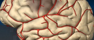
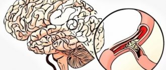
- Atherosclerotic lesion of cerebral vessels. The death of brain cells begins when atherosclerotic deposits, causing a narrowing of the lumen of blood vessels, cause a decrease in the trophism of neurons, and subsequently, as the disease progresses, their death. The process is disseminated. Brain atrophy caused by atherosclerotic vascular damage is one of the special cases of ischemic atrophy.
- Chronic intoxication effects. The death of nerve cells in the brain in this form of the disease is caused by the harmful effects of toxic substances on them. Alcohol, drugs, some pharmaceuticals, and nicotine can have a similar effect. The most striking examples of this group of diseases can be considered alcoholic and drug encephalopathy, when atrophic changes in the brain are represented by a smoothing of the relief of the convolutions and a decrease in the thickness of the cerebral cortex, as well as subcortical formations.
- Residual phenomena of traumatic brain injuries. Hypotrophy and atrophy of the brain as a long-term consequence of head trauma are, as a rule, local in nature. The death of nerve cells occurs in the damaged area of the brain; in their place, cystic formations, glial foci or scars subsequently form. This atrophy is called post-traumatic.
- Chronic cerebrovascular insufficiency. The most common causes of this condition are the atherosclerotic process, which reduces the patency of cerebral vessels; arterial hypertension and age-related decrease in the elasticity of blood vessels of the cerebral capillary bed.
- Degenerative diseases of nervous tissue. These include Parkinson's disease, Alzheimer's disease, Pick's disease, cerebral degeneration with Lewy bodies and others. Today there is no clear answer about the reasons for the development of this group of diseases. These diseases have a common feature in the form of gradually developing atrophy of various parts of the brain, are diagnosed in elderly patients and in total account for about 70 percent of cases of senile dementia.
- Intracranial hypertension. Compression of the brain substance with a long-term increase in intracranial pressure can lead to atrophic changes in the brain substance. A clear example is the cases of secondary hypotrophy and atrophy of the brain in children with a congenital form of hydrocephalus.
- Genetic predisposition. Today, clinicians know several dozen genetically determined diseases, one of the features of which is atrophic changes in the brain substance. One example is Huntington's chorea.
Dementia (dementia)
Dementia is a clinical syndrome that is part of at least 50 different diseases. About 80% of patients with dementia have Alzheimer's disease. Other causes include a number of disorders.
Primary degenerative diseases of the central nervous system:
- Huntington's disease;
- Parkinson's disease;
- other conditions.
Group of various brain injuries:
- metabolic diseases;
- vascular changes;
- hyperthermia;
- hypoxia;
- tumors (cancer);
- post-traumatic conditions;
- hydrocephalus;
- encephalitis;
- brain damage from toxins, drugs, alcohol, medications.
Age-related brain atrophy itself is not a cause of dementia. The previously used term "senile dementia" does not correlate with any morphologically or clinically distinct entity. Old age and dementia are not synonymous.
Also, a certain connection between dementia and vicarious organ manifestations was previously suggested. Today it has been proven that there is no association between these conditions.
Manifestations
As the pathological process progresses, it goes through several stages
- Stage of minimal changes. A patient with this pathology is able to work; usual activities do not cause him any difficulties. Signs of atrophy at this stage are often attributed to age-related changes and are limited to a slight decrease in cognitive functionality. From time to time, memory loss and some difficulties in solving complex problems are noticeable. Changes in gait, episodes of dizziness and headaches are possible. Psycho-emotional deviations are expressed in a decrease in the affective background, tearfulness, and irritability. There are no restrictions on life activity with grade 1 brain atrophy.
- At the stage of moderate atrophy, aggravation of symptoms is observed. Now the sick person needs help in solving complex problems and demonstrates a progressive disturbance of behavioral reactions (up to the loss of the ability to control his actions). Discoordination and movement disorders increase; social adaptation suffers.
- Severe cerebral atrophy of the brain is characterized by a progression of symptoms, the rate of which depends on the number of dying nerve cells. Severe motor and mental disorders appear, including dementia. Memory disorders progress, reaching the point of forgetting the names and purposes of objects. Autonomic functions are also impaired; urination and swallowing disorders are typical. A sick person in a state of dementia is not socially adapted, loses the ability to self-care, and needs constant monitoring and care.
In addition to general manifestations, brain atrophy is accompanied by specific symptoms, depending on the intensity and location of the pathological process.
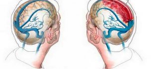
Frontal region syndrome
Atrophy of the frontal lobes of the brain manifests itself in the form of the following symptom complex:
- reduced ability to self-control;
- gradual decline in spontaneous and creative activity;
- severe irritability;
- selfish character traits appear or worsen;
- a sick person is prone to impulsiveness, affective breakdowns, and rudeness;
- decreased intelligence and memory, not reaching the level of dementia;
- tendency towards apathy and lack of desires;
- hypersexuality, primitive jokes.
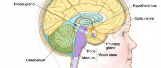
Cerebellar atrophy
Cerebellar atrophy is accompanied by the following symptoms:
- severe motor and speech disorders;
- loss of writing ability;
- cranialgia;
- nausea and vomiting;
- hearing disorder;
- visual disturbances.
Degenerative diseases of the central nervous system
This is a fairly broad and disparate group. At the forefront of changes are neuronal destruction of inflammatory, toxic or metabolic origin. Many cases are familial in origin and their etiology is unknown. They appear predominantly in adulthood, often in the presenile period. The disorders affect one or more functional neural systems while other systems remain intact.
Degenerative diseases affecting the cerebral cortex
A group of diseases affecting the cerebral cortex includes Alzheimer's and Pick's diseases.
Alzheimer's disease (presenile dementia)
The main structural change is a decrease in the number of neurons, changes in neurofibrils, granutocuolar bodies in the cytoplasm of neurons (vacuoles 3-5 μm, with a central fine-grained shape).
The brain tends to shrink. Its weight can be less than 900 g. The convolutions narrow, the grooves widen. Maximum changes occur in the occipital and frontal lobes. Damage occurs to the white matter, hippocampus, and basal ganglia. The cerebellum and brainstem remain largely unchanged. Atrophy is accompanied by expansion of the lateral ventricles (hydrocephalus).
Alzheimer's disease is the most common cause of dementia. As a rule, it begins before the age of 50 and is characterized by slow progression (10 years or more). Death usually occurs from intercurrent infection, usually bronchopneumonia. Women get sick more often than men; the incidence is sporadic. Autosomal recessive inheritance has also been documented with a genetic defect on chromosome 21.
In Down syndrome, in people over 40 years of age, Alzheimer's disease is the rule. The cause is unknown; Biochemically, a deficiency of choline acetyltransferase and acetylcholinesterase was detected in the affected areas. The effect of chronic aluminum intoxication taken into account has not been reliably demonstrated.
Pick's disease
The main structural change is the gradual bihemispheric (both hemispheres - hemispheres are affected) extinction of neurons, observed during the proliferation of astrocytes. The adjacent white matter demonstrates demyelination, loss of nerve fibers. In the cytoplasm of persistent neurons there are Pick bodies, which are round argyrophilic formations. They are formed by a plexus of atypical neurofibrils; their origin is unclear.
Typically, atrophy manifests itself in the frontal and temporal lobes. The affected gyri are reduced, the whole lobe has a hard consistency (lobar sclerosis).
Atrophy of the cerebral cortex can also appear irregularly, rarely diffusely.
The disorder may occur in the basal ganglia, hippocampus, and other areas of the central nervous system.
Pick's disease is a disease of middle and old age. It occurs sporadically, less often in families. Causes about 5% of all cases of dementia. Affects men more.
Degenerative diseases affecting the extrapyramidal system
The extrapyramidal system includes:
- basal ganglia;
- subthalamic nucleus;
- substantia nigra;
- red core;
- thalamic nucleus;
- brain stem.
Damage to the extrapyramidal system leads to a number of disorders, including:
- akinesis;
- violations of the postural reflex;
- disruption of normal muscle tone;
- partial paresis (paralysis).
Changes in the brain can be either limited, focal, or diffuse.
Huntington's disease
The main structural change is the death of brain cells in the caudate and basal ganglia. Changes begin with a decrease in the number of small, later large, neurons, with a simultaneous increase in the number of glial cells. Irreversible loss of neurons is observed to varying degrees in other nuclei of the extrapyramidal system, in the cortex, and cerebellum. Atrophy of the basal ganglia, especially the reduction of the caudate nucleus to a thin layer, leads to significant dilation of the lateral ventricles. Hydrocephalus often leads to a cerebrospinal fluid cyst.
Unlike primary cortical and subcortical (subcortical) degenerations, the gyri and cortical layers are not significantly expanded. Brain weight is reduced by up to 30%. Gray matter atrophy correlates with a decrease in white matter volume.
The disease usually begins between 30 and 50 years of age, rarely at a younger age. It is characterized by an autosomal dominant type of inheritance with localization on the 4th chromosome. The disease progresses over 15-20 years.
Parkinson's disease
The main structural change in cortical cerebral atrophy accompanying Parkinson's disease is the gradual disappearance of neurons in the pigmented nuclei of the substantia nigra and locus, and to a lesser extent in the dorsal motor nucleus of the vagus nerve. Neuronal necrosis is accompanied by the proliferation of astrocytes. Lewy bodies are present in the cytoplasm of permanent neurons, often several in one cell. Numerous Lewy bodies may also be found in the hypothalamus and other areas of the central and peripheral nervous system; greater cortical involvement is part of the clinical manifestations of dementia. The severity of clinical signs correlates with the loss of dopamine neurons. Dopamine is also significantly reduced in other parts of the central nervous system, mainly in the striatum.
Parkinson's disease does not occur simultaneously in identical twins, highlighting the influence of external factors.
Degenerative diseases of the cerebellum
With atrophy of the cerebellum, its cortex, efferent and afferent brain pathways. They include a large number of rare units with varying topography and degrees of degenerative changes. Morphological changes can be focal, diffuse, affecting some systems of brain pathways (olivopontocerebellar atrophy, dentoalveolar atrophy, optic-cholesterolent degeneration). Cerebellar degeneration is also part of spinal degenerations (morbus Friedreich) and paraneoplastic changes.
Signs
What are the signs of brain atrophy? At the initial stage of the disease, the symptoms are hardly noticeable and can only be detected by close people. The patient develops apathy, lack of desires, aspirations, lethargy and indifference. A lack of moral principles and increased sexual activity are often observed.
As brain cell death progresses, the following signs are observed:
You will be interested in: The drug "Novopassit": analogues, indications, instructions for use, reviews, contraindications and side effects
Then, with atrophy, a deterioration in well-being appears and thought processes decrease. A person does not recognize familiar things and forgets about the rules for their use. Elimination of one's behavioral characteristics causes the appearance of the “mirror” syndrome, in which a person begins to copy other people. Then senile insanity and absolute degradation of personality are observed.
The changes in behavior that have arisen do not allow an accurate diagnosis to be made, so to establish the causes of the changes, a list of studies must be carried out. But thanks to the doctor, it will be possible to determine which part of the brain has undergone destructuring. In case of destruction in the cerebral cortex:
Symptoms of changes in the subcortical substance are determined by the functions that the affected section performs, so limited atrophy has its own characteristics. With necrosis of the tissues of the medulla oblongata, respiratory failure, digestive failure are observed, and the cardiovascular and immune systems suffer.
If damage to the cerebellum is observed, muscle tone is disrupted and coordination of movements is impaired. With the destruction of the midbrain, there is no reaction to external stimuli. When the cells of the intermediate section die, a violation of the body's thermoregulation and a metabolic failure appear.
With damage to the anterior section, all reflexes are lost. When neurons die, the function of independent maintenance of life is lost, which usually leads to death. Often, necrotic changes appear from injury or long-term poisoning by toxins.
Multiple sclerosis
Multiple sclerosis is an autoimmune inflammatory, demyelinating and neurodegenerative disease caused by an abnormal immune response directed against the nervous system, brain and spinal cord. Knowledge of the processes and mechanisms leading to damage or disappearance of the neuroaxonal cytoskeleton is gradually growing.
The clinical picture is initially dominated by inflammation and demyelination. But there is evidence that even in the early period, especially at a progressive stage, persistent dysfunctions develop that do not respond to treatment. Progressive neurodegenerative changes are correlated with this condition. MRI demonstrates increasing generalized cerebral atrophy; spinal cord damage occurs.
Histopathological studies show significant pathology of the gray matter of the brain along with damage to the white matter (especially in the cortex, thalamus and other parts of the brain, collectively called dark gray matter). These structures undergo the same changes as the white matter, but with the dominance of activated microglia and the presence of myelin-containing macrophages.
Vitamin deficiency
Long-term lack of vitamin B12 in the diet can lead to brain atrophy. A study conducted by researchers at the University of Oxford found that people with lower levels of B12 had greater volume loss than study volunteers with higher levels of the vitamin.
A deficiency of B12 in the diet is dangerous for pregnant women. It can cause low birth weight in the newborn and insulin resistance in the baby. Those. The child is at increased risk of developing diabetes.
Depression
Researchers from Yale University have concluded that severe depression can destroy neurons and shrink the human brain. During times of extreme stress, a single transcription factor that controls several genes may switch.
Brain atrophy caused by severe depression and chronic stress can lead to emotional and cognitive problems. Yale University scientists have identified one of the reasons for this phenomenon. They compared gene expression in depressed and non-depressed brain tissue. The samples were obtained from a brain bank where donated organs are stored for scientific research.
The brains of depressed people showed signs of reduced expression of genes needed to maintain the structure and function of the synapses that connect neurons, transducing nerve impulses. At least 5 of these genes are regulated by a single transcription factor, GATA1. Its activation turns off genes needed to maintain synapses. Their number decreases, synapses weaken, which in turn leads to loss of brain mass and the development of a subatrophic state. Its activation in experimental rodents caused symptoms of depression. This suggests that GATA1 is involved not only in brain volume reduction, but also in the development of depressive symptoms. Scientists also suggest that its genetic variants may affect a person's susceptibility to stress and depression.
Severity
According to the international classification, there are different degrees of brain atrophy and locations of pathology. Each stage of treatment for the disease has its own symptoms:
With further development, complete destruction occurs and vital systems fail. At this stage, it is better to hospitalize the patient in a psychiatric clinic, since he is difficult to control.
Depending on the location of the affected cells, there are the following types of ailments:
With moderate brain atrophy, changes in personality will be barely noticeable.
Neuroleptics
Impaired coordination of movements, tremors, “restless” limbs... These are side effects that may accompany the first stage of treatment for schizophrenia. They also appeared in healthy adult volunteers who took part in a study of the side effects of the drug Haloperidol, usually prescribed to schizophrenics. Within 2 hours after the administration of this substance, volunteers developed problems with motor skills. Brain MRIs showed that they were associated with a decrease in the volume of gray matter in a region called the striatum, which is responsible for controlling movement.
But the effect of the drug was temporary - a few days after the experiment, the brain volume of the volunteers returned to its original level. According to scientists, this result may reassure people who are panicky that drugs will destroy their brain cells.
Dead neurons in the brain are not restored, therefore, when destroyed by the drug, a return to the original volume is impossible. Therefore, scientists believe that the reason for the reduction in volume is a temporary decrease in the number of synapses (functional connections between neurons). The BDNF protein, which is involved in synapses and disappears after the use of antipsychotic agents, is most likely responsible for this.
Folk remedies for brain atrophy
The destruction of nerve cells is fraught with consequences such as dementia and death. With proper and timely help, people can usually live another 5-10 years. But quality of life also matters. It worsens not only for the patient, but also for his family members.
It is very difficult to coexist with a person with altered consciousness. And it’s even more difficult to constantly listen to angry speeches and grumbling. Therefore, to calm and relax the patient, he is offered to drink teas and herbal tinctures prepared at home.
The following medicinal plants are used:
- rye;
- oregano;
- chickweed;
- motherwort;
- mint;
- Melissa;
- nettle;
- horsetail
The components can be brewed separately or combined to taste. You can drink a cup of this tea 3 times a day. He will be able to relax the patient, reduce tension and normalize mood, put emotions in order.
Alcohol
While drinking alcohol in moderation can be good for the heart, drinking too much is bad for the brain.
Although mild brain atrophy is common in old age, studies show that in alcoholics this process is significantly accelerated and the consequences are more dramatic.
Additionally, alcohol-induced neuronal death is more pronounced in women. No wonder. It is known that women react to alcohol differently than men. They are more sensitive to it and absorb it faster.
The greater vulnerability of women to alcohol consumption is also confirmed by the fact that they have cirrhosis of the liver, damage to the heart muscle, and nerves develop much faster than in male alcoholics. The same rule applies to brain damage.
Treatment
Unfortunately, there is no targeted treatment for brain atrophy. As a rule, therapy is prescribed by a doctor after diagnosing the underlying disease based on examination findings (CT, lumbar puncture, MRI, etc.). It aims to make the patient's life easier, relieve symptoms and potentially cure the underlying cause.
Conditions leading to dementia also lack effective therapeutic measures; Only drugs to reduce symptoms and support (specialized and home) are available. Today, only the developmental technique of using stem cells can stop some diseases. Perhaps, thanks to the latest methods, a person will be able to live with a healthy brain into old age.
Diagnostics and therapy
In order to determine pathological changes, the following is carried out:
- Computed tomography. With its help, you can detect disturbances in blood circulation, identify aneurysms and neoplasms.
- Magnetic resonance imaging. This is the most informative method for detecting structural changes in brain tissue.
After evaluating the research results, treatment is prescribed.
Therapy is carried out to relieve symptoms and slow down the development of the pathological process. This effect is achieved using:
- Nootropic drugs.
- Sedatives and antidepressants.
- B vitamins.
- Means to improve blood circulation.
- Diuretics.
- Antiplatelet agents.
The care of loved ones plays an important role in the treatment process. Therefore, patients are not kept in a hospital, as this will only worsen the situation.
To improve brain function, you need to follow doctors' recommendations regarding nutrition: eat more foods with polyunsaturated fatty acids. It is also necessary to avoid flour products, fried and fatty foods.
Drinking alcohol, smoking and drugs is strictly contraindicated.