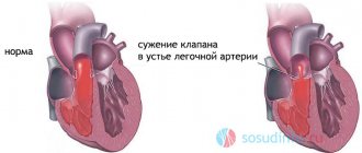
The main symptom of pulmonary hypertension is an increase in pressure in the pulmonary artery (the norm in some cases is exceeded two or more times). In most cases, this pathology is a secondary condition. However, if specialists are unable to determine the cause of its development, pulmonary hypertension is considered primary. This type of disease is characterized by vasoconstriction with subsequent hypertrophy. An increase in pressure in the pulmonary artery causes increased load on the right atrium, resulting in disturbances in the functionality of the heart.
Many people are interested in what the pressure in the pulmonary artery should be normally?
Description
Most often, pulmonary hypertension is manifested by such clinical symptoms as chest discomfort, severe dizziness, shortness of breath with overexertion, recurrent fainting, and fatigue. Diagnosis of the disorder involves measuring the pressure in the pulmonary artery. Pulmonary hypertension is treated with vasodilators. In some of the most severe cases, the patient is indicated for surgical intervention.
We will consider the norm and pressure gradient in the pulmonary artery below.
Symptoms
A newborn with pulmonary hypertension immediately after birth or several hours later:
- breathes heavily, with shortness of breath;
- when inhaling, the chest is drawn inward;
- has pronounced cyanosis (blue discoloration) of the skin and mucous membranes;
- responds poorly to oxygen therapy: the condition does not improve as expected.
Read about other symptoms of this disease (not only in children) and its treatment here.
Possible pathologies
Quite often, pulmonary hypertension is a complication of certain diseases. Possible pathologies:
- Hypoventilation of the lungs.
- Cirrhosis of the liver.
- Myocarditis.
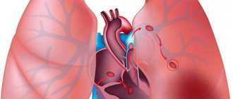
- Thrombosis of veins, arteries, branches of the lungs.
- Impaired lung function.
- Pathological changes in the interatrial septum.
- Congestive heart failure.
- Mitral valve stenosis.
In this regard, if the pressure in the pulmonary artery deviates from normal, it is important to immediately consult a doctor and undergo a full examination.
Stages of progression
The danger of pulmonary hypertension is that it does not have clinical manifestations at an early stage of development, when its growth can be slowed down. During this period, children only experience shortness of breath during intense exercise, which is mistakenly attributed to signs of low fitness of the body.
The second stage begins with a decrease in the release of blood into the arterial network. This is the phase of advanced symptoms - hypoxia, shortness of breath, cyanosis, fainting. The pressure in the vessels of the lungs is steadily increased.
The third stage is heart failure. It is characterized by a significant drop in pressure in the systemic circulation and an increase in the small circle. The duration of each stage is very variable, from 5 months to 5 years until complete decompensation and death. A severe and rapid course occurs with inadequate treatment and the absence of surgical correction of heart disease.
Norm
Normal blood pressure readings in the lungs are as follows:
- The average pressure in the pulmonary artery is normal - from 12 to 15 mm. rt. Art.
- Diastolic – from 7 to 9 mm. rt. Art.
- Systolic pressure in the pulmonary artery is normal - from 23 to 26 mm. rt. Art.
These indicators must be stable.
What is the normal estimated pressure in the pulmonary artery? In accordance with the recommendations adopted by WHO, normally the calculated systoles should be a maximum of 30 mm. rt. Art. In this case, the maximum diastolic pressure is 15 mm. rt. Art. Pulmonary hypertension is diagnosed when the deviation exceeds 36 mm. rt. Art.
Few people know the norm for estimated systolic pressure in the pulmonary artery.
How is the treatment carried out?
Drug therapy
Sodium nitroprusside can be used to treat patients older than infants.
If persistent pulmonary hypertension has been diagnosed in newborns, then therapy must be carried out in intensive care settings. Doctors normalize blood pressure in the lungs as quickly as possible and relieve vascular spasms. If the disease is observed in older children, treatment is mainly carried out in a hospital. They resort to the help of medications that have a relaxing effect on the vascular walls and relieve spasms. Tolazoline and sodium nitroprusside can be used.
Therapy is not complete without intravenous administration of medications to prevent the onset of heart failure. Adrenaline and Dopamine are used for these purposes. If the pathology is caused by infection of the body, they resort to antibiotics. Sometimes diuretics and anticoagulants may be used. It is important to remember that the correct medication and the duration of its use should be prescribed exclusively by a physician, taking into account the severity of the disease and the general condition of the small patient.
Other methods
If there is a high probability of death, the child is prescribed extracorporeal membrane oxygenation. This technique allows you to saturate the blood of a small patient with oxygen using a special device. It is connected with catheters to the patient and the blood is purified and also saturated with oxygen.
It is useful for such patients to attend massage treatments.
To avoid the development of complications, doctors recommend that parents take their children for a massage and engage in physical therapy with them. The set of exercises is selected by the doctor, who takes into account the physical characteristics of the little patient. In addition, once a year a visit to a sanatorium-resort treatment is required, which will improve the overall health of the child. It is important not to forget about systematic visits to the attending physician to monitor the condition of the baby with congenital heart disease.
Symptoms of pulmonary hypertension
The pressure in the pulmonary artery can only be determined by instrumental methods, since in a moderate form of the pathology, symptoms practically do not appear - characteristic signs appear only when the disease becomes severe.
At the initial stages, deviations from normal pressure in the pulmonary artery are manifested by the following symptoms:
- Shortness of breath appears, which bothers a person in the absence of intense physical activity and even at rest.
- Body weight gradually decreases, and this does not depend on the quality of a person’s diet.
- An asthenic disorder occurs, depression, severe weakness develops, and there is no ability to work. It is worth noting that this condition does not depend on the time of day or changes in weather conditions.
- Cough occurs regularly, with no discharge from the respiratory system.
- Hoarseness occurs.
- Discomfort appears in the abdominal cavity. A person experiences a feeling of pressure from within, heaviness. The reason for this symptom lies in congestion in the portal vein, which transfers blood to the liver.
- The brain is affected by hypoxia, which causes frequent dizziness and fainting.
- Tachycardia gradually becomes noticeable and palpable in the neck.
Diagnostics
To determine the condition, a number of hardware examinations are used:
- ECG: overload appears on the right, characterized by pathological enlargement of the ventricle and proliferation of the atrium. The onset of various forms of extrasystole and atrial fibrillation is characteristic;
Echocardiography: the method is very informative - it allows you to calculate the average pressure in the pulmonary artery
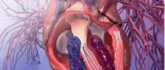
INTRACARDIAC PRESSURE
- pressure in the cavities of the heart that arises during its rhythmic activity. The value of V. d. is different for each chamber of the heart and changes at different moments of the cardiac cycle. It depends on the degree of blood filling of the chambers, the contractile function of the myocardium and the resistance value of the blood outflow tract, as well as a number of other cardiac and extracardiac factors - the radius of curvature of the chambers of the heart, the degree of tension of the connective tissue base of the heart, intrathoracic pressure.
Registration of changes in blood pressure over time in the form of pressure curves in the cavities of the heart allows one to characterize the state of intracardiac hemodynamics and blood circulation in general and obtain the necessary information about the degree and nature of disorders of the pumping function of the heart in various pathological conditions (see Blood circulation).
Most authors distinguish three positive waves on the atrial pressure curve - a, c and v and two negative waves (collapse) - x and y (Fig.). The onset of wave a coincides in time with the middle or last third of the P wave on the ECG. In case of disturbances in the systolic activity of the atria, which occurs, for example, in patients with atrial fibrillation, wave a is absent in the pressure curve. Wave c occurs at the moment of closure of the atrioventricular valves, i.e. at the beginning of ventricular systole. The cause of wave c is the jerk-like protrusion of the mitral valve into the atrium at the beginning of isometric contraction of the ventricle. Wave x is associated with a decrease in pressure and corresponds in time to the period of relaxation of the atrium myocardium. The main reason for the appearance of wave x is considered to be an increase in atrial volume due to relaxation of muscle fibers. During wave x, the pressure in the atrium reaches atmospheric pressure or decreases by several millimeters of mercury. Subsequently, wave x is replaced by wave v, which is caused by an increase in blood flow into the atria from the pulmonary and vena cava. The y wave follows the peak of the v wave and its beginning coincides in time with the opening of the atrioventricular valves and the beginning of diastolic filling of the ventricles. During this period, intraatrial pressure decreases in parallel with a decrease in pressure in the left ventricle to end-diastolic pressure (end-diastolic pressure is the pressure in the ventricular cavities immediately before the closure of the atrioventricular valves). The y wave is followed by a gently increasing portion of the intraatrial pressure curve. To fill the left atrium with blood, whose elastic properties are higher than those of the right atrium, higher pressure is required. According to the data available in the literature, obtained by probing the heart of healthy people, in the left atrium the average value of the a wave is 10-11 mm Hg. Art., waves v -12 -14 mm Hg. Art. The average pressure in the left atrium, equal to the integral value of all fluctuations, is in the range of 8-9 mm Hg. Art., in the right atrium - is 3 mm Hg. Art.
Experiments have established that the curve of the dependence of intraatrial pressure on the volume of atrial filling remains linear up to 9-11 mm Hg. Art. With a further increase in volume, the increase in intraatrial pressure occurs to a much greater extent.
To analyze the cardiac cycle, we mainly examine the duration of its individual phases and determine the average and maximum rate of change in pressure in the cavities of the ventricles during periods of isometric stress and relaxation. Ventricular systole begins with a phase of asynchronous contraction, during which there is a sequential involvement of individual sections of the myocardium of the left and right ventricles in the contractile process. During this phase, the configuration of the ventricular cavity changes with a slight increase in intraventricular pressure. The isometric contraction phase begins from the moment the atrioventricular valves close. The rise in the intraventricular pressure curve in this phase is steepest and is interrupted by a slight bend or notch, which reflects the opening of the corresponding semilunar valves and coincides in time with the beginning of the period of expulsion of blood into the aorta or pulmonary artery. The next period, the period of expulsion, is divided into phases of maximum and reduced expulsion. In the phase of maximum expulsion, which normally begins from the moment the corresponding semilunar valves open and the duration corresponds to 1/3 of the entire period of expulsion, 2/3 of the stroke volume of blood is ejected.
Signs of disease progression
Over time, the disease progresses, pulmonary hypertension intensifies and causes the following symptoms:
- When you cough, you produce sputum mixed with blood, which indicates swelling in the lungs.
- Attacks of angina appear, accompanied by pain in the sternum, an unreasonable feeling of fear, and severe sweating. Such symptoms indicate the development of myocardial ischemia.
- Atrial fibrillation develops.
- The patient experiences pain in the right hypochondrium. This condition occurs due to the development of a number of pathologies of the circulatory system.
- The lower extremities swell greatly.
- Ascites develops (a significant amount of fluid accumulates in the abdominal cavity).
At the terminal stage of the pathology, blood clots form in the arterioles, which can lead to increasing suffocation and heart attack.
First signs
At the initial stage of pathological changes in the vascular system, the first signs indicating the presence of the disease are absent, since compensatory and replacement mechanisms cope with the load. Upon reaching critical levels of 25-30 mm Hg. Art. the first signs of pulmonary hypertension appear. At this stage, the patient experiences a deterioration in general well-being after physical overexertion. With further progression of degenerative changes in the vascular system, susceptibility to physical activity decreases and clinical symptoms increase.
Symptoms characteristic of the initial stage of the disease:
- difficulty breathing after exercise;
- pain in the chest of various types;
- dizziness;
- fainting;
- bouts of dry cough;
- the appearance of blood streaks in the sputum;
- causeless fatigue;
- swelling in the area of the legs and feet.
Important! In most cases, patients complain of fatigue, severe shortness of breath, progressing after physical activity. Shortness of breath may be accompanied by atypical discomfort in the chest area, and after physical exertion, attacks of dizziness and fainting are observed. These manifestations are caused by insufficient cardiac output.
In severe cases, PH is accompanied by the following clinical manifestations:
- peripheral edema;
- congestive processes in the liver;
- protrusion of the right ventricle;
- angina attacks;
- heart rhythm disorder.
The appearance of pain in the right hypochondrium is caused by the occurrence of venous stagnation, when the systemic circulation is involved in the process, the size of the liver has increased, and the capsule surrounding the organ has stretched, resulting in pain.
The terminal stage is manifested by the presence of blood clots in the pulmonary arterioles, which leads to the death of active tissues. At this stage, patients experience hypertensive crises and attacks of pulmonary edema, mainly at night. The attacks begin acutely with a feeling of lack of oxygen, followed by a severe dry cough, followed by the release of bloody sputum. The skin becomes bluish and there is strong pulsation in the carotid artery. The patient is in an excited state, loses self-control, and has chaotic movements. A favorable outcome of a crisis is involuntary urination or loss of feces; an unfavorable outcome is death. The cause of death is blockage of the pulmonary artery by a thrombus, with further development of heart failure.
Diagnosis of pulmonary hypertension
To identify the condition, a series of hardware tests should be carried out. These include:
- Radiography. Allows you to identify excess capacity in the pulmonary fields, displacement of the cardiac edges to the right, and enlargement of the roots.
- ECG. Allows you to identify overloads on the right side, which are characterized by a pathological increase in the size of the ventricle and proliferation of the atrium. Characteristic is the onset of various forms of atrial fibrillation and extrasystole. Deviation of pressure in the pulmonary artery from normal on EchoCG can be seen.
- Echocardiography (EchoCG). It is the most informative method for diagnosing arterial hypertension and allows us to identify most of all disorders in the heart. In addition, echocardiography can show arterial hypertension even in the initial stages of development.
- Respiratory system tests, analysis of the level and amount of gases in the blood. This method is used to determine the severity of the pathology and the degree of respiratory failure.
- MRI. CT. These research methods allow you to obtain a high-quality image, and the introduction of a contrast agent allows you to assess the condition of the respiratory and cardiovascular systems.
- Centigraphy. Indicated for thromboembolism. In 90% of cases, the technique allows us to obtain reliable data.
- Ultrasound. This diagnostic technique allows you to determine the parameters of the heart chambers and wall thickness.
Diagnostic methods
When examining a child with PH, there may be no abnormalities in the respiratory system: the chest has the correct shape, percussion sound does not change color, breathing over the lungs is vesicular, wheezing in the absence of diseases of the bronchi and alveoli is not audible.
When examining the heart, they reveal:
- increased pulsation in the epigastric region or 2 - 3 intercostal space;
- expansion of the borders of the heart to the right (sometimes the large right ventricle pushes aside the reduced left one, then the left border also expands);
- increased tone 2 above the pulmonary artery, it acquires a metallic tint;
- systolic murmur (when the tricuspid valve leaflets are not closed), diastolic (due to an enlargement of the pulmonary artery trunk).
Pulmonary hypertension in childhood
Deviation of pressure in the pulmonary artery in children from the norm occurs against the background of congenital pathologies of blood vessels and heart. The disease manifests itself in children with rapid breathing and cyanosis. At an older age, the disease begins to progress, which is accompanied by the occurrence of circulatory failure - the liver enlarges, tachycardia develops, and shortness of breath appears.
The most common factors for abnormal systolic pressure in the pulmonary artery in childhood are the following congenital pathologies of the heart and blood vessels:
- One common cardiac ventricle.
- Patent atrioventricular canal or ductus Botall.
- Combination of septal defect with transposition of the pulmonary artery and aorta.
- Large hole in the interventricular septum.
In addition, pulmonary hypertension in children can develop due to pulmonary hypoplasia due to a hernia of the diaphragm or due to the penetration of amniotic fluid or intestinal contents into the respiratory tract at the time of birth.
Description and statistics
Persistent fetal circulation in a newborn is a kind of signal from the baby’s body about the impossibility of fully adapting the blood circulation in the lungs to life outside the womb.
In the prenatal period of development, the lungs undergo a number of changes that prepare them for functioning in the air, but at this time the placenta “breathes” for them. After the birth of a child, “real” breathing should begin , but sometimes for a number of reasons it occurs with pathology.
With pulmonary hypertension, there is a sharp jump in pressure inside the vascular bed of the lungs, as a result of which the child’s heart begins to experience enormous stress.
The newborn’s body, trying to avoid impending heart failure, produces an emergency decrease in pressure in the lungs by reducing the volume of blood circulating in them - the blood is “dumped” through the open foramen ovale in the heart or the open ductus arteriosus that babies have.
This in turn leads to an increase in the amount of venous blood in the general bloodstream, a decreased oxygen content in the blood and cyanosis in the child.
According to statistics, the pathology occurs in 1-2 babies out of 1000 . Approximately 10% of newborns requiring intensive care suffer from this disease. Moreover, most of them are full-term or post-term babies.
Fetal circulatory syndrome occurs much more often in children born by cesarean section - in approximately 80-85% of cases.
The overwhelming number of relevant diagnoses (97%) were made in the first three days of life of small patients - such early diagnosis allows us to significantly reduce the number of deaths, since without timely medical care, 80% of sick children can die.
Provoking factors
The following factors contribute to an increase in pressure in the pulmonary arteries:
- Maternal preeclampsia, maternal use of various medications, toxicosis in late pregnancy.
- Infections of the newborn or fetus.
- Autoimmune pathologies.
- Labor hypoxia.
- Pneumonia.
- Vascular thrombosis.
- Bronchospasm.
- Hereditary predisposition.
The classic manifestations of childhood arterial hypertension are the following: rapid heartbeat, fainting, chest pain, cyanosis of the skin, poor weight gain, decreased appetite, tearfulness, irritability, lethargy, difficulty breathing.
If primary signs of pulmonary hypertension are detected, the child should be immediately shown to a specialist, since this pathology is very dangerous in childhood.
Prognosis for patients
The appearance of pulmonary hypertension in primary or secondary form is considered as one of the extremely unfavorable signs. It significantly aggravates the condition of children with congenital developmental pathologies, and with rapid progression, it reduces the chances of successful surgical treatment of heart disease.
Without surgery, such patients have a fairly low chance of maintaining health and life. Children who were able to survive have deviations in physical and mental development; they are susceptible to chronic diseases of the pulmonary system, ENT organs, and heart.
And here is more information about triscuspid regurgitation.
Pulmonary hypertension in children occurs with developmental defects or intrauterine infections, hypoxia. The primary form has an unknown origin and a rapidly progressive course. With secondary PH due to heart and respiratory diseases, the initial stages are asymptomatic, and manifestations occur with irreversible changes.
Treatment includes drug therapy, surgical correction, and lung transplantation. The pathology is characterized by an unfavorable prognosis.
Treatment in the form of surgery may be the only chance for patients with atrial septal defect. It can be a congenital defect in a newborn, manifest in children and adults, or secondary. Sometimes it closes on its own.
If pulmonary hypertension is diagnosed, treatment should be started as soon as possible to alleviate the patient's condition. Drugs for secondary or high hypertension are prescribed in a comprehensive manner. If the methods do not help, the prognosis is unfavorable.
Cor pulmonale develops after diseases in the chest. Symptoms in children and adults appear the same. The course can be acute or chronic. Diagnostics will help to identify the problem in a timely manner and begin treatment. How long do people live with cor pulmonale?
During fetal formation, pulmonary artery hypoplasia and agenesis may develop. Reasons: smoking, alcohol, toxic substances and other harmful factors. The newborn will have to undergo surgery to be able to live and breathe normally.
Treatment of pulmonary hypertension
To stabilize the pressure in the pulmonary artery, drug therapy is prescribed, first of all. Drugs and treatment regimens should be determined by a doctor individually and only after a complete examination.
Pulmonary hypertension is treated with medication and non-drug methods. Drug treatment involves the use of drugs from the following groups:
- Calcium antagonists. These substances can normalize heart rhythm, relieve spasms in blood vessels, relax the muscles of the bronchi, and give the heart muscle resistance to hypoxia.
- Diuretics. The drugs help remove excess fluid from the body.
- ACE inhibitors. The effect of these drugs is aimed at narrowing blood vessels, reducing the load on the heart muscle, and lowering blood pressure.
- Antiplatelet agents. Allows you to eliminate the adhesion of red blood cells and platelets.
- Nitrates. Their use reduces the load on the heart. The effect occurs as a result of the expansion of the veins located in the legs.
- Indirect anticoagulants. Helps reduce blood clotting.
- Direct anticoagulants. They help prevent blood thickening and, as a consequence, the development of thrombosis.
- Endothelin receptor antagonists. Drugs in this group have a pronounced vasodilating effect.
- Antibiotics. Indicated for use in case of bronchopulmonary infection.
- Bronchodilators. Helps normalize lung ventilation.
- Prostaglandins. They have a number of positive effects on the body. For example, they promote vasodilation, slow down the formation of connective tissues, reduce damage to endothelial cells, and prevent the adhesion of blood elements (erythrocytes, platelets).
Right atrial pressure
Right atrium pressure is the same as central venous pressure (as assessed by jugular venous pressure during physical examination of the patient) due to the absence of valves to prevent blood from returning to the right atrium.
Normally, during diastole, the pressure in the right atrium is equal to the pressure in the right ventricle, since the right side of the heart functions as a common chamber when the tricuspid valve is open. Mean right atrial pressure decreases as intravascular volume decreases. It increases with right ventricular failure, right-sided valve disease, and cardiac tamponade (in which the heart chambers are surrounded by high-pressure pericardial fluid
Some abnormalities cause characteristic changes in individual components of pressure in the right atrium (and, as a consequence, in the jugular veins) (Table 3.3).
For example, the two main causes of a prominent a wave are tricuspid valve stenosis and right ventricular hypertrophy. Under these conditions, when the right atrium contracts, it has to overcome resistance from both the stenotic tricuspid valve and the rigid right ventricle. As a result, a pronounced wave a is formed. A pronounced V wave is observed with tricuspid valve insufficiency, since the normal filling of the right atrium is disrupted by reverse blood flow.
Right ventricular systolic pressure increases with pulmonary valve stenosis or pulmonary hypertension. Right ventricular diastolic pressure increases when the right ventricle is subjected to pressure or volume overload. Such an increase in pressure may be a sign of right ventricular failure.
Operation
If the described methods are ineffective, life-threatening pathology can be eliminated through surgical intervention performed in three ways:
- Atrial septostomy. Involves creating a small opening between the atria. As a result, the pressure in the atrium and pulmonary arteries decreases to normal.
- Thrombendarterectomy. Involves removing blood clots from blood vessels.
- Lung transplantation (lungs and heart). The main indications for such a procedure are hypertrophic changes in the muscles of the heart, insufficiency of the heart valves.
Stages of pathology
The disease develops in stages with a gradual deterioration in the condition of the little patient.
In a child, pulmonary hypertension is divided into 4 stages. The first is the easiest and, with appropriate therapy, can completely cure a small patient. If the disease is not diagnosed in a timely manner, it goes into stage 2. If effective treatment is not carried out, the disease progresses and progresses to stages 3 and 4. They are characterized by irreversible disturbances in the functioning of the heart and lungs.
Normal values
An increase in systolic and diastolic pressure in the pulmonary artery occurs for three main reasons: 1) with left ventricular heart failure, 2) with parenchymal diseases of the lungs (for example, chronic bronchitis or severe emphysema) and 3) with diseases of the pulmonary vessels (for example, with pulmonary embolism artery, primary pulmonary hypertension).
Under normal conditions, the diastolic pressure in the pulmonary artery coincides with the pressure in the left atrium due to the low resistance of the pulmonary vascular system that separates them. If pressure in the left atrium increases, for example, due to left ventricular heart failure, then systolic and diastolic pressure in the pulmonary artery necessarily increases to maintain pulmonary blood flow. This situation leads to passive pulmonary hypertension.
However, under some conditions, systemic pulmonary vascular resistance becomes abnormally large, resulting in increased pulmonary artery diastolic pressure compared with left atrial pressure. In this case, they speak of active pulmonary hypertension. When obstructive pulmonary vascular disease develops as a complication of a chronic cardiac shunt (eg, atrial or ventricular septal defect), the disease is called Eisenmenger syndrome.
If the catheter is directed into the right or left pulmonary artery, its tip will eventually reach one of the small branches of the pulmonary artery and temporarily block the flow of blood behind it. During this time, a stagnant column of blood forms between the tip of the catheter and parts of the capillary system, as well as distant segments of the pulmonary venous system (Fig. 3.14).
This column of blood acts as a continuation of the action of the catheter, and the pressure that is recorded through the catheter reflects the pressure inside the chamber - the left atrium (LA). This pressure is called pulmonary artery wedge pressure or pulmonary capillary wedge pressure (PCWP) and it accurately reflects the left atrium pressure in a large number of people (hence this pressure measurement is equivalent to the left atrial pressure measurement as presented in Fig. 2.1).
In addition, while the mitral valve is open during diastole, the pulmonary veins, left atrium, and left ventricle are at the same pressure. Thus, pulmonary capillary wedge pressure (PCWP) is also used to estimate left ventricular diastolic pressure and to measure ventricular preload. Therefore, measuring PCWP using a right-sided catheter is a key point in monitoring critically ill patients in intensive care units.
An increase in the average PCWP is observed in left ventricular heart failure and mitral stenosis. In addition, individual components of PCLC may be abnormally high; wave a can be increased with reduced elasticity of the left ventricle during its hypertrophy or with acute myocardial ischemia (Table 3.3). The v wave exceeds its normal level when the left atrium is overfilled, which happens, for example, with mitral regurgitation.
Pulmonary artery wedge pressure (PAWP) is the most important hemodynamic characteristic. In most cases (although not always!) it corresponds to left atrial pressure and left ventricular end-diastolic pressure (LVEDP) and thus reflects the state of pulmonary capillary blood flow and the risk of pulmonary edema in patients with left ventricular heart failure.
It is believed that when one of the central branches of the pulmonary artery becomes jammed, the blood flow in its basin is completely stopped. From the tip of the catheter to the corresponding vein of the same name, a stationary column of blood now passes through the entire intercalary microvasculature. The contact of this static blood with the preserved main blood flow occurs at the so-called “J” point (from the English joint - connection, articulation). It is located at the level of the pulmonary veins, in close proximity to the orifice of the left atrium.
Theoretically, the pressure at the catheter tip in the wedged position corresponds to the pressure at the “J” point (Pj). In turn, Pj is identical to the pressure in the cavity of the left atrium (Rlp). And finally, Rlp normally does not differ from the pressure in the left ventricle at the very end of its diastole (LPV).
• diastolic filling of the left chambers of the heart;
• hydrostatic pressure in the pulmonary veins.
The diagnostic concept can be formulated as follows. With PAWP less than 6 mm Hg. Art. According to the experience of clinical observations, filling of the left ventricle is considered insufficient. Heart performance will obviously be limited by such a low preload. In this situation, it is necessary to intensify the administration of fluid. When PAWP is more than 12 mm Hg. Art.
forced infusions are considered inappropriate. An increase in filling pressure above this value, as a rule, does not lead to an increase in heart function. Moreover, the danger of volume overload of the pulmonary circulation is aggravated. Thus, PAWP in the range of 6–12 mm Hg. Art. is considered a certain physiological optimum, towards maintaining which one should direct one’s efforts.
Increased pressure in the pulmonary artery, the norm of which can be exceeded several times, is the main sign of pulmonary hypertension. In almost all cases, this disease is a secondary condition, but if doctors cannot determine the cause of its occurrence, then pulmonary hypertension is considered primary. This type is characterized by narrowing of blood vessels and their subsequent hypertrophy. Due to increased pressure in the arteries of the lungs, the load on the right atrium increases, which often leads to dysfunction of the heart.
Typically, pulmonary arterial hypertension is manifested by symptoms such as fatigue, possible fainting, shortness of breath with exertion, severe dizziness and discomfort in the chest area. Diagnostic measures involve measuring pulmonary pressure. Hypertension is treated with drugs with a vasodilating effect, and in particularly difficult cases, surgical intervention is required.
10 Pressure jam
The idea of relativity dominates in the exact sciences
Pulmonary capillary wedge pressure (PCP) is traditionally used in the practice of critical care medicine, and the term “wedge pressure” has already become quite familiar to doctors. Despite the fact that this indicator is used quite often; it is not always thought through critically. This chapter identifies some of the limitations of the use of the PCI and discusses misconceptions that arise when using this indicator in clinical practice.
DZLK is used to determine pressure in the left atrium. The information obtained allows us to assess intravascular blood volume and left ventricular function.
Dk - Dlp = Q x Rv
Rice. 10-1. The principle of measuring DZLK. The lungs are divided into 3 functional zones based on the ratio of alveolar pressure (Palv), mean pressure in the pulmonary artery (avg. Dla) and pressure in the pulmonary capillaries (Pk). DZLK allows you to accurately determine the pressure in the left atrium (LAP) only when Dc exceeds Ralv (zone 3). Further explanations in the text.
where Dk is the pressure in the pulmonary capillaries, Dlp is the pressure in the left atrium, Q is the pulmonary blood flow, Rv is the resistance of the pulmonary veins.
If Q = 0, then Dk - Dlp = 0 and, therefore,
The pressure at the tip of the catheter at the time of balloon occlusion of the pulmonary artery is called LVP, which, in the absence of an obstruction between the left atrium and the left ventricle, is considered equal to the left ventricular end-diastolic pressure (LVEDP).
LVEDP (LVEDP) is a reliable indicator of preload only when left ventricular compliance is normal (or unchanged{amp}gt;.
The assumption that ventricular compliance is normal or unchanged in adult patients in intensive care units is unlikely. At the same time, the prevalence of impaired diastolic function in such patients has not been studied, although in some conditions their ventricular compliance is undoubtedly altered. This pathology is most often observed due to mechanical positive pressure ventilation, especially when inspiratory pressure is high (see Chapter 27).
DPLC is used as an indicator of hydrostatic pressure in the pulmonary capillaries, which allows one to assess the possibility of developing hydrostatic pulmonary edema. However, the problem is that PCWP is measured in the absence of blood flow, including in capillaries. Features of the dependence of the DPLC on hydrostatic pressure are presented in Fig. 10-2.
How to settle
Increased pressure is regulated by receptors on the vascular wall, branches of the vagus nerve and the sympathetic nerve. The largest receptor zones are located in large and medium-sized arteries, veins and their branching areas. When arteries spasm, the supply of oxygen to the blood is disrupted. And tissue oxygen starvation stimulates the release of substances into the blood that increase tone and enhance the pulmonary pressure gradient.
When irritated, the fibers of the vagus nerve increase blood flow through the lung tissue, and the sympathetic nerve, on the contrary, has a vasoconstrictor effect. If the pulmonary pressure is normal, the interaction of the nerves is balanced.
Cardiac index
Cardiac output (CO) (along with CO, the concept of “cardiac minute volume” - MCV) is often used. If ventricular filling is maintained at a sufficient level, then the magnitude of cardiac output at any stroke volume depends on the heart rate (HR). Calculation formula: SV or MOS = (SV • HR) l/min. Thus, CO is a function of stroke volume and heart rate. An increase in CO during tachycardia requires more efficient diastolic filling of the heart.
As the heart rate increases, the relative time of diastole decreases compared to the duration of systole. However, in a normally functioning heart, which contracts within 170 beats/min, its filling does not decrease due to shortening of diastole.
In an intact heart, during tachycardia, the process of relaxation of the heart muscle is accelerated, which ensures faster and more complete filling of the heart with blood during shortened diastolic periods. This effect is partially mediated through stimulation of p-receptors by catecholamines, which increase the relaxation of cardiomyocytes due to the accelerated removal of intracellular Ca2 from them. With excessive tachycardia (more than 170 beats/min), such complete diastolic relaxation may not occur, and therefore a further increase in CO.
Cardiac index (CI). In modern medicine, the CO indicator is normalized in order to give it the comparability properties necessary to compare the results of its measurement in different individuals and under different conditions of cardiac functioning. The normalized indicator was called the “cardiac index”, i.e. SI is a calculated indicator, the size of which in healthy people depends on gender, age, and body weight.
where S is the surface area of the body, m2; B—body weight, kg; P—height, cm; 0.007184 is a constant coefficient.
Essentially, SI is a measure of blood flow from the heart and as such is a primary indicator of its pumping function. In a healthy person at rest, the index is considered normal within the range of 2.5-3.6 l/min/m2. A decrease in the ability of the heart to perform its pumping function in various forms of pathology leads to a decrease in SI.
Thus, the SI indicator more adequately than SV characterizes the hemodynamic capabilities of a specific (and not some virtual) healthy organism even in conditions of the development of heart failure. It is this indicator that is used to objectively assess the degree of its severity. As such, SI is one of the main classification criteria for heart failure.
This indicator characterizes the degree of efficiency of the heart during systole. It is generally accepted to measure EF of the left ventricle, the main component of the heart pump. EF is expressed as a percentage of the volume of blood in the ventricle at its maximum filling during diastole. For example, if there were 100 ml in the left ventricle, and during systole 60 ml of blood entered the aorta, then the EF is 60%.
where EDV is end-diastolic volume, ESV is end-systolic volume.
Along with the calculation of EF, hardware methods for its determination are used: echocardiography, radiocontrast or isotope ventriculography.
The normal value of left ventricular EF is 55-75%. With age, there is a tendency for this indicator to decrease. It is generally accepted that an EF value below 45-50% indicates insufficiency of the pumping function of the heart.
The EF indicator for various cardiovascular diseases is not only diagnostically, but also prognostically significant. However, it has certain limitations, because depends on myocardial contractility and other factors (pre-, afterload, frequency and rhythm of heart contractions).