© Author: Sazykina Oksana Yuryevna, cardiologist, especially for SosudInfo.ru (about the authors)
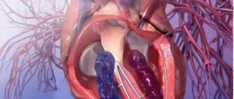
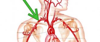
The pulmonary artery (PA) is one of the largest vessels in the human body, carrying blood from the heart to the vessels of the pulmonary tissue, where the blood is enriched with oxygen, and thus the formation of the pulmonary circulation is completed. In another way, this vessel is called the pulmonary trunk.
If the lumen of the vessel becomes smaller, then they speak of stenosis, or pathological narrowing, of the pulmonary artery.
picture: valve form of pulmonary artery stenosis
Stenosis occurs due to congenital or acquired causes, and is characterized by the following hemodynamic processes in the heart:
- The right ventricle experiences stress as it pushes blood through the narrowed lumen of the pulmonary artery.
- The load on the ventricle may vary depending on the severity of the stenosis.
- Less blood enters the lungs than normal, as a result of which less blood is saturated with oxygen, and the body receives less oxygen overall, which leads to the development of hypoxia (oxygen starvation) of the internal organs.
- Constant load on the right ventricle leads to gradual wear and tear of the heart muscle, which is initially compensated by an increase in myocardial mass (right ventricular hypertrophy), and later leads to the development of severe cardiac right ventricular failure.
- Due to the constantly increased final volume of blood, which cannot be completely released into the artery, tricuspid regurgitation develops, that is, a reverse flow of blood is formed into the right atrium, which leads to stagnation of venous blood and disruption of microcirculation in the vessels of the internal organs - hypoxia is aggravated.
- Severe stenosis leads to the development of severe heart failure, which can cause death if left untreated.
Depending on the location of the lesion, supravalvular, subvalvular and valvular forms of stenosis are distinguished, that is, the narrowing is located above, below or at the level of the valve, respectively. Valvular pulmonary artery stenosis is more common than other forms.
forms of pulmonary artery stenosis by location
General description of the pathology
Before looking at the causes, symptoms and effective treatments, it is worth reviewing general information about stenosis. The pulmonary arteries are paired, fairly large branches of the pulmonary trunk that arise from the right ventricle.
The main function of this vessel is to transport venous (oxygen-poor) blood from the right atrium to the capillary system of the lungs, where gas exchange occurs and the blood is saturated with oxygen. In fact, this is where the pulmonary circulation begins. At the point where the pulmonary trunk exits the right atrium there is a tricuspid valve, which regulates blood flow.
Pulmonary artery stenosis is said to occur if there is a narrowing of the vessel itself or a malfunction of the valve (for example, if the valve leaflets are fused at the edges, then blood cannot circulate normally). Impaired blood flow negatively affects the condition of the entire body. First of all, the heart suffers - the right parts of the myocardium often hypertrophy in order to compensate for problems with blood ejection and cope with increased pressure in the right ventricle.
As statistics show, this pathology in most cases is congenital. Timely diagnosis and properly administered therapy help avoid possible complications.
Which doctor should I contact?
All newborn children in maternity hospitals are examined by a neonatologist, who, if a congenital heart defect is suspected, will draw up the necessary examination plan.
If similar symptoms appear in an infant or older child, you should immediately consult a pediatrician.
The adult population should seek help from a general practitioner or cardiologist.
In any case, the examination plan is approximately the same and includes the following diagnostic methods:
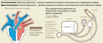
- Echocardioscopy, or ultrasound of the heart, allows you to visually assess the structure of the valve ring, as well as measure the pressure in the right ventricle and the pressure gradient (difference) between the cavities of the right ventricle and the pulmonary artery. The higher the pressure in the right ventricle and the greater the pressure difference between the ventricle and the artery, the more pronounced the narrowing of the lumen of the vessel. According to these data, the degree of stenosis is divided into:
- moderate (P liquid - less than 60 mm Hg, degrees P - 20-30 mm Hg),
- pronounced (R liquid – 60-100 mm Hg, degrees R – 30-80 mm Hg),
- sharply expressed (P yellow - more than 100 mm Hg, degree P - more than 80 mm Hg) and decompensated (severe disturbances of intracardiac hemodynamics, myocardial contractile function sharply decreases).
- ECG, if necessary - conducting an ECG after dosed physical activity (walking on a treadmill, bicycle ergometry).
- X-ray of the chest organs - determines the degree of enlargement of the heart due to myocardial hypertrophy.
- Right heart catheterization allows you to more accurately measure pressure in the right ventricle and pulmonary artery.
- Ventriculography is the injection of a radiopaque substance into the vessels, entering the right half of the heart and using X-ray images to display the anatomical nuances of the stenosis.
Reasons for the development of stenosis
Pulmonary artery stenosis in children can develop under the influence of various factors. Unfortunately, the exact mechanisms of the development of the disease have not yet been fully studied. Nevertheless, it is worth familiarizing yourself with the list of prerequisites.
- Genetic inheritance plays an important role. The risk of developing stenosis increases if the child’s parents or close relatives have been diagnosed with congenital heart defects.
- The presence of metabolic and systemic pathologies in the mother.
- Potentially dangerous are infectious diseases (especially of viral origin) suffered by a woman in the early stages of pregnancy.
- The development of stenosis can be caused by taking medications that have teratogenic properties.
- The risk of this pathology increases with multiple pregnancies.
- Various chromosomal diseases are dangerous.
- Risk factors include disturbances in fetoplacental blood flow.
- The likelihood of developing such congenital defects in a child increases if the mother is over 38 years old and this is her first pregnancy.
Often the development of stenosis is associated with the influence of several unfavorable factors at once.
Prevention
To prevent the development of pulmonary stenosis, experts recommend the following:
- It is mandatory to register at the antenatal clinic department when confirming pregnancy;
- at each stage of gestation, consult with an obstetrician and gynecologist (the frequency of visits to a specialist increases as the duration of pregnancy increases);
- if you need to take medications, you must first consult with your doctor;
- before planning a child, it is necessary to get vaccinated against rubella;
- It is recommended to stop drinking alcoholic beverages and smoking;
- if among your relatives there are patients with stenosis, you should be regularly examined in the hospital;
- If you have concomitant diseases, you should immediately consult a doctor.
These measures help minimize the risk of developing pathology. If there is any suspicion of stenosis, self-medication is prohibited. To do this, it is better to visit a specialist who will conduct an examination and select the optimal therapeutic regimen.
Types of stenosis
Depending on the location of the narrowing of the vessel, several types of pathology are distinguished.
- Supravalvular pulmonary artery stenosis in children is accompanied by a slight narrowing of the pulmonary artery, the formation of an incomplete or complete membrane in its lumen. In addition, the formation of multiple peripheral narrowings of the pulmonary artery is possible.
- Pulmonary valve stenosis in children is a very common pathology. In this case, the valve flaps are partially fused. The valve itself takes on a dome shape with a small hole in the middle. Of course, such a structure makes normal blood circulation impossible.
- Subvalvular pulmonary artery stenosis in children is often associated with various defects of the septum between the ventricles of the heart. The pathology is characterized by a funnel-shaped outflow tract of the right ventricle or an incorrect location of the muscle bundle, which disrupts the release of blood into the pulmonary circulation.
Complications and prognosis
Cardiologists take all necessary measures to prevent the development of this pathology and note that refusal of surgical intervention increases the likelihood of death due to the disease.
The most common complications of stenosis:
- the disease sometimes causes the development of heart failure: the heart muscle in this case is not able to pump a sufficient amount of blood, so the body gradually weakens due to lack of oxygen;
- often the pathology is accompanied by a bacterial form of endocarditis;
- sometimes pneumonia appears with stenosis of the pulmonary valve;
- disruption of oxygen supply to brain tissue can cause a stroke.
Important information: Alcoholic drinks dilate or constrict blood vessels in the brain
Stages of pathology development
It is customary to distinguish four stages of development of this disease.
- The first stage, or moderate pulmonary stenosis, in a child is rarely accompanied by any noticeable external symptoms. There is a slight overload of the right ventricle, which can be noticed during the ECG. The baby, as a rule, feels quite normal.
- The second stage is a pronounced form of the disease, which is already accompanied by noticeable disorders.
- The third stage, or severe stenosis, is characterized by a severe deterioration in the condition of the small patient. The pressure in the pulmonary valve increases. You may notice signs of serious circulatory problems.
- The stage of decompensation is accompanied by severe disruption of blood flow. Right ventricular failure develops, and degenerative processes begin in the myocardial tissues.
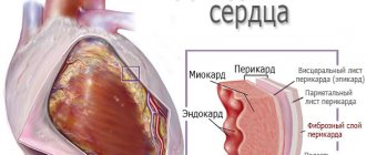
General characteristics of the pathology
Pulmonary artery stenosis (PAS) is characterized by lesions that collectively are associated with obstruction of the right ventricular tract of the heart. As a result, the movement of biological fluid in the area of the pulmonary artery (PA) valve is disrupted, which affects the condition of the entire body. This defect is considered a complex pathology.
Causes
Most often, congenital changes are diagnosed. The appearance of this congenital heart defect (CHD) can be triggered by reasons that during pregnancy can affect the formation of the main organ in the fetus. These include:
- inhalation of toxic fumes by a pregnant woman, for example, while working with toxic substances in hazardous industries;
- failures of embryonic evolution due to taking medications that negatively affect the fetus (taking psychotropic drugs, drugs, corticosteroids, cytostatics, antibiotics);
- previous diseases of viral etiology during pregnancy - influenza, rubella, herpes viruses;
- gene mutation in one of the parents (especially on the mother’s side);
- living in an area with polluted air, for example, next to large-scale production.
Therefore, expectant mothers need to protect themselves as much as possible from the effects of these unfavorable factors. Only in this case, congenital pulmonary artery stenosis does not threaten the fetus.
The causes of acquired narrowing of the orifice of the pulmonary artery include:
- various infectious lesions (rheumatism, syphilis, tuberculosis);
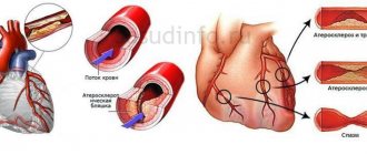
- systemic pathology – atherosclerosis of large vessels;
- aneurysm of the cardiac aorta - a local expansion of a section of the main artery due to the weakness of its walls;
- calcification of the pulmonary artery, aortic valves - disturbance of the electrolyte balance of the blood caused by diseases of the kidneys, endocrine system and other factors;
- hypertrophic cardiomyopathy – primary isolated myocardial damage, characterized by ventricular hypertrophy and a decrease in heart volume;
- endocarditis, pericarditis, myocarditis - complications of many infectious diseases;
- myxoma of the heart is a benign neoplasm;
- Carcinoid tumor is a slow-growing type of malignant tumor.
In some cases, the valvular type of disease causes enlargement of the lymph nodes, which compress the pulmonary column.
Clinical classification
Experts distinguish 4 stages of pulmonary valve stenosis, which are classified according to the level of blood pressure and the pressure gradient (difference) that occurs in the parts of the heart.
| Stage | Short description | Systolic blood pressure (mmHg) | Gradient (mmHg) |
| First (moderate) | Clinical symptoms of moderate stenosis are mild, and no changes are recorded on the ECG. Blood pressure is within normal limits | 60 | 20-30 |
| Second (pronounced) | The first symptoms of the disease appear | 60-100 | 30-80 |
| Third (sharply expressed) | Blood circulation is severely impaired, clinical manifestations are extremely pronounced | Above 100 | Above 80 |
| Fourth (decompensation) | Insufficient contractility of the ventricle occurs, and as a result, dystrophy of the heart muscle (myocardium) develops. | Decreasing | Decreasing |
Attention! Decompensation is especially dangerous when the risk of death increases.
The main classification of stenosis occurs according to the location of the narrowing, which can form in the following places:
- valve (frequency 90%);
- subvalvular (5%);
- supravalvular (5%);
- combined (a combination of several types of stenosis or with other heart defects).
In medical practice, isolated valvular stenosis of the pulmonary artery is more often diagnosed. The remaining cases occur in subvalvular and supravalvular types of stenosis.
Clinical manifestations
The intensity of clinical manifestations depends on the stage of the pathology. As a rule, a mild form of the disease is not accompanied by any symptoms. Nothing bothers the person, only sometimes slight shortness of breath may occur. It is usually associated with lack of sleep and emotional stress.
Severe symptoms occur when blood pressure levels increase and the narrowing is more significant. The most common manifestations accompanying pulmonary stenosis:
- rapid fatigue;
- constant weakness, drowsiness;
- muscle weakness;
- pulsation, swelling of veins in the neck;
- pain in the chest area, radiating to the shoulder, neck;
- increased heart rate;
- attacks of shortness of breath with minimal exertion;
- dizziness, loss of coordination;
- swelling of the lower extremities (usually in the evening);
- blueness of skin, lips.
Attention! In children, the disease manifests itself as frequent colds, dizziness, loss of consciousness, and delayed physical and mental development.
Symptoms appear gradually and usually begin to bother a person when right ventricular failure develops.
Pulmonary stenosis in children: symptoms
Of course, the signs of the disease directly depend on the stage of its development. In the initial stages, there may be no symptoms at all. But as the pathology progresses, the first sign appears - shortness of breath. First, breathing problems appear during physical activity, and then at rest.
The child gets tired quickly, suffers from weakness and dizziness. The little patient becomes drowsy. Sometimes you can notice severe swelling and pronounced pulsation of the veins in the neck. Older children sometimes complain of a burning sensation and pain in the chest. The child often loses consciousness. The doctor may hear a heart murmur during auscultation. More serious circulatory disorders are accompanied by pallor - the skin often acquires a bluish tint (the color is most pronounced in the area of the nasolabial triangle).
Clinical picture
The symptoms of the disease are not always obvious. For example, when systolic pressure is in the range from 50 to 70 millimeters, symptoms are not determined.
The following symptoms are characteristic of severe pulmonary artery stenosis:
- angina pectoris;
- heart murmurs;
- fainting;
- drowsiness;
- pulsation, swelling of the veins in the neck;
- heart hump;
- painful sensations in the heart;
- excessive fatigue associated with physical activity, even minor ones;
- dyspnea;
- constant weakness;
- dizziness.
Signs of pulmonary stenosis
Note! If a child experiences these symptoms, it is necessary to immediately consult a doctor and undergo appropriate examination.
Pulmonary artery stenosis in children: how dangerous is the pathology? Possible complications
This is a very dangerous pathology that should never be ignored. A child with stenosis is more predisposed to developing various inflammatory diseases of the respiratory tract.
Impaired blood flow leads to gradual degeneration of the myocardium, which can result in a heart attack, stroke, or severe right ventricular failure. The most dangerous complication of isolated pulmonary artery stenosis in children is septic endocarditis. That is why a child with such a diagnosis must be constantly under vigilant supervision.
Features of the course of the disease
When pulmonary stenosis is observed, the size of the opening may vary. It is he who largely influences the well-being of a sick child. If the size is normal and ensures blood flow, such an ailment may not be detected immediately. Sometimes diagnosis reveals the disease after a few years of life, under random circumstances, for example, during a routine examination. It is worth noting that the characteristic signs do not provide a clear clinical picture: a specialist can determine that the pressure in the left side is higher when compared with the right side. In this case, even the characteristic trembling that occurs over the heart cannot be considered an obvious symptom.
When the hole has a minimum diameter of less than one millimeter, the baby must undergo urgent surgery. In this case, the outflow of blood is so seriously impaired that only surgical intervention saves the child from death.
Complications in the development of pulmonary artery stenosis are associated with a significant load placed on the right ventricle of the heart muscle. Ultimately, the internal cavity of the ventricle expands, and the wall noticeably thickens. If the baby is already suspected of developing this heart defect, he needs to be constantly monitored and undergo regular examinations. A characteristic picture of the development of the disease is given by pressure, which is measured directly at the heart valve. The critical data of the examinations are as follows: fifty millimeters is the difference established during the examination between the pulmonary artery and the right ventricle. In this case, there is a need for immediate surgical intervention. In accordance with statistical data from medical practice, operations for established arterial stenosis are usually performed on children under 9 years of age.
Isolated pulmonary stenosis
In adults, this defect is most often not observed, since it is operated on in infants or in childhood. However, if the defect is eliminated due to standard dissection of the valves, then the correct geometric structure is no longer restored. In the future, the patient may begin to suffer due to disturbances in the functioning of the heart valves. That is, blood flows through normally, but the valves will no longer be able to close completely. Because of this, adults who suffered stenosis as children may need a special heart prosthesis instead of a valve. This complication is becoming a common consequence of this disease.
It is important. Nowadays prosthetics are installed in many hospitals. The operations are successful, there is no threat to life.
Diagnostic measures
As a rule, such a disease is diagnosed immediately after the birth of a child or in the first months of life. During examination, a systolic murmur is heard in the second intercostal space (on the left side of the sternum).
An obligatory part of the diagnosis is an electrocardiogram. If we are talking about the initial stage of development of the disease, then some changes may be absent. However, in most cases, signs of right ventricular hypertrophy can be detected during the procedure. Sometimes the appearance of supraventricular arrhythmias is observed.
Echocardiography is also informative. During the procedure, the doctor may notice dilation of the pulmonary artery and dilatation of the right ventricle of the heart. Sometimes Doppler ultrasound is additionally performed - this procedure allows you to determine the difference in pressure between the right gastric and pulmonary trunk.
Diagnosis necessarily includes a chest x-ray. In the image you can see the expansion of the pulmonary artery trunk. A very characteristic symptom is depletion of the pulmonary pattern.
Possible complications, prognosis
Chronic failure of the right ventricle of the heart is the main complication of narrowing of the artery if it is not treated. This pathological condition can lead to sudden death of the patient. Dangerous consequences also include:
- myocardial infarction (heart attack);
- acute cerebral circulatory disorder - stroke;
- pneumonia - pneumonia;
- infective endocarditis;
- disruption of myocardial function.
Acute heart failure is a postoperative complication that is often associated with surgical errors.
The prognosis for slight and moderate narrowing is conditionally favorable even without surgical intervention. It is necessary to prevent further development of the disease. Therefore, the prognosis is quite favorable if surgery to eliminate the stenosis is performed in a timely manner. When the defect is congenital, it must be done as early as possible.
Attention! Some parents believe that surgery is very dangerous for their child. But delaying treatment is even more dangerous. Typically, a sick child dies within 3-5 years if surgery is not performed on time.
The outcome of the disease is unfavorable in the absence of surgical treatment. In this case, the risk of death increases several times.
Is drug treatment possible?
The treatment regimen is selected individually, since much depends on the stage of development of the pathology. If the narrowing is small and the blood flow is not impaired, then special treatment may not be required - sometimes a mild degree of stenosis goes away on its own with age.
However, the only way to correct the narrowing is surgery. Drug therapy is carried out only as preparation for surgery. In particular, antibiotics are prescribed to young patients to protect against bacterial infection and prevent septic endocarditis.
Treatment
Patients must be fully monitored, which includes:
- diagnosis of streptococcal infection, concomitant sanitation carried out in chronic lesions;
- conducting echocardiography;
- preventive measures to prevent infective endocarditis;
- prophylactic antibiotic therapy.
Treatment of pulmonary stenosis
Therapy is carried out medicinally and therapeutically. The main method of combating this serious illness is surgery. However, the patient must be prepared for surgical intervention, as an extreme and labor-intensive measure. Most often, surgery is performed when stenosis develops in the second or third stage. In addition, treatment is carried out to improve the general condition of the patient if the stage is no longer operable.
When is surgery necessary?
Surgical treatment of pulmonary artery stenosis in children can be carried out in different ways - in this case, the location of the narrowed area is of great importance.
The most effective and efficient procedure is endovascular balloon valvuloplasty. A special balloon is inserted through the venous wall into the affected area of the vessels. Using a catheter, air is pumped into the balloon, as a result of which the device inflates and expands the walls of the vessel, thereby eliminating the narrowing.
Open valvuloplasty is performed much less frequently. In this case, the patient must be connected to a heart-lung machine. The doctor requires direct access to the heart and blood vessels in order to carefully examine the affected area and excise the fused commissures.
Sometimes a small patient needs systemic-pulmonary bypass. If supravalvular stenosis occurs, reconstruction is performed using a patch from the patient's own tissue. In some cases, installation of a prosthesis is required.
In most cases, surgery is performed when the child reaches 5-10 years of age. However, in the presence of severe stenosis, surgery may be prescribed in infancy. In most cases, the procedure is successful. Nevertheless, even after successful correction of stenosis, the child (and then the adult patient) must undergo regular examinations by a cardiologist and carefully monitor their health.
How is the treatment carried out?
For minor stenosis, drug therapy is prescribed.
The main groups of funds used are indicated in this table:
| Group | Drugs |
| Potassium-containing products | Asparkam, Orocamag, Potassium orotate, Panangin |
| Vitamin complexes | Vitamins A, B, C, P |
| Cardiac glycosides | Digoxin, Korglykon, Celanide, Strophanthin |
Medicines are prescribed to maintain the functioning of the heart and large vessels. A radical way to treat stenosis is to perform surgery.
The purpose of the procedure is to normalize blood flow in the right side of the heart. The type of procedure is determined taking into account the localization of pathological foci.
Among them:
- Balloon valvuloplasty. It is effectively used when the patient has valvular venous stenosis. To carry it out, a special balloon is inserted into the mouth of the affected vessel. After this, a stent is installed in it, with the help of which the narrowing is eliminated.
- In the case of narrowing of the supravalvular localization, the most effective is resection of part of the vascular walls that have undergone stenosis. Instead, patches taken from the patient's pericardium are applied.
- The essence of surgical therapy for subvalvular stenoses is to eliminate areas of cardiac muscle hypertrophy in the area of the right atrium.
- To treat a combined type of disease, a combination of the above methods is used.
After the narrowing is eliminated surgically, blood circulation in the area of the mouth of the pulmonary trunk is normalized over time. Symptoms of the pathology gradually decrease, the patient’s general condition stabilizes.
Prevention: how to avoid danger?
Unfortunately, there is no specific prevention, since the development of the disease is associated with factors that are not always possible to influence. It is very important for the expectant mother to monitor the progress of pregnancy, maintain normal body function, eat right, avoid contact with infectious patients and not take any medications without a doctor’s permission. Of course, you need to regularly undergo routine examinations and ultrasounds - this makes it possible to diagnose certain pathologies in the fetus even before its birth.
Treatment of PA stenosis
Treatment for PA stenosis depends on its severity and the age of the patient. Typically, moderate narrowing in one or two branches of the pulmonary artery does not require treatment. In more severe cases it is used:
- Expansion using a balloon. With this method, a special catheter with a balloon is inserted into the narrowing of the pulmonary artery. Once positioned, the balloon is gently inflated, causing the narrowed area to expand. After this, the balloon is deflated and removed. Although most children improve after balloon dilatation, in 15–20% of cases the pulmonary artery at that location narrows again over time.
- Balloon expansion and stent implantation. To improve the results of balloon expansion, it is supplemented with stent implantation. A special stent system is inserted into the narrowed area of the pulmonary artery, after which the balloon is inflated to the recommended pressure. This inflated balloon expands the stent and implants it into the vascular wall at the site of narrowing. Similar procedures are carried out, for example, for diseases such as renal artery stenosis and coronary heart disease.
- Surgery. Various techniques are used to surgically repair the pulmonary artery. The choice of the appropriate surgical method depends on the characteristics of the stenosis, the properties of nearby blood vessels and other anatomical structures.
Forecasts for young patients
Valve pulmonary artery stenosis in a child (as well as any other form of narrowing) is a pathology that should never be ignored. Fortunately, timely diagnosis allows doctors to detect the disease in time and take appropriate measures.
Minimal narrowing of the pulmonary artery has virtually no effect on the quality of life or development of the baby. Nevertheless, children with this diagnosis should be registered with a cardiologist and periodically undergo comprehensive examinations. If we are talking about valvular pulmonary artery stenosis in a child, then there is a possibility that in adulthood the patient will need surgery to replace the valve.
Diagnostic Basics
Instrumental diagnostics plays an important role in determining pulmonary artery stenosis. First of all, the doctor listens to the heart very carefully. A rough systolic murmur in the second intercostal space and between the shoulder blades is a sign of stenosis. The second tone in the first and second stages of the disease is heard practically unchanged, but with pronounced narrowing of the PA it can disappear completely. The doctor will then order an electrocardiogram. If the disease is mild, then no changes are recorded on the ECG. In subsequent stages, hypertrophy of the right ventricle is observed, and sometimes signs of arrhythmia are detected.
The complex of diagnostic measures also includes the following instrumental studies:
- radiography;
- Ultrasound (ultrasound examination);
- dopplerography;
- Ventriculography.
All of these methods allow you to assess the condition and size of the heart, the structure of the valve ring. In addition, Dopplerography of the veins of the lower extremities, catheterization of the cardiac cavities, computed tomography (CT), magnetic resonance imaging (MRI), clinical and biochemical analysis of biological fluid, and urinalysis will be required. The most important thing in diagnosis is to differentiate pulmonary stenosis from other heart diseases that have a similar clinical picture.
Manifestations in children and adults
Symptoms of pulmonary artery stenosis in children may appear immediately after the baby is born, or, if the pathology is not sufficiently pronounced, they may not appear for a long time. Sometimes, until the child enters adulthood. However, basically, the disease makes itself felt immediately, or during the first months of life.
Pulmonary artery stenosis in children manifests itself in different ways and correlates with the degree of narrowing of the lumen. The first degrees of pathology may not appear at all for several years. Typically, such pulmonary valve stenosis is detected during a routine examination.
Starting from the third degree, symptoms appear, which are as follows:
- blue discoloration of certain areas of the skin;
- the appearance of shortness of breath in a calm state, during feeding;
- severe fatigue;
- uncharacteristic restlessness in newborns;
- growth retardation;
- little weight;
- shortness of breath during the development of motor skills.
Such clinical manifestations concern children only with severe stenosis. The fourth form is considered the most severe and requires immediate surgical intervention.
In adults, pulmonary valve stenosis has completely different symptoms. It may not appear for many years if it was not diagnosed in childhood. The patient may not be aware of the problem throughout his life.
Clinical manifestations begin to concern patients only if right ventricular failure develops. Symptoms are usually gradual, growing rather slowly.
Symptoms include the following:
- dizziness;
- reduced performance;
- weakness;
- dyspnea;
- severe swelling.
If such clinical manifestations are present, you should visit a doctor as soon as possible. You need to contact your local doctor and cardiologist.
Pulmonary stenosis in newly born babies is most often diagnosed in the maternity hospital. During the examination, the neonatologist draws up an action plan that will help accurately diagnose the child. The children are then observed by a pediatrician.
When the first symptoms of stenosis appear, if it has not been identified previously, you should consult a doctor as soon as possible. After certain procedures to diagnose the disease, a specialist will be able to make a diagnosis and prescribe treatment.
Why does pathology occur and how to diagnose it?
Narrowing of the pulmonary artery is a congenital or acquired pathology that can occur as a result of the following reasons:
- while carrying the baby, the mother suffered a dangerous illness (rubella);
- the expectant mother took medications other than as prescribed by the doctor;
- As a result of illnesses suffered by a person throughout his life, compression of the artery can develop.
The disease can be diagnosed only through a comprehensive examination, which includes:
- Listening to heart murmurs. As stenosis develops, a rough systolic murmur is heard. Mainly heard between the shoulder blades. The noise appears only at the third stage of the disease, and may be absent at the first two.
- An ECG (electrocardiogram) does not show abnormalities if the stenosis is minor. However, in severe forms of the pathology, deviations appear, expressed in hypertrophy of the right ventricle of the heart muscle and arrhythmia.
- Doppler allows you to identify differences between the ventricles (right and left), which indicates the presence of disorders.
- X-rays show changes in the pulmonary pattern, including an enlarged pulmonary artery.
After a full examination, the specialist makes a diagnosis. In accordance with the form of stenosis, treatment is prescribed and prognoses are made.
Let's sum it up
Pulmonary artery stenosis is a truly complex disease that cannot be treated simply by taking pills or mixtures. To eliminate the disease, the only effective and efficient method today is surgery, which belongs to the category of the most complex surgical interventions in the circulatory system.
To prevent the diagnosis from becoming a death sentence, it is important not to panic, but to immediately seek qualified help and agree to an operation if it is recommended by specialists. Optimistic outlooks on life, following doctors’ recommendations and effective treatment when used in combination will help prolong the patient’s life and significantly improve its quality.
Symptoms
The severity of symptoms depends on how narrowed the artery is. Often the disease manifests itself in newborns or young children, much less often in adolescents and adults.
A heart defect can be hidden, not showing any symptoms throughout life, or have minor manifestations that the patient can confuse with symptoms of other ailments. The defect can be accidentally detected on an ultrasound of the heart. The main and most characteristic symptoms are:
- fast fatiguability;
- general weakness;
- pain in the sternum;
- rapid onset of shortness of breath during physical activity;
- bluish skin tone;
- periodic fainting and dizziness.
Methodology for the treatment of pulmonary artery stenosis
Pulmonary artery stenosis is a disease that is considered a life-threatening defect and requires only surgical treatment. Drug therapy is provided only in cases where it is necessary to prepare the patient for surgery, or if the disease is at the last, inoperable stage. In such situations, patients need the strictest control of doctors, consisting of the following measures:
- Systematic echocardiography to monitor the condition of the artery and right atrium.
- Antibiotic therapy aimed at preventing infectious diseases.
- Prevention of infectious myocarditis.
- Carrying out procedures to improve the patient's health.
Surgeries to eliminate stenosis are indicated for patients diagnosed with category 2 and 3 disease. With mild stenosis, the patient is not prepared for surgery, however, he is under regular supervision of cardiology specialists. In the fourth phase of the disease, the feasibility of surgery is decided by doctors. Often only supportive therapy is provided, since surgery can worsen the situation and hasten cardiac arrest.
In any case, if specialists offer surgery, it makes sense to agree to the procedure, since this is the only chance to save life and capacity.
There are several types of operations used to eliminate the defect by modern medicine:
- Pulmonary artery valvuloplasty. The specificity of the operation lies in the introduction of a special device through the left intercostal approach to the heart, with the help of which the newly formed membrane is dissected, and a probe is installed in the opening to expand it.
- Catheter-type valvotomy. A special probe equipped with knives is inserted through the vein to make cuts at the site of stenosis, as well as a medical balloon, which is installed to restore the blood flow line.
- Open valve valvotomy is considered the most difficult and at the same time effective. The operation is performed by opening the patient's chest, after first connecting the patient to equipment that provides blood circulation in the body artificially. In this case, valvular stenosis of the pulmonary artery can be examined from the inside, and incisions can be made precisely according to the location of the muscle tissue. Open surgery allows you to visually examine the pathological formation and effectively eliminate the defect.
Pulmonary artery valvuloplasty
Preventive actions
As preventive measures, it is necessary to protect the woman from negative external factors that may affect her during pregnancy. If a baby is suspected of having an illness, he should be registered with a cardiologist. In this case, it will be possible to prevent the development of infective endocarditis.
Against the background of the defect, myocardial dystrophy additionally develops. Some patients also experience inflammation in the upper respiratory tract. The situation can lead to septic endocarditis. Only in advanced cases does the patient experience stroke, heart failure and heart attack.
Additionally, it should be noted that it is necessary to eliminate the pathology as early as possible. For this purpose, surgery is performed in early childhood. Only in rare cases do parents refuse surgery (for religious reasons). This leads to an increased risk of death. Even if the fetus is diagnosed with pulmonary stenosis, parents should not despair. Today there are many methods and ways to eliminate the defect.
Manifestations in children and adults
Pulmonary artery stenosis in adults coincides with childhood symptoms in only one point - it can also remain in a “dormant” state for quite a long period, without tormenting the body with excessive nutritional deficiencies. A person can live a long life and not even suspect that anything in his structure differs from general norms.
However, if the acquired defect begins to manifest itself clearly, the development of the disease does not stop until it can be stopped with radical treatment methods.
Symptoms of pulmonary artery stenosis in adults are caused by:
- Loss of performance, dizziness and nausea;
- Symptoms of shortness of breath, acute lack of air with minor physical effort, and, during the development of the disease and during periods of relaxation;
- Swelling of the legs at the very primary stage of heart failure.
- The next stage is the accumulation of fluid in the abdominal and chest cavities.