General information
Thrombosis is the formation of a blood clot in a blood vessel that interferes with blood flow.
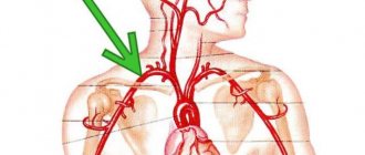
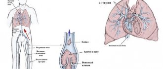
Its danger lies in the fact that there is a danger of a blood clot or its fragment moving through the bloodstream - an embolism and blockage of another vessel occurs. For example, supplying the brain or heart. Pulmonary embolism often occurs, which leads to severe organ dysfunction or death. What is pulmonary embolism in medicine? Pulmonary embolism is an acute occlusion (blockage) of the pulmonary artery trunk or its branches (main, lobar or segmental). Occlusion occurs more often due to embolization of a thrombus from the right half of the heart or veins of the lower extremities. The incidence of this disease increases with age and the age of such patients is 62 years.
Pulmonary artery thrombosis is an emergency in cardiac resuscitation and is often the cause of patient death. Seriously ill patients with a high risk of pulmonary embolism and death are patients with cardiac arrhythmias, cancer pathology , and thrombosis of the veins of the lower extremities . Diagnosis is often difficult, so the disease is often not recognized. Early diagnosis and initiation of intensive treatment are important in the prognosis of this disease. The code for pulmonary embolism according to ICD-10 is I 26.
Treatment
Timely diagnosis of pulmonary embolism significantly increases the chances of a successful outcome, because mortality is reduced to approximately 1-3%. Today, pulmonary embolism is still a problem for treatment, this is due to the possibility of almost immediate death of the patient.
Anticoagulant therapy can be prescribed to the patient at the stage of diagnosing the disease before the final test results. The main goal of this method is to reduce mortality among people at high risk of the disease, as well as if there is recurrent pulmonary embolism. Anticoagulant therapy is suitable for the prevention of pulmonary embolism.
Treatment of pulmonary embolism with anticoagulants lasts on average about 3 months, although according to doctor's indications, the duration of therapy can be increased. Thrombolytic therapy is based on the use of medications, the exact dosage and administration of which is calculated by the doctor based on the patient’s weight and current condition. Here is a list of the most popular drugs that a history of pulmonary embolism requires for use:
- Unfractionated heparin;
- Enoxaparin;
- Rivaroxaban;
- Warfarin.
During the treatment of symptoms of pulmonary embolism, the patient also takes special medications. Unlike anticoagulants, which only slow down growth and are often taken as a prophylactic agent, thrombolysis involves dissolving the embolus. This method is considered more effective, however, recommendations for the treatment of pulmonary embolism allow its use only in life-threatening cases. Thrombolytic therapy is combined with anticoagulants. This method of treatment is not completely safe, since the dissolution of blood clots often leads to bleeding, among which the greatest harm is caused by intracranial bleeding.
- The fastest way to solve the problem is surgical thrombectomy. The operation will help even in cases where the pulmonary embolism develops quickly, but the patient will be promptly taken to the cardiac surgery department. This method involves cutting both pulmonary arteries and removing the clot.
- Another option to address the problem of repeated episodes of pulmonary embolism is venous filters. The technique is mainly used for contraindications to anticoagulants. The essence of the filters is that they prevent detached emboli from reaching the pulmonary artery along with the bloodstream. Filters can be installed for several days or for a longer period. However, such treatment for pulmonary embolism usually comes with many risks.
- It is worth noting the peculiarities of treatment in specific patients. Pulmonary embolism can occur in pregnant women, but diagnosing them is difficult. At moderate or low risk, a blood test for D-dimer is practically useless, since during this period its values will in any case differ from normal. Carrying out CT scans and other diagnostic procedures involves exposure of the fetus to radiation, which often negatively affects its development. Treatment is carried out with anticoagulants, since most of them are absolutely safe both during pregnancy and lactation. It is not possible to use vitamin K antagonists (warfarin) alone. When treating pulmonary embolism, the doctor pays special attention to the delivery.
- If the cause of the blockage is not a blood clot, but another clot, a pulmonary embolism will be treated based on the reasons for its formation. A foreign body can only be removed surgically. However, if the clot that formed after an injury consists only of fat, treatment does not require surgical intervention, since the fat will resolve on its own over time; it is only necessary to maintain the patient in normal condition.
- Removing air bubbles from the bloodstream is done by inserting a catheter. An infectious embolus is removed with intensive treatment of the disease that caused it. Unfortunately, the most common cause of infectious embolism is intravenous drug administration through an infected catheter. Complications of pulmonary embolism in this case manifest themselves not just in the form of an obstructing clot, but also in sepsis.
Pathogenesis
The main thing in the pathogenesis of pulmonary embolism is occlusion of the pulmonary artery and its branches by embolism, which leads to disruption of hemodynamics and gas exchange. During occlusion, the pressure in the pulmonary artery increases by almost 50% due to vasoconstriction associated with the release of serotonin and thromboxane A2 . A sharp increase in pulmonary resistance causes expansion of the right ventricle, stretching of myocytes and tension of the ventricular walls. Lengthening the time of its contraction towards diastole causes protrusion of the septum into the left ventricle. Therefore, left ventricular filling becomes more difficult, cardiac output decreases, and systemic hypotension .
Massive infiltrates are found in the ventricular myocardium, which further destabilizes hemodynamics. Right ventricular failure due to pressure overload is a leading cause of death. Respiratory failure is also associated with hemodynamic disturbances. A decrease in cardiac output causes desaturation of venous blood. The appearance of zones of reduced blood flow causes a mismatch between perfusion and ventilation. Small emboli in the periphery do not affect hemodynamics, but cause pulmonary hemorrhage with hemoptysis, pleurisy and pleural effusion (“pulmonary infarction”).
Treatment of pulmonary embolism
In the acute period of PE, the fundamental issue in the treatment of the patient is to preserve the patient’s life, and in the long-term period, treatment is aimed at preventing possible complications and preventing recurrent cases of PE.
The main directions in the treatment of pulmonary embolism are the correction of hemodynamic disorders, removal of thrombotic masses and restoration of pulmonary blood flow, prevention of recurrent thromboembolism.
In a situation where PE of segmental branches is diagnosed, accompanied by minor hemodynamic disturbances, anticoagulant therapy is sufficient. Anticoagulant drugs have the ability to stop the progression of existing thrombosis, and small thromboemboli in the lumen of segmental arteries are independently lysed.
In hospital settings, the use of low molecular weight heparins is recommended, which are free of hemorrhagic complications, have high bioavailability, do not affect the functioning of platelets and are easy to dose when used. The daily dosage of low molecular weight heparins is divided into two doses, for example Fraxiparine is used subcutaneously in 1 monodose up to 2 times a day. The duration of heparin therapy is 10 days, after which it is advisable to continue anticoagulant therapy using indirect anticoagulants in tablet form for 6 months (Warfarin 5 mg once a day).
All patients receiving anticoagulant therapy should undergo a screening test of laboratory parameters:
- fecal occult blood test;
- blood clotting indicators (aPTT daily throughout heparin therapy). A positive effect of anticoagulant therapy is considered to be an increase in aPTT by 2 times compared to the initial value;
- a complete blood test with determination of the platelet count (indication for discontinuation of heparin therapy is a decrease in the platelet count by more than 50% of the initial value).
Absolute contraindications to the use of indirect and direct anticoagulants for pulmonary embolism are severe cerebrovascular accidents, cancer, any form of pulmonary tuberculosis, chronic liver and kidney failure in the decompensation stage.
Another effective direction in the treatment of pulmonary embolism is thrombolytic therapy, but for its use there must be convincing indications:
- massive pulmonary embolism, in which more than 50% of the blood volume is excluded from the bloodstream;
- severe disturbances of pulmonary perfusion, which are accompanied by severe pulmonary hypertension (pressure in the pulmonary artery more than 50 mmHg);
- decreased contractility of the right ventricle;
- severe hypoxemia.
The drugs of choice for thrombolytic therapy are: Streptokinase, Urokinase and Alteplase according to developed regimens. Scheme for using Streptokinase: during the first 30 minutes, a loading dose of 250,000 units is administered, and then the dose is reduced to 100,000 units per hour for 24 hours. Urokinase is prescribed at a dosage of 4400 IU/kg body weight for 24 hours. Alteplase is used at a dose of 100 mg for 2 hours.
Thrombolytic therapy is effective in lysing the thrombus and restoring blood flow, but the use of thrombolytics is dangerous due to the risk of bleeding. Absolute contraindications for the use of thrombolytic agents are: early postoperative and postpartum period, persistent arterial hypertension.
To assess the effectiveness of thrombolytic therapy, the patient is recommended to undergo repeated scintigraphy and angiography, which are screening diagnostic methods in this situation.
There is a method of selective thrombolysis, which involves the introduction of a thrombolytic into a blocked pulmonary vein using a catheter, but this manipulation is often accompanied by hemorrhagic complications at the site of catheter insertion.
After completion of thrombolysis, anticoagulant therapy using low molecular weight heparins is always carried out.
If there is no effect from the use of drug treatments, the use of surgical treatment is indicated, the main goal of which is to remove the embolus and restore blood flow in the main trunk of the pulmonary artery.
The most optimal method of embolectomy is to perform the operation via transsternal access under conditions of assisted venoarterial circulation. Embolectomy is performed by fragmenting a thrombus using an intravascular catheter located in the lumen of the pulmonary artery.
Classification of pulmonary embolism
The 2008 classification of the European Society of Cardiology identifies:
- Low risk pulmonary embolism.
- Intermediate.
- Tall.
Clinical classification takes into account the caliber of the pulmonary arteries and the percentage of pulmonary involvement:
- Massive (cessation of blood flow in less than 50% of the pulmonary bed). It is of great clinical importance because it is accompanied by shock or decreased pressure, pulmonary hypertension. With occlusion of the trunk, pronounced cardiorespiratory disorders develop. Under such conditions, the right ventricle cannot perform the function of a pump and its cavities quickly expand, and tricuspid valve insufficiency develops. The septum between the ventricles shifts to the left (towards the left ventricle), which is accompanied by poor filling in diastole. Due to the cessation of blood flow, the pulmonary parenchyma is not supplied with blood, but is ventilated. In the affected area, obstruction of the bronchi occurs, the alveoli collapse and surfactant is not formed in them, which contributes to the development of atelectasis (collapse) of the lungs on days 1-2 of embolism. Such hemodynamic disturbances and impaired pulmonary function often lead to the death of the patient.
- PE of small branches of the pulmonary artery (cessation of blood flow is noted in less than 30% of the pulmonary bed). In this case, there is occlusion of lobar and segmental small branches. The disease is not severe without hemodynamic disturbances and patients only need anticoagulant therapy. The pulmonary circulation has compensatory capabilities and there is a possibility of independent dissolution of blood clots when fibrinolysis .
- Submassive—thromboembolism of the branches of the pulmonary artery (cessation of blood flow in less than 30-50% of the bed). It manifests itself as right ventricular failure, and hemorrhagic infarctions form in the lungs.
Diagnostic measures
Early and correct diagnosis makes it possible to identify thrombosis and prescribe adequate treatment for the disease.
Emergency cardiac care has a developed algorithm for diagnostic measures, which include certain examinations that make it possible to recognize the disease in the shortest possible time. They are usually divided into 3 stages:
- Stage before hospitalization. It consists of monitoring the manifestations of symptoms and collecting anamnestic data.
- Stage 2 of diagnosis is carried out in a hospital setting using non-invasive (hardware) techniques.
- Stage 3 involves identifying the location of blood clots, as well as ways to eliminate them.
Stages 2 and 3 of diagnostic studies are very important, since only with their help can the presence of the disease be accurately determined.
They include the following examinations:
- ECG. Using electrocardiographic studies, sinus arrhythmia and the peak of the P wave are determined. These factors indicate congestion of the right atrium, which leads to pulmonary embolism.
- CT scan. With its help, you can clearly monitor the presence of thrombosis in the arteries of the lungs.
- Ultrasound of veins. Allows you to determine the onset of embolism at the very first stages of the disease.
- Dopplerography. With its help, areas of arteries with impaired blood flow speed are determined.
- Radiography. A very accurate study that allows not only to identify the location of the blood clot, but also its size.
- ECHO-CG of the heart. With its help, you can examine the entire vascular system of the heart, and also see a clear enlargement of the right ventricle.
No less important diagnostic factors are the results of laboratory tests, which, together with hardware tests, make it possible to accurately determine the disease. After confirming the diagnosis, treatment is prescribed, and in the case of an acute or subacute course, emergency care is provided.
Causes of pulmonary embolism
Considering the causes of pulmonary embolism, it is necessary to highlight a number of diseases and conditions that are accompanied by the formation of blood clots, which are the basis of embolism:
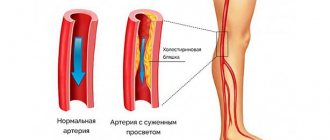
- Varicose veins and phlebothrombosis of the deep veins of the lower extremities. This pathology causes thromboembolism in 90% of cases. The threat is posed by floating blood clots, which are freely located in the lumen of the vessel and are connected to the vein wall in only one part.
- Congestive heart failure , overstretching of the right ventricle, which creates conditions for the formation of blood clots in the cavity of the right ventricle.
- The use of oral contraceptives and pregnancy , which are associated with an increased risk of blood clots.
- Antiphospholipid syndrome (autoimmune thrombotic vasculopathy ), manifested by venous and arterial thrombosis . Vessels of different sizes may be affected - capillaries and large arterial trunks. Deep vein thrombosis of the lower extremities is a typical manifestation of antiphospholipid syndrome. Repeated pulmonary embolisms are typical.
- Central venous catheterization.
- The presence of malignant diseases.
- Carrying out chemotherapy .
- Spinal cord injury.
- Forced immobilization after surgery, during a stroke , fractures of the pelvis and limbs.
- Hip replacement.
- Atrial fibrillation.
- Polytrauma and extensive surgery.
- Hereditary predisposition to thrombosis ( thrombophilia ), which is caused by mutations of certain genes, deficiency of antithrombin III, deficiency of protein C and S.
Considering pulmonary thrombosis , it is necessary to note the risk factors for this condition:
- Damage to the venous endothelium.
- Obesity.
- Age.
- Hypercoagulation.
- Slowing of venous blood flow.
- Infection.
- Blood transfusions.
- Migraine.
- The combination of several factors is accompanied by a high risk of thrombus embolism .
What is thromboembolism
Diseases of the legs are increasingly disrupting a person’s normal life, because pathologies of the vascular system are one of the first places in patient complaints in medical institutions. One of the most dangerous disorders in the functioning of veins and blood vessels is thromboembolic disease.
Let's figure out what it is - thromboembolism, as well as what symptoms it has and why it is dangerous? A detailed study of this topic will allow you to detect pathology in time and take immediate action.
A thrombus is a blood clot that has accumulated in a vessel and blocks the blood flow, thereby disrupting the normal functioning of veins and blood vessels, making it difficult for the supply of the required amount of oxygen.
Risk factors
Risk factors for vein thrombosis and pulmonary embolism are:
- a long-term state of immobility (bed rest, frequent and long flights, trips, paresis of the limbs), chronic cardiovascular and respiratory failure, accompanied by a slowdown in blood flow and venous stagnation.
- taking large amounts of diuretics (massive loss of water leads to dehydration, increased hematocrit and blood viscosity);
- malignant neoplasms - some types of hematological malignancies, polycythemia vera (a high content of red blood cells and platelets in the blood leads to their hyperaggregation and the formation of blood clots);
- long-term use of certain medications (oral contraceptives, hormone replacement therapy) increases blood clotting;
- varicose veins (with varicose veins of the lower extremities, conditions are created for stagnation of venous blood and the formation of blood clots);
- metabolic disorders, hemostasis (hyperlipid proteinemia, obesity, diabetes mellitus, thrombophilia);
- surgery and intravascular invasive procedures (eg, central catheter in a large vein);
- arterial hypertension, congestive heart failure, strokes, heart attacks;
- spinal cord injuries, fractures of large bones;
- chemotherapy;
- pregnancy, childbirth, postpartum period;
- smoking, old age, etc.
The clinical manifestations of pulmonary embolism are nonspecific; they can be observed in other pulmonary and cardiovascular diseases; their main difference is the sharp, sudden onset in the absence of other visible causes of this condition (cardiovascular failure, myocardial infarction, pneumonia, etc.). The classic version of PE is characterized by a number of syndromes:
Treatment of pulmonary embolism
Once the diagnosis of pulmonary embolism is confirmed, therapy begins immediately. If there is a very high probability of pulmonary embolism, medical therapy may be started even before the diagnosis is confirmed.
Blood thinners - anticoagulants .
The main treatment for pulmonary embolism is the use of anticoagulant blood thinners to prevent further clotting.
Blood thinners commonly used to treat PE are either intravenous heparin , or a heparin derivative that can be given by subcutaneous injection, such as Arixtra or Fondaparinux .
The heparin family of drugs provides an immediate anticoagulant effect and helps prevent further blood clot formation.
Thrombolytic therapy.
When PE is severe and causes cardiovascular instability, anticoagulation therapy is often insufficient. In these situations, powerful clot-busting agents called thrombolytics are used. These drugs include fibrinolytic agents such as streptokinase , which are designed to dissolve a blood clot that is blocking a pulmonary artery.
Thrombolytic therapy carries significantly greater risks than anticoagulant therapy, including a high risk of serious complications. If the pulmonary embolism is severe enough to be life-threatening, the potential benefits of this treatment may outweigh the side effects of this class of drugs.
Surgery.
Surgery is a method that can directly remove the clot. The most common surgical procedure, called surgical embolectomy , is quite risky and not always effective, so it is reserved for people who have a very low chance of survival without surgery.
Surgery is a method that can directly remove the clot. The most common surgical procedure, called surgical embolectomy , is quite risky and not always effective, so it is reserved for people who have a very low chance of survival without surgery.
Damage to the upper and lower extremities
Thromboembolism of the arteries of the legs (femoral) or upper extremities (brachial, ulnar and radial) occurs against the background of the same pathological changes. However, the frequency of such phenomena is low. Diagnosis is not particularly difficult, since embolism almost always affects only one limb. In addition, the disease occurs after a massive fracture, with injury to large vessels, and this is worth remembering. Characteristic signs may indicate a diagnosis even at the prehospital stage:
- severe, unbearable pain in the limb, below the site of thrombosis;
- changes in skin color, from marbled to whitish, gray and sallow;
- decreased limb temperature;
- lack of pulse;
- swelling and cyanosis may occur with venous thromboembolism;
Such patients require emergency surgical care, and here, its timely provision should be extremely important. After removal of the blood clot and a period of rehabilitation, the basic functions of the limb can be restored. A vascular incident that has occurred should draw the doctor’s attention to the causes of this phenomenon. Namely, to the diagnosis of mitral valve pathology.
Diagnosis of thromboembolism should be based on determining the causative diseases and assessing risk factors. Considering the serious consequences, the pathology must be identified and stopped before the thrombus begins to migrate along the vascular bed. Particular attention should be paid to older patients with rheumatic heart disease and valvular defects. The list of diagnostic tools can cover various methods of almost every type of clinical medicine:
Thromboembolism of the mesenteric arteries
This condition is rare. Diagnosing the disease before surgery is difficult. Most often, patients with this disease are admitted to the hospital with a diagnosis of peritonitis. The main symptom is severe abdominal pain. In this case, perietal symptoms are observed.
- loss of consciousness;
- pale skin;
- chills;
- shallow breathing;
- convulsions;
- cyanosis of lips, skin, limbs;
- cough;
- decrease in blood pressure;
- pulse is frequent and difficult to hear;
- convulsions;
- massive bleeding.
Symptoms of pulmonary embolism (PE)
Making a diagnosis in the early stages is difficult, only in the acute course the symptoms of pulmonary artery thrombosis appear manifestly and suddenly: shortness of breath , chest pain , tachycardia , decreased blood pressure. In this case, the patient has risk factors for thromboembolism - varicose veins and phlebothrombosis .
Hypotension and shock indicate central thromboembolism . Severe acute shortness of breath also develops with central pulmonary embolism. With the same localization, chest pain is angina-like in nature . Massive thromboembolism is accompanied by signs of right ventricular overload - this is the bulging of the jugular veins in the neck and the right ventricular gallop rhythm.
Signs of PE in subacute cases are not very specific. Respiratory and right ventricular failure progresses, infarction pneumonia and hemoptysis appears. With a recurrent course, repeated attacks of shortness of breath , there are signs of pneumonia and periodic fainting occurs. If the thrombus blocks small arteries in the periphery, shortness of breath is minor and transient. Only in the case of chronic lung pathology or heart failure does shortness of breath worsen. Sometimes PE is asymptomatic.
The clinical picture depends on the caliber of the blocked vessel and, accordingly, the degree of involvement of the pulmonary vascular bed. Massive pulmonary embolism (most often thrombosis of the main branch ) occurs with symptoms of shock or a decrease in pressure by 40 mm Hg. Art. for a short time that is not associated with arrhythmia , sepsis , or decreased blood volume. Characteristic shortness of breath , cyanosis , and sometimes fainting .
Submassive pulmonary embolism (obstruction of lobar or segmental branches) is manifested by right ventricular failure (swelling of the neck veins, pallor, cyanosis), but without arterial hypotension. The patient develops shortness of breath , tachycardia , pulmonary infarction (fever, cough, pulmonary-pleural pain, sputum streaked with blood).
In non-massive cases, there are no signs of right ventricular failure, and the pressure is normal.
Symptoms
The clinical picture depends on a lot of factors: the exact localization of the source of obstruction, the size of the blocking agent, and the duration of the pathological process.
We are not talking about a total blockage. Because the last thing a person manages to feel is acute pain in the chest, lack of air. Then comes fainting and death.
In other situations, clear symptoms of pulmonary thromboembolism are detected. Verification of the diagnosis is carried out urgently, using instrumental methods.
The pulmonary embolism clinic includes the following manifestations:
- Pain syndrome in the chest. Pulling, pressing. It is painful for the patient because the resistance differs. Although at medium intensity. The duration of discomfort is indefinitely high.
There is almost no spontaneous regression. When you inhale, especially deeply, the pain increases sharply. Therefore, the patient tries to control the natural process, which leads to hypoxia and aggravation.
- Cough. Unproductive in the sense that there is no phlegm. But blood flows abundantly in the form of foamy scarlet clots.
This is an indication of increased pressure in the small circle, ruptures of blood vessels. Unfavorable sign. In almost all cases, it indicates thromboembolism of the pulmonary arteries.
It is not difficult to exclude other diseases, such as tuberculosis or cancer - signs of pulmonary embolism develop suddenly and are complemented by aspects atypical for oncology or infection.
- Increased body temperature. Up to febrile levels (38-39 degrees), other levels are also found.
Symptom relief with antipyretic medications does not provide significant effectiveness, because the process is of central origin. Provocateurs are not infectious agents. This is a reflex reaction of the body.
- Shortness of breath at rest. During physical activity it reaches critical levels, making it impossible even to move at a slow pace or change body position.
A decrease in exercise tolerance develops rapidly, which is typical for pulmonary thromboembolism and atypical for other cardiovascular diseases.
- Tachycardia. The body increases the number of heartbeats to provide oxygen to all tissues.
Quality is replaced by quantity: the blood moves faster in order to somehow be enriched with oxygen in small parts.
The system is running out of steam. Very soon the process is reversed. Heart rate decreases. At first it is insignificant, a slight bradycardia is detected (about 70 beats), then it goes into a critical phase.
- Falling blood pressure levels. Also gradual, but pronounced. It is discovered as decompensation progresses.
- Headache. An alarming manifestation. Indicates a gradual disruption of nutrition of cerebral structures. In the short term it may result in a major stroke and death of the patient.
- Vertigo. Inability to navigate in space. The person takes a forced lying position and moves less so as not to provoke an increase in the symptom. Also called dizziness.
- Nausea, vomiting. Are relatively rare
- Paleness of the skin and mucous membranes.
- Cyanosis of the nasolabial triangle. Formation of a bluish ring around the mouth. A typical symptom of cardiac dysfunction and oxygen starvation of the body.
If you look at the problem from a temporal perspective and monitor its progression, the average time from the onset of the first symptoms of pulmonary embolism to death ranges from 3 to 12 days. Rarely more. Perhaps less.
Tests and diagnosis of pulmonary embolism
Instrumental diagnostics of this condition includes:
- Chest X-ray . In the lungs, discoid atelectasis , a raised dome of the diaphragm, or pleural effusion . All these signs are nonspecific, but exclude various causes of pain and shortness of breath.
- ECG.
- Echocardiography . It is of key importance in diagnosis, especially in patients with unstable hemodynamics. This test directly detects blood clots in the right side of the heart, and blood clots in the large arteries of the lungs (trunk and main branches). An indirect sign of pulmonary embolism detected by echocardiography is overload of the right chambers. An indirect sign of pulmonary artery thrombosis is identified - overload of the right ventricle: expansion of the ventricular cavity, unusual movement of the septum between the ventricles and a D-shaped left ventricle.
- Ultrasound of the deep veins of the extremities.
- A special research method is ventilation-perfusion scintigraphy . This is a safe study that involves the intravenous administration of albumin microspheres that are labeled with technetium. Albumin microspheres block pulmonary capillaries and lung perfusion is assessed based on this feature. This study is complemented by a ventilation study, which increases the specificity of the examination (in case of thrombosis, ventilation in segments of the lungs in which there is poor blood supply remains normal). That is, a discrepancy between ventilation and perfusion is detected. Radioactive radiation during scintigraphy is lower than during CT angiography. Perfusion scintigraphy alone can be performed in patients with a normal radiograph.
- CT angiography . This study has become the method of choice for diagnosing pulmonary vascular pathology. The pulmonary arteries can be viewed down to the segmental branches. The specificity of the method is 96%. The technique is optimal for diagnosing a patient whose condition is stable. In another case, the patient cannot be transported from intensive care.
- Pulmonary angiography is considered the gold standard for diagnosing PE. However, it is rarely performed since a less invasive test, CT angiography, has become available. Diagnosis is based on identifying a blood clot that causes a defect in the filling of the pulmonary artery branch or is completely absent. The method allows you to obtain images of peripheral pulmonary arteries and detect blood clots of 1-2 mm in the smallest arteries. A contrast agent is injected for the study.
ECG signs of PE and radiographic signs
Blood tests:
- D-dimer is a fibrin degradation product. Its increase in plasma is observed during acute thrombosis - this is explained by the activation of fibrinolysis and coagulation. A normal D-dimer level makes the diagnosis of PE unlikely. Also, this indicator is nonspecific, since fibrin overproduction is observed during inflammation, bleeding, myocardial infarction , aortic aneurysm , oncological processes, trauma, and surgery. The specificity of D-dimer for thrombosis decreases with age. This indicator is paid attention to during treatment, since an increase in its level after the end of anticoagulant treatment indicates a risk of relapse.
- of brain natriuretic peptide increases , which is associated with distension of the right ventricle. The degree of increase is proportional to the severity of the patient. However, an increase in this marker is nonspecific, since it can be observed with myocardial ischemia , left ventricular hypertrophy , sepsis and tachycardia . However, the absence of a significant increase indicates a favorable prognosis for pulmonary embolism.
Diagnostics
If pulmonary embolism is suspected, a complex of laboratory and instrumental examination is prescribed, including:
- X-ray of the chest organs - signs of pulmonary embolism are: atelectasis, congestion of the roots of the lungs, amputation symptom (sudden rupture of the vessel), Westermarck's symptom (local decrease in pulmonary vascularization);
- ventilation-perfusion scintigraphy of the lungs - signs of a high probability of pulmonary embolism are: normal ventilation and decreased perfusion in one or more segments (the diagnostic value of the method is reduced in cases of previous episodes of pulmonary embolism, lung tumors and chronic obstructive pulmonary disease);
- angiopulmonography is a classic method for diagnosing pulmonary embolism; The criteria for diagnosis are the detection of a thrombus outline and sudden rupture of a branch of the pulmonary artery;
- electrocardiography (ECG) - allows you to identify indirect signs of pulmonary embolism and exclude myocardial infarction.
Source: stopvarikoz.net
Differential diagnosis is carried out with non-thrombotic pulmonary embolism (tumor, septic, fat, amniotic), psychogenic hyperventilation, rib fracture, pneumonia, bronchial asthma, pneumothorax, pericarditis, heart failure, myocardial infarction.
In children
Thrombosis in children occurs much less frequently. For their occurrence, a combination of acquired and congenital factors is necessary. Of the hereditary ones, it is worth noting the hereditary decrease in the activity of antithrombin III and proteins C, S, which are natural anticoagulants. Acquired factors:
- severe somatic diseases
- infectious diseases;
- systemic connective tissue diseases;
- malignant tumors;
- connective tissue dysplasia ;
- hemolytic anemia;
- antiphospholipid syndrome.
The most important factor in thrombosis is the use of an intravascular catheter. Up to 80% of thromboses are associated with catheterization. In children, pulmonary embolism is often associated with deep vein thrombosis. Venous thrombosis in children is preceded by infection, thrombocytopenic purpura , antiphospholipid syndrome , and in some, excessive insolation. Recurrent pulmonary embolism also occurs, but the source of thrombosis cannot be identified. PE in children is often mistakenly regarded as pneumonia .
Causes of development and factors that increase risk
Typically, blockage of the pulmonary artery occurs due to the entry of a large blood clot that has broken off from the wall of one of the venous vessels. The blockage occurs unexpectedly and the symptoms of this condition develop very quickly. Also, an obstacle to blood flow is created by:
- harmful microorganisms;
- air bubbles that entered the vessel during surgery or intravenously;
- a foreign body in the form of a bone marrow clot or adipose tissue that enters the artery during mechanical damage;
- blood clots penetrating into the lungs from foci of infectious disease in other organs;
- accumulation of cancer cells;
- amniotic fluid.
In pregnant women, the constantly enlarging uterus puts pressure on the venous vessels. This increases the risk of a blood clot breaking off and entering the systemic circulation. An additional danger is posed by hormonal changes that increase the likelihood of thrombosis in the deep veins of the legs. In addition, blockage of the artery can be caused by amniotic fluid entering the bloodstream during childbirth.
When infected emboli penetrate small branches of the pulmonary artery, a septic pulmonary embolism occurs. It is a consequence of sepsis, which develops in organs with infectious infection. Most often, this condition occurs in people who administer narcotic drugs intravenously.
Although there are many causes for pulmonary embolism, most often this condition develops due to a detached blood clot from the venous vessels of the legs or from the inferior vena cava. The bloodstream carries the embolus to the pulmonary artery. If it is small in size, the embolism occurs without obvious symptoms.
The risk of developing a pulmonary embolism, with or without mention of a heart attack, increases if a person does not exercise much. In such people, blood stagnation becomes a provoking factor. Therefore, people who, due to their profession, are forced to sit for a long time or spend a lot of time in an upright position, regularly undergo preventive examinations for the condition of their veins. To prevent thromboembolism from developing, such people need to play sports and lead a more active lifestyle.
The use of certain medications increases the likelihood of developing pulmonary embolism. The disease is one of the side effects of taking diuretics. They dehydrate the body and cause blood thickening. Other dangerous drugs are various hormonal drugs. They provoke changes in hormonal levels and an increase in blood clotting.
Important! Hormonal medications include contraceptives, medications for the treatment of infertility and certain thyroid diseases.
Prevention of pulmonary embolism
- Primary prevention of embolism after surgery on the pelvic organs and orthopedics.
- If the risk of thromboembolism is low, prevention involves the use of compression stockings or elastic compression with an elastic bandage of the lower extremities.
- enoxaparin or fondaparinux or dabigatran until discharge .
- If the risk is high, these recommendations should be followed for a month.
- New oral anticoagulants are registered for primary prevention after orthopedic surgery.
There are also non-surgical patients (chronic heart failure class III-IV), who are also at risk for pulmonary embolism. Their choice of anticoagulant is the same. The standard of treatment is enoxaparin .
Secondary prevention is the prevention of recurrent thromboembolism. Without prophylaxis, recurrence of pulmonary embolism within the first three months is observed in 20-45% of patients. With prevention, the number of relapses is reduced to 1%. Warfarin is used for prevention , and for the first six months, cancer patients are prescribed low molecular weight heparins ( Fraxiparin , Fraxiparin Forte , Clexane , Enixum , Fragmin , Daltep ), then they are transferred to oral medications ( vitamin K antagonists - Warfarin , Warfarex ).
How long should I take them? To do this, the risk of bleeding is assessed based on risk factors (age 65 years or older, history stroke malignant tumor , renal and liver failure , diabetes mellitus , low platelet levels, anemia and others). The risk of bleeding is high if more than two risk factors combine.
It is also possible to use “new” anticoagulants for secondary prevention, which are included in the American Guidelines for the prevention of pulmonary embolism. Rivaroxaban ( Xarelto ) is superior in long-term treatment and can be prescribed for 6-12 months with a low risk of bleeding, providing protection against relapse.
Prevention
No specific methods have been developed. But you can minimize the risks if you adhere to certain points:
- Avoid stress. If it is impossible to master relaxation techniques. In order not to cause a hormonal “explosion” in every situation at work, study, etc.
- Eat consuming less animal fat and salt (no more than 7 grams).
- Sleep at least 7 hours per night.
- Adhere to an adequate individual physical activity regimen. Don't overexert yourself and don't sit still.
- Treat all pathologies in a timely manner, regardless of type and location.
- Regularly visit at least a therapist if you have problems with the cardiovascular system - a cardiologist. At least once a year for preventive examinations. Then the doctors will advise you on what to do if necessary.
Venous thromboembolism is a dangerous immediate or gradually increasing pathological process of partial or complete blockage of a blood vessel.
Requires urgent medical attention. Otherwise, dangerous complications cannot be avoided.
Consequences and complications
- Hemodynamic disturbances (increased vascular resistance in the pulmonary artery system and increased afterload on the right ventricle).
- Respiratory failure.
- Hypoxia.
- Pulmonary-pleural complications ( atelectasis , pleurisy , lung abscess , pulmonary infarction , pneumonia , pyopneumothorax , pleural empyema ).
- Post-embolic pulmonary hypertension .
Diagnosis of the disease
The range of diagnostic measures aimed at confirming pulmonary embolism and searching for the source of the embolus is as follows:
- Multislice (multi-slice) CT, with a slice thickness of 2.5 mm, makes it possible to establish thrombosis at the level of subsegmental arteries.
- CT with pulmonary angiography to assess the condition of the pulmonary artery and its branches using a contrast agent. A complication of CT with a contrast agent is nephropathy, which is characterized by an increase in creatinine. On CT, pulmonary embolism can be determined directly: a defect in the filling of the artery, its local expansion and sudden break, as well as indirectly: characteristic signs of pulmonary infarction, accumulation of exudate in the chest cavity and cardiac sac, disc-shaped collapses (atelectasis) of the lung tissue, avascular zones.
- CT phlebography for diagnosing thrombosis of the femoral veins and veins of the legs.
- MRI: the method has high sensitivity to vascular and parenchymal tissue, does not provide radiation exposure compared to CT, but the lengthy process of performing the procedure does not allow it to be performed in an emergency.
- determination of the partial pressure of oxygen and carbon dioxide (oxygen pressure is less than 80 mm Hg, carbon dioxide pressure is reduced or normal);
MSCT and CT with pulmonary angiography in the diagnosis of pulmonary embolism are methods No. 1.
Differential diagnosis:
- acute myocardial infarction;
- attack of bronchial asthma;
- attack of cardiac asthma;
- accumulation of blood or serous effusion in the heart sac (hemo- and hydropericardium);
- acute pneumonia;
- chronic pulmonary embolism.
List of sources
- Vatutin N.T., Sklyannaya E.V., Eshchenko E.V. /Pulmonary embolism. Review of recommendations of the European Society of Cardiology for diagnosis and treatment (2014 // Practical Angiology. - 2020. - No. 1 (68). - P. 5-18.
- Russian clinical recommendations for the diagnosis, treatment and prevention of venous thromboembolic complications // Phlebology. — 2020.— No. 4, issue 2. 46 p.
- Vatutin N.T. Emergency cardiology. - Donetsk, 2011. - 236 p.
- Vatutin N.T., Kalinkina N.V., Perueva I.A. /Pulmonary embolism // Practical angiology. – 2011. – No. 2. – P. 32-40.
- Vatutin N.T., Kalinkina N.V., Eshchenko E.V. and others. Pulmonary embolism (basic information and own observation) // Heart and Vessels. – 2013. – No. 1. – P. 120-123.
Symptoms depending on location
Thromboembolic disease mainly affects the arteries of the body, creating a colossal danger at the moment, since there is practically no time left to respond.
Death is likely in a matter of minutes or the development of dangerous complications.
The signs of thromboembolism are not the same and depend on where exactly the blockage occurred and are characterized by ischemic and hypoxic phenomena.
Damage to the abdominal arteries
They are also called mesenteric. The pathological process is accompanied by the development of a clinical picture of acute abdomen.
Among the typical points:
- Pain in the abdominal area. Intense, pressing, burning, cutting. Accompanied by movement back and forth, wandering, localization is not clear, so the patient cannot say exactly where the discomfort is concentrated.
- Nausea and severe vomiting. Possibly with blood if the disorder progresses.
- Flatulence, increased gas formation.
- Against the background of bloating, defecation disturbance is detected. A person may experience painful urges (tenesmus) with an inability to relieve themselves. It is possible to develop intestinal obstruction (probability about 70%).
- Collaptoid state. An increase in blood pressure, and then its rapid drop, increased sweating, tachycardia, increased heart rate, pallor of the skin, shallow breathing, depression of consciousness. An extremely worrying sign.
- Increase in body temperature (hyperthermia at a level slightly above 37.5 degrees).
Thromboembolism of the abdominal arteries is extremely dangerous. The development of intestinal necrosis and death from peritonitis in a short time is possible.
Blockage of arteries in the brain
Classified as ischemic or hemorrhagic stroke. Both forms carry great danger. Accompanied by general and focal neurological symptoms.
The first ones are almost always the same, with different intensities:
- Unbearable headache. Baling, pressing. Localized in the back of the head, crown. The temples or has a diffuse, diffuse character; it is impossible to determine exactly where it is located.
- Nausea, vomiting. Reflex, short-term.
- Impaired consciousness. Syncope.
- Dizziness. Vertigo. With the inability to navigate in space.
- Weakness, drowsiness, asthenia. The opposite effect is possible with the development of psychomotor agitation.
Focal signs depend on the specific location of tissue necrosis. If the occipital region is involved, vision suffers, the temporal region suffers from hearing, consciousness and memory, the frontal region affects behavior, intelligence, the parietal region suffers from smell, cognitive abilities, and so on.
Symptoms of the pre-stroke condition, depending on the location, are described in this article.
MRI helps to put an end to the question. Although not in all cases, survey data are sufficiently informative. Especially with the development of massive damage to brain structures (major stroke).
Involvement of the arteries of the extremities in the process
Accompanied by a critical malnutrition of the arms or, much more often, legs.
The clinical picture is typical, so it is almost always possible to detect the problem directly during the initial examination.
Among the symptoms:
- Pain in the leg area on the affected side.
- Severe numbness up to complete loss of sensitivity.
- General critical condition. Collapse.
- Paleness, bluishness of the skin layers, a feeling of goosebumps. The appearance of a vascular pattern on marble-colored skin.
In the absence of high-quality immediate surgical treatment, gangrene cannot be avoided. And then death (especially if the femoral artery is blocked).
Pulmonary artery damage
The most dangerous option in terms of the likelihood of death of the patient. Affects a key vessel of the pulmonary circulation.
When the blockage is more than 80%, sudden death occurs, without preliminary symptoms. The person does not have time to understand anything.
In less complex situations, the clinical picture develops in a matter of minutes. Doctors have about half an hour to provide assistance.
The following points indicate an emergency:
- Chest pain. Expressed. Unbearable. Bursting.
- Paleness of the skin.
- Dyspnea. Reaching asphyxia. When the body position changes to lying from sitting, threatening complications develop. A person may suffocate and die.
- Cough. At first unproductive, then with the release of bloody, foamy sputum of a pinkish hue, with scarlet streaks. Negative sign. Indicates the development of cardiac asthma.
- Impaired consciousness.
- Collaptoid state. Coma in difficult cases.
- An increase in body temperature to significant levels of more than 38 degrees.
Fatal complications, respiratory failure, increase quite quickly. Doctors have very little time to provide assistance and even transport them to the hospital.
Renal artery disease
It develops slowly and also progresses slowly over several days.
Among the typical manifestations:
- Lower back pain. From the side of the lesion, movement to the back and spine is possible.
- Discomfort when urinating.
- Delayed urine output.
- Increasing phenomena of oliguria. Reducing daily diuresis to 300-500 ml, and in critical cases nothing is separated at all.
- Blood in urine. Macrohematuria. The shade changes from straw yellow to pinkish and even red.
- Increase in body temperature.
- Collapathoid state.
Malignant hypertension may develop within a few days. With critical indicators of blood pressure, destruction of target organs (heart, brain, etc.).
Attention:
The progression time from the first manifestation to the death of the patient is 2-3 days.
Involvement of the peritoneal veins in the process
The symptoms of the lesion are approximately the same as described earlier (mesenteric arteries). The difference is that the time frame for progression of the pathological process is higher.
On the one hand, this is good, since there is much more time to provide medical care. On the other hand, blockage of veins poses a significant danger.
It is this type of pathological process that most often ends in necrosis of intestinal loops and the need for mutilating surgery to remove dead areas.
There are other, unnamed forms of the disorder. Thus, when the coronary arteries are damaged, an extensive infarction develops, most often ending in the death of the patient. Other veins in the body may be involved.
Symptoms of thromboembolism are specific, which is why doctors suspect the disease during the initial examination. The clinic plays an important diagnostic role.