Left atrium dilatation: what is it?
Dilatation of the left atrium is an expansion of the walls of the heart muscle caused by increased stress. This happens due to the fact that blood enters the organ in a volume exceeding the permissible norms. Pathology in the left atrium is observed many times more often than on the right side.
This is a dangerous symptom that can cause heart disease. The difficulty is due to the fact that in the early stages of development it passes almost without a trace. It is important that immediately after your health worsens, consult a cardiologist and undergo the necessary examination. The sooner a diagnosis is made and treatment is prescribed, the greater the chance of protecting yourself from irreversible consequences.
Preventive measures
It is easier to prevent a disease than to treat it for a long time. Both healthy people and those who have already encountered the disease should adhere to preventive measures. Doctors recommend:
- adhere to proper nutrition;
- quit smoking and alcohol forever;
- maintain a daily routine;
- engage in only feasible sports;
- spend more time in the fresh air, take walks;
- avoid stress and emotional overstrain.
Dilation of the left atrium cavity is a significant deviation that can seriously affect a person’s life. It is almost always associated with the development of dangerous life-threatening consequences. If dilatation is detected, you must follow all the recommendations of the cardiologist. By eliminating the cause in time, you can stop the further development of the problem.
Kinds
LA dilatation comes in 2 types:
- Tonogenic. It occurs due to an excess of blood filling in a muscle organ, which is why the chamber wall does not change at first, but gradually the pressure in it increases. With this type of pathology, the size of the heart remains practically unchanged, and fluid stagnation does not occur.
- Myogenic. The cause of the disease is a change in the heart muscles, because of this the ability to contract the myocardium gradually decreases. The tone of the organ decreases, muscle fibers lengthen and stretch.
Both deviations are a sign of the appearance of significant disturbances in the functioning of the body; if treatment is ignored, a person’s well-being will constantly deteriorate, and there is a risk of life-threatening complications.
Why does heart dilatation occur and how is it treated?
The widening of one or more chambers of the heart is called dilatation.
The main factors leading to an increase in the internal volume of the atria or ventricles are the action of high blood pressure and a decrease in the tone of the heart muscle.
This condition is not an independent disease and requires clarification of the cause of its occurrence. Once it is eliminated, the size of the heart can return to normal. An advanced form of dilatation is accompanied by heart failure.
Types of heart dilatation
Dilatation of the chambers of the heart can be:
- Tonogenic, caused by increased pressure in the cavity. Occurs when the valve openings are narrowed or there is increased pressure in the aorta or pulmonary artery. May precede myocardial hypertrophy. The tone of the heart muscle and contractility are preserved; it becomes the myogenic type as it progresses.
- Myogenic. The cause is a decrease in the contractility of the heart muscle due to dystrophic disorders. A persistent and irreversible process, the myocardial fibers are stretched and elongated. Occurs in myocarditis and atherosclerosis.
Why does the right and left sections, ventricles, cavities, chambers change?
Changes in each part of the heart occur due to disruption of the outflow pathways from it or increased load on the incoming increased volume of blood.
Left atrium dilatation
Expansion of the cavity can occur when the left atrioventricular opening is narrowed, impeding the passage of blood into the ventricle.
Increased intake due to arterial hypertension may subsequently increase wall stretching.
We recommend reading the article about dilated cardiomyopathy. From it you will learn about the causes of the pathology and the mechanism of its development, the symptoms of cardiomyopathy, the provision of diagnosis and treatment.
And here is more information about combined heart defects.
Left ventricular dilatation
The causes of increased left ventricular volume are associated with the following diseases:
Right atrium dilatation
Most often, the etiological factor is diseases of the bronchi and lung tissue, occurring with chronic obstruction (difficulty in patency). In addition, enlargement of the right atrium can be caused by:
- unclosed duct of Botallus,
- vices of Fallot,
- atrial septal defect,
- narrowing of the pulmonary trunk,
- valve insufficiency or narrowing of the opening between the right parts of the heart.
Right ventricular dilatation
Occurs with pulmonary hypertension, a hole in the interventricular septum, narrowing of the pulmonary artery, damage to the valves of the right heart, a heart attack that has spread to the right ventricle, prolonged cardiac stimulation.
Diseases leading to changes
Dilatation of the chambers of the heart may occur for an unknown reason. In this case, it is called idiopathic. Among the most common diseases of dilation are the following:
- congenital heart defects;
- rheumatism;
- bacterial endocarditis;
- viral infections;
- violation of immune defense, autoimmune processes;
- hypertension;
- intoxication with alcohol, drugs, toxic compounds, medications;
- hormone imbalance;
- deficiency of protein and B vitamins, selenium in the diet.
Symptoms of heart dilatation
For a long time, the expansion of the cavities of the heart may not manifest itself clinically due to the compensatory work of the heart muscle. As its reserve capabilities weaken, the following symptoms arise and progress:
- difficulty breathing during physical activity, and subsequently at rest;
- attacks of suffocation;
- cough, sputum streaked with blood;
- cyanosis of lips, fingertips;
- swelling in the legs in the evening;
- pain and heaviness in the right hypochondrium;
- weakness, dizziness, fainting;
- memory impairment;
- atrial fibrillation, extrasystole, blockade of impulses.
Watch the video about left ventricular hypertrophy, causes of pathology and treatment methods:
What are the health risks for children and adults?
With advanced dilatation, signs of circulatory decompensation increase:
- attacks of suffocation,
- inability to breathe in a horizontal position,
- wheezing over the surface of the lungs,
- swelling of the face and limbs,
- accumulation of fluid in the abdominal cavity and chest.
Ineffective cardiac output in children leads to developmental delays, including mental retardation.
Diagnostic methods
When making a diagnosis, the history of the occurrence of signs of circulatory failure is taken into account - shortness of breath, tachycardia, wheezing in the lungs, swelling of the extremities, expansion of the boundaries of the heart during percussion. To clarify the diagnosis, instrumental examination methods are required:
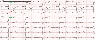
- ECG - signs of overload of one or more chambers of the heart, rhythm disturbances in the form of atrial fibrillation, conduction blockades.
- Ultrasound of the heart is the main way to detect dilatation of the atria or ventricles, as well as the presence of intracardiac hemodynamic disorders - reverse blood flow, pressure overload.
- X-ray – the size of the heart is increased due to one of the cavities.
- Scintigraphy to assess changes in the myocardium.
- MRI and CT scan to detect the cause of cardiac dilatation.
Treatment of dilatation
Therapy for enlargement of the heart chambers involves correction of manifestations of circulatory failure and arrhythmia.
In case of decompensation, patients are placed on bed rest and the intake of water and table salt is reduced. The following groups of drugs are used:
- ACE inhibitors – Enap, Capoten;
- diuretics - Veroshpiron, Triampur;
- antiarrhythmic - Bisoprolol, Carvedilol;
- long-acting nitrates (reduce blood flow to the right side of the heart) - Isoket, Nitrogranulong.
Cardiac glycosides are prescribed with caution. In extremely severe cases, a heart transplant is indicated.
Prevention
Prevention of heart enlargement consists of adequate treatment of rheumatism, endocarditis, viral and bacterial infections, which may be accompanied by myocarditis, identification and timely surgical treatment of congenital and acquired heart defects.
In case of diseases that are potentially dangerous in relation to dilatation of the atria or ventricles, it is necessary to completely abstain from alcohol and ensure a sufficient amount of protein and vitamins in the diet.
We recommend reading the article about right ventricular dysplasia. From it you will learn about the causes of the development of pathology and its symptoms, diagnosis and treatment, and prognosis for patients.
Read more about restrictive cardiomyopathy here.
Cardiac dilatation occurs when the myocardium is overloaded with excess blood volume or high blood pressure. The condition of the myocardium itself plays a major role in the development of pathology: during dystrophic or prolonged inflammatory processes, muscle fibers weaken, lose contractility and stretch.
Manifestations of heart dilatation include circulatory failure, arrhythmia, and in children there is a delay in growth and development. Medications are used for treatment; severe forms are an indication for heart transplantation.
Source: https://CardioBook.ru/dilataciya-serdca/
Degrees
Like any disease, dilatation of the left atrium cavity is classified into degrees. Depending on the affected area, it can be 1, 2, 3 or 4.
- The first degree is the easiest. If at this stage a diagnosis is made and treatment is prescribed, then it will be possible to get rid of the pathology almost without a trace. It can only be detected by doing an ECHO of the heart. The person continues to feel well and does not notice signs of pathology.
- The second degree is moderate dilatation. At this stage, primary symptoms of dysfunction of the muscular organ appear in the form of shortness of breath, rapid breathing and pain in the chest area. It is no longer possible to be cured completely even with timely diagnosis; a person can live quite happily with this disease if he follows the treatment and is under the supervision of a cardiologist.
- The third degree is severe pathology. It is impossible not to notice it, since by this time the patient is no longer able to perform the usual work and exercise. The muscular organ is already noticeably worn out, so a complete cure is excluded.
- The fourth degree is the terminal phase. The patient’s life is at risk, all treatment is aimed at making the person feel acceptable. The prognosis in this case is sad. The life span of a patient with extreme degree of left atrium dilatation does not exceed 3-4 years.
The pathology develops quickly. Complications from grade 1 to 4 usually occur within 4-8 years if the person does not adhere to the therapy prescribed by the cardiologist. Sometimes it is less, it depends on the individual characteristics of the human body.
Attention! It is important to always listen to your body and visit a clinic when the first symptoms of illness appear. In this case, it will not be possible to get rid of the disease, but it will be possible to increase life expectancy and normalize well-being.
Classification
Depending on the reasons for the expansion of the cavity, there are 2 types of left atrium dilatation:
- Tonogenic. It develops due to a persistent increase in pressure in the chambers of the heart, which leads to their excessive blood supply. This type is often combined with hypertrophy of the heart muscle (myocardium).
- Myogenic. This type of dilatation develops against the background of heart disease and leads to a weakening of the contractile activity of the myocardium. This condition is considered irreversible.
There are 4 degrees of development of the disease:
- Mild dilatation has no clinical manifestations. Identification is a matter of chance.
- It is a little easier to recognize a moderate degree of pathology, because... a person has symptoms of left atrium dilatation. With timely treatment, the problem can be successfully kept under control.
- Severe left atrium dilatation is characterized by clear clinical manifestations. The effectiveness of therapy is extremely low. A person's life expectancy does not exceed 5-6 years from diagnosis.
- Without treatment, a dangerous terminal phase occurs. In this case, they talk about palliative care, when therapy is aimed at maintaining an adequate standard of living.
Experts also distinguish between congenital and acquired forms.
Attention! The modern classification of pathology must be taken into account when determining the treatment regimen.
Symptoms
Dilatation of the left atrium is a disease in which it is impossible to postpone a visit to the doctor. You should be alert to the following signs of deterioration in health:
- general weakness, a feeling of heaviness and lethargy in the upper and lower extremities;
- decreased endurance and performance, the appearance of difficulties in performing usual household or production tasks;
- the appearance of shortness of breath, first after moderate physical activity, then in a calm state;
- swelling of the legs and arms;
- increased heart rate in a calm and relaxed state;
- pale skin and distinct expression of the nasolabial triangle.
The danger of dysfunction of a muscular organ is due to the fact that in the first and second degrees the disease goes away almost without a trace. A person may experience shortness of breath or an increased frequency of bullets after walking the usual distance, but he will not attach any importance to this. At this time, the pathology becomes more complicated, and a person’s life is at risk.
How to recognize the disease?
To determine the disease, the doctor needs to conduct a thorough diagnosis of the patient's cardiovascular system. Only after studying the examination results can a specialist announce the final verdict. Having learned what atrial dilatation is, patients are interested in how the diagnosis will affect their future lifestyle. If you regularly take prescribed medications and undergo examinations, then people are often not in danger. The length of life of a person with such a diagnosis depends entirely on how timely treatment was started.
Diagnostic methods:
- Chest X-ray. When studying the image, the doctor can see a bulging of the atrium on the left, its appendage. In addition, the pattern of the arteries will be strengthened, and the trunk of the left bronchus will shift slightly upward.
- Electrocardiogram (ECG). The technique will show an increase in the volume of the atrium on the left with a P wave, it looks wide and high. There are other signs of such a disorder that can be seen on the cardiograph tape.
- Echocardiography (EchoCG). It is the most informative way to diagnose dilatation, since it can be used to distinguish the valve apparatus, its condition, as well as the thickness of the myocardial muscle fibers and the size of the heart chambers. Echocardiography with Doppler more accurately assesses this situation.
After examining all the data obtained from the survey, it is sometimes necessary to apply additional measures. Blood tests are ordered to determine hormone levels or blood sugar levels. The attending physician decides this individually in each case.
Causes
The causes of stretching of the walls of the left atrium include:
- excessive physical activity, athletes whose activities are aimed at increasing muscle mass are at risk;
- improper functioning of the mitral valve, which is accompanied by backflow of blood;
- hereditary factor, the more people in the patient’s family who suffer from heart disease, the higher the risk that he will develop this pathology;
- anatomical changes in cardiac structures – arterial hypertension;
- benign or malignant tumors;
- lack of blood discharge into the main artery of the muscular organ, which causes congestion in the ventricles.
Development mechanism
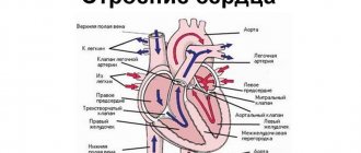
Normally, all 4 chambers of the heart (2 ventricles and 2 atria) function continuously and rhythmically. The release of blood into the systemic circulation is ensured by the alternating contraction of all organ structures. The functioning of the left atrium is closely related to the pulmonary circulation.
Under the influence of some unfavorable factors, the volume of biological fluid entering the cavity exceeds the permissible norm. Impaired blood outflow leads to stretching of the walls (dilatation) and a change in the size of the organ. Most often, one left atrium undergoes dilatation.
This condition is dangerous because it does not manifest itself in any way at the initial stages of development. Usually a person learns about the presence of a disease by chance, during unplanned diagnostic tests. A cardiologist should tell you more about what it is – dilatation of the left atrium.
Diagnostics
One of the first decisions when your health worsens is to visit a cardiologist. He will take an anamnesis based on the patient’s complaints, and then prescribe the following types of diagnostic measures:
- pressure measurement;
- determination of heart rate;
- ECG;
- ECHO;
- MRI - if the doctor suspects the development of a tumor in the heart muscle.
The main types of diagnostics for stretching the walls of the left atrium are ECHO and ECG. In the first case, the presence or absence of organic defects and their degree are determined. The ECG results will allow us to determine the degree of deviations in the functioning of cardiac structures.
Attention! During dilatation, it is prohibited to carry out test loads. This is associated with a high probability of cardiac arrest and sudden death.
Root causes of deviation
Dilatation of the right parts of the heart and its left part is caused by a violation of blood transportation through the atrioventricular openings. They are involved in the interaction between the left and right atrium. The deviation requires immediate investigation.
Dilatation of the LA (left atrium) occurs due to prolonged persistence of elevated pressure in the systemic circulation. Expansion of the cavity can occur with atrial fibrillation.
The deviation is often associated with regular and excessive physical activity.
Pathology can develop against the background of left atrium dilatation
The expansion of the right atrium (RA) is caused by a long-term increase in pressure in the pulmonary circulation. Pathology can make itself felt when:
- diseases of the bronchi and lungs of a chronic type, in which there is spasm of certain muscles;
- pathologies of blood vessels;
- infectious disorders of the heart muscles;
- pulmonary hypertension;
- heart defects that are present from birth or acquired at any age.
In some cases, the cause of the disease is pulmonary hypertension.
Normal heart weight is presented in the table.
| Gender of a person | Normal heart weight |
| In men | 335 grams |
| Among women | 225 grams |
The table shows approximate indicators. With dilatation, the volume of the organ chambers increases, but the thickness of their walls remains unchanged. The deviation is often present in hypertensive patients. With increased blood pressure, the heart muscles begin to contract more intensely. Pathological expansion occurs.
Excessive alcohol consumption can cause similar heart problems
Most often, patients have enlargement of the left atrium. The disorder is possible in people with alcohol addiction. Dilatation often occurs during an inflammatory process in the heart muscle. The long-term presence of pathology may be due to:
- fungal and viral infections;
- the presence of parasitic microorganisms;
- intoxication of the body;
- side effects after taking medications.
Various infections can cause pathology
Treatment
Therapeutic measures are aimed at reducing the load on the muscle organ, preventing the development of complications and improving the patient’s well-being. A cardiologist prescribes treatment with drugs from the following groups:
- diuretics – ensure proper fluid outflow, prevent the formation of stagnation;
- antiarrhythmic - normalize heart rate;
- glycosides – necessary to stabilize myocardial contractions;
- antihypertensives - prescribed only for systematic high blood pressure;
- antithrombic medications to prevent the development of blood clots due to fluid stagnation.
In addition to drug therapy, surgical intervention may be prescribed, in which it is necessary to remove the tumor or eliminate the pathology that caused the disease. In case of severe arrhythmia, a pacemaker is installed to support the functioning of the muscular organ. The last resort is transplantation, which is rarely performed due to the lack of suitable transplant material and the complexity of the procedure.
Treatment and prognosis
To eliminate the pathology, you need to find and make every effort to combat the underlying disease that led to dilatation. If the root cause can be identified and eliminated, then the size of the heart decreases or may even return to normal levels.
Until this happens, heart function may gradually deteriorate (dilated hypertrophy is formed), therefore standard therapy for heart failure with a reduced ejection fraction is carried out. Drug therapy is prescribed (ACE inhibitors; beta blockers, receptor blockers, nitrates, diuretics and digoxin); for atrial fibrillation, the use of anticoagulants and other drugs is indicated.
Surgery may be appropriate as long as the underlying cause of the chamber dilatation is addressed, otherwise the patient's survival will not be improved. In extreme cases (if the failure worsens), a heart transplant is recommended.
Possible complications
At the initial stage of stretching the walls of the atrium, human health is safe. As the pathology progresses, there is a risk of the following complications:
- blood stagnation, which disrupts muscle nutrition and causes inflammation of the myocardium;
- death caused by simultaneous atrial and ventricular fibrillation and severe arrhythmia;
- formation of blood clots and blockage of large vessels;
- heart failure;
- heart attack caused by acute lack of nutrition of cardiac structures;
- reverse outflow of blood, movement of fluid from the stomachs to the atrium;
- cardiogenic shock;
- stopping the work of the heart muscle.
If a person does not adhere to treatment, then his condition becomes critical. Death can occur at any time, and the risk increases after stress or physical exertion.
How dangerous is the pathology?
The condition threatens the development of a group of complications, including:
- Myocardial inflammation. They are formed as a result of blood stagnation and muscle malnutrition.
- Atrial fibrillation and then ventricular fibrillation. Characterizes severe arrhythmia, the functioning of cardiac structures is disrupted. The parallel course of the two processes determines the increased risks of death.
- Formation of blood clots, blockage of large vessels with a blood clot. A typical consequence of dilatation, since stagnation of blood contributes to its thickening.
- Congestive heart failure. In essence, this is dysfunction of a muscular organ of varying severity. It is determined by a group of species that differ in clinical manifestations, prognosis, and recovery prospects.
- Heart attack. Acute malnutrition of cardiac structures and, as a result of tissue necrosis, replacement of functionally active myocyte cells capable of contracting and conducting electrical impulses with rough scars. It is essentially dead tissue. The more it is, the worse the heart works.
- Paroxysmal arrhythmia. Usually of the type of tachycardia (acceleration of the activity of cardiac structures).
- Mitral insufficiency. It suggests possible regurgitation (reverse flow of blood from the ventricles into the atria), disruption of the functional activity of the organ and blood output, and a drop in hemodynamics. Lethal outcome is a matter of time.
- Cardiogenic shock. Acute, urgent condition. Requires urgent assistance, but the chances of the patient returning are almost negligible. Even if you are lucky, death will occur within a few years. Exceptions can be counted on one hand throughout the entire solid practice of doctors around the planet.
- Heart failure. Asystole.
Preventing the development of complications is one of the goals of treatment at any stage. It is best addressed early.
Prevention
The appearance of congenital pathology cannot be prevented. But, the risk of complications is minimal, since it is diagnosed on time and the patient is under the supervision of a cardiologist and adheres to treatment. The occurrence of a secondary disease can be prevented by following preventive measures:
- proper nutrition, adding nutrients to the diet;
- rejection of bad habits;
- regular exercise, only moderate physical activity is allowed;
- rational alternation of work and rest.
If there are many people in your family who suffer from heart disease, then you should periodically visit a cardiologist and conduct a diagnostic examination. This will allow you to identify the disease at an early stage of its development and promptly intervene, preventing the development of complications.
Preventive measures for cardiodilation
Dilation of the left atrium, right atrium, left and right ventricles, and aortic walls can appear from birth (congenital form) or during life (acquired form). Therefore, the general recommendations of doctors look like this:
- Give up bad habits: smoking, alcohol abuse and use of narcotic (psychotropic) substances.
- Follow your daily routine, eat right, eat enough fresh fruits and vegetables that contain a lot of useful vitamins and microelements, don’t overdo it. Nutrition should be moderate, but at the same time complete.
- Try to do everything to remove yourself from stress, negative emotions and significant physical activity. But this does not mean that physical exercise is prohibited. On the recommendation of the attending physician, light loads are permissible.
Left Atrial Dilatation and Surgery
This is important! If you follow these simple recommendations, you will be able to reduce the number of complications of cardiac pathology and the severity of the disease.
Forecast
The prognosis is favorable in one case, if a disorder of the cardiovascular system is detected at stage 1 or 2. The patient will be on maintenance treatment, so for the next few decades he will continue to feel well and not experience the symptoms characteristic of this disease.
If the disease is diagnosed already at stages 3-4, then the prognosis is unfavorable. Even with continued treatment, the heart muscle continues to wear out, leading to death. The maximum lifespan with this pathology is 10 years, but even with proper treatment, only a quarter of patients survive until this time.
Left half extension
The function of the left atrium is to pump blood from the lungs into the cavity of the left ventricle. From here, oxygen-enriched fluid enters the aorta and all organs. A valve is located between these chambers.
Atrial lesion
The cause of left atrium enlargement may be narrowing of the mitral valve leaflets or its deformation. Sometimes dilatation occurs with atrial flutter.
There are several degrees of expansion. With a mild degree, clinical manifestations do not yet occur. Detection of dilatation at this stage is most often a diagnostic finding during examinations for another reason. Initial organic changes can be diagnosed using echocardiography.
Moderate dilatation is detected more often. The patient develops the first nonspecific complaints - shortness of breath, arrhythmias, chest pain. It is more difficult to achieve a complete recovery at this stage, but you can control the process and get a stable remission with good health.
Severe dilatation is accompanied by severe symptoms and instability to physical activity. The skin becomes pale or bluish, breathing is heavy. Persistent changes are observed in other organs - the liver, etc. The prognosis at this stage is unfavorable - even with high-quality treatment, life expectancy is no more than 3-4 years.
The terminal stage occurs if the patient does not receive proper treatment. Symptoms depend on the disease that was the cause. Treatment is mainly palliative, aimed at improving well-being.
Ventricular dilatation
As the left ventricle expands, the pressure in its cavity increases. At the initial stages, this increase is compensated by hypertrophy of the heart muscle. For some time, compensatory mechanisms keep circulatory functions normal, the patient does not feel discomfort. At this time, pathology can be discovered by chance.
The causes of left ventricular dilatation are:
- Aortic valve narrowing or insufficiency.
- Aortic stenosis (narrowing) or aneurysm (widening and thinning of the walls).
- Myocarditis of various origins.
- Arterial hypertension.
- Rheumatism and other infectious diseases.
- Myocardial ischemia.
If, during examination of the patient, all of the listed causes were excluded, it is customary to speak of dilated cardiomyopathy.
With the acute development of the process, severe manifestations of heart failure, pulmonary edema, and cardiac asthma are observed.
Dilation of the atria and ventricles of the heart: treatment and prognosis
Dilatation is the physiological or pathological stretching of any hollow organ of the human body, in which hypertrophy of the walls is not observed.
The heart is an organ with four sections: two ventricles and the same number of atria. Each section of a muscular organ is capable of dilatation for various reasons. The main problem with this process is the inability to pump the required volume of blood. Stagnation is observed, as a result of which the pathological process progresses.
Timely diagnosis of the disease is important to prevent a fatal outcome.
Types of dilatation and reasons for its occurrence
There are two types of dilatation of the heart chambers:
- tonogenic dilatation develops in the chambers due to high blood supply. At the initial stages, the muscle wall remains non-hypertrophied;
- myogenic dilatation appears due to significant thickening of the heart wall, for this reason the contractile function of the myocardium suffers.
Common causes of dilatation of the heart chambers
The causes of overstretching of the chambers of the heart may be previous inflammatory diseases, when an infectious agent attacks the heart muscle.
There are frequent cases of fungal, parasitic and viral invasion, which negatively affect not only the contractile function of the myocardium, but also significantly enlarge the physiological cavities of the heart.
The toxic effect of alcohol and drugs, as well as some medications, have a negative effect on the main pump of the human body. Cases of dilatation of the heart chambers due to autoimmune, endocrine and oncological diseases have been reported.
Left atrium dilatation
The main role of the left atrium is to pump oxygenated blood to the left ventricle, from where it enters the aorta and is distributed to all human organs.
The most common cause of left atrium enlargement is a narrowing or other pathology of the valve located between the left chambers of the heart.
If the opening between them is insufficient, a large volume of blood cannot be quickly transferred from the left atrium, forming stagnation, which subsequently leads to stretching of the chamber walls. In addition, reverse blood flow is possible through the valve, which also provokes expansion of the atrium.
Another cause of left atrium enlargement is atrial flutter - atrial fibrillation. However, arrhythmia can develop secondarily, joining dilatation.
Main symptoms of left atrium dilatation
The symptoms of this disorder do not have their own distinctive signs, since they exist only in connection with other diseases. A person may complain of arrhythmia or valve stenosis. Among them are severe or moderate shortness of breath, pallor and cyanosis of the skin.
Left ventricular dilatation
The reason that causes dilatation of the left ventricle of the heart is the narrowing of the aortic valve, which connects it to the aorta.
The left ventricle performs the main pumping function, pumping blood to all organs of the human body. Therefore, even minor changes in the aortic valve will lead to severe distension of the ventricle.
Also, the cavity of this chamber can change greatly due to arterial hypertension, coronary heart disease and myocarditis.
Right atrium dilatation
The right atrium carries venous blood with carbon dioxide coming from organs and tissues. It is a link in the pulmonary circulation, and the reasons that provoke the development of dilatation of the right atrium are lung diseases. Congenital and acquired valve defects also play an important role in the occurrence of pathology.
Right ventricular dilatation
The main reason why the right ventricle expands is pulmonary hypertension, which forms when there is excessive load on the pulmonary circulation. Valvular changes as a result of bacterial infection (infective endocarditis) or fungal infection, as well as rheumatic heart disease, cause significant right ventricular dilatation.
Diagnosis of cardiac dilatation
Minor enlargements of the heart cavities do not lead to the patient’s characteristic complaints. Therefore, initial changes are often an incidental finding during routine preventive examinations.
The main diagnostic method is ultrasound examination of the heart. It allows you to evaluate the functioning of the organ in full: the size of the chambers, the condition of the vessels, valves.
Electrocardiography is used to exclude concomitant pathology of the cardiovascular system, type of rhythm and conduction disturbance.
Using chest x-ray, you can evaluate the size of the heart and its chambers and identify the presence of signs of pulmonary hypertension.
Stress tests provide valuable information about the functionality of the cardiovascular system.
Coronary angiography is prescribed to determine the tactics of surgical treatment.
Treatment of left heart dilatation
The main tactics for dilatation of the left chambers of the heart is the therapeutic correction of diseases that led to dilatation (coronary heart disease, myocarditis, defects, arrhythmia). Moderate dilatation is a reason to prescribe metabolic drugs, the action of which is aimed at improving metabolic processes in the cells of the organ.
Treatment of right heart dilatation
As in the case of the left side of the heart, the main method of correction is the treatment of underlying diseases. Without eliminating the primary causes, dilatation will only progress.
In cases where therapeutic methods do not give the desired result, the question is raised about solving the problem in a radical way, requiring surgery to transplant a donor organ.
Regardless of the affected part of the heart, almost every patient shows signs of heart failure.
Correction of this condition comes down to the prescription of medications from different pharmacological groups: aldosterone antagonists, diuretics, beta blockers, cardiac glycosides, antiplatelet agents.
The dosage of drugs is determined individually in accordance with the stage of the disease, the presence of concomitant ailments, the age and weight of the patient.
An integral part of the treatment is non-drug therapy: following a diet containing a small amount of salt, water and the complete exclusion of alcohol; daily physical activity, the intensity of which is determined by the stage of the disease.