Many people are interested in what this is - an enlargement of the left atrium. This means that this condition is pathological in nature, and pathology is often diagnosed in people of different ages. An increase in the size of the atrium on the left is regarded as a harbinger of quite serious diseases that are associated with the work of the heart vessels and its muscles. It is very necessary to identify this deviation in the patient as quickly as possible, and also to effectively eliminate it.
Description of the organ
The left ventricle is a special chamber of the heart that begins the largest circle of blood circulation.
Due to the fact that this ventricle contracts, it pushes out the main amount of blood, which is supplied to various organs and, in fact, the brain, also feeding the entire heart. That is, this chamber plays an extremely important role in the full functioning of the heart, experiencing considerable stress. Therefore, it is the left atrium that begins to suffer the consequences first when any problems occur.
The essence of the disease
An enlargement of the cavity of the left atrium is also called hypertrophy, a disease caused by thickening of the left side of the ventricle, which causes loss of elasticity of the surface. With uneven enlargement of the septum, a person may also additionally experience impaired functioning of the mitral and aortic heart valves.
An increase in the left atrium is indicated by a thickening of the myocardium, which reaches at least 1.5 cm. This disease is called the fundamental cause of myocardial infarction and the preliminary death of young people who are very actively involved in various power loads.
Left atrial volume enlargement that is not treated in time may progress. Adapting to more difficult working conditions, cardiomyocytes begin the process of proliferation; the walls of the ventricle, located on the left side of the heart, become denser and may lose their elasticity.
The insides of the chamber do not change, and the septum, which is located between adjacent ventricles, can gradually expand, which sometimes causes disruption in the functioning of the aortic and mitral heart valves.
Due to the enlargement of the left atrium in a child and an adult, the vessels of the myocardium, which require an increased amount of oxygen, may contract.
It is noteworthy that the signs of this pathology are not always pronounced, which, of course, aggravates the general condition of the sick person. With timely treatment, even advanced disease can be successfully cured. After you have found out what it is - an enlargement of the left atrium, the signs of which are not always visible, it is worth talking about what causes them.
What it is?
Right atrial hypertrophy, translated from medical terminology, means thickening of the walls of the atrium due to an increase in their muscle mass. It can be congenital or acquired in adulthood.
In children, pancreatic hypertrophy can be physiological in nature, for example in infants in the first days of life, when the load on the right side of the heart increases. Some symptoms of the disease do not appear immediately, but after a year or several years.
According to the degree of increase in the right side of the organ, the GPP can be: moderate, medium, sharp in severity. The disease does not occur in isolation; it is usually observed in combination with changes in other parts of the heart and neighboring organs.
Mechanical obstacles when pumping blood or returning part of the blood from the right ventricle lead to overload of the myocardium and a compensatory increase in its mass.
For example, narrowing (stenosis) of the tricuspid valve makes it difficult to push blood into the right ventricle. Increased pressure in the pulmonary artery in pulmonary diseases also leads to hypertrophy of the right atrium and right ventricle.
Hypertrophy can develop over several weeks or months; in some people, the compensatory abilities of the myocardium last for decades.
In any case, sooner or later there will come a time when the heart and its functions weaken or become completely exhausted. This condition is dangerous due to the development of severe pathology - a decompensated form of heart failure.
Reasons for the increase
The risk of thickening of the atrium on the left increases significantly if relatives have previously been diagnosed with heart disease.
Among the main causes of slight enlargement of the left atrium, the following are very common:
- diabetes;
- overweight;
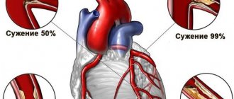
- ischemia and atherosclerosis;
- hypertension;
- heavy loads;
- peripheral vascular diseases;
- stressful situations;
- muscle dystrophy;
- aortic valve stenosis;
- Farby's disease;
- bad habits;
- mental instability;
- inactivity of a person;
- insufficient sleep.
The above factors provoke an increase in the pulsation of blood mass and an increase in the heart muscle, as well as excessive compaction of the left ventricular septum.
Other reasons
The severity of this condition depends on the loads that force the myocardium to work much harder. Factors that provoke enlargement of the left ventricle include the following:
- atherosclerosis of the aortic valve;
- arterial hypertension (very common);
- glomerulonephritis.
The listed reasons can provoke the development of acquired enlargement of the left cardiac ventricle. Doctors also identify a number of hereditary anomalies, which causes the growth of the muscle layer of the left atrium:
- reduced aortic size;
- mutation of genes that control protein synthesis;
- defect of the septum between the ventricles;
- fusion of the pulmonary artery;
- aortic valve stenosis;
- mitral heart failure.
Forecast, preventive measures
The prognosis depends on the course and severity of the disease that caused hypertrophy. With timely diagnosis and proper treatment, the outcome is favorable. The normal dimensions of the atrium are restored. The patient continues to lead his usual lifestyle. When heart defects occur, irreversible changes in the functioning of the myocardium are observed, and the prognosis worsens.
To prevent the development of HPP, you need to lead a healthy lifestyle, you must:
- eat right, follow a diet;
- monitor body weight;
- prevent obesity;
- spend more time in the fresh air;
- avoid stress and emotional experiences;
- go swimming;
- maintain a daily routine;
- stop smoking, alcohol;
- limit coffee consumption;
- take walks;
- take any medications with the permission of a doctor;
- treat any diseases, especially infectious ones, in a timely manner.
Normal sleep, diet, and feasible physical activity will help prevent heart problems.
Overload of the right atrium is the result of respiratory problems and cardiac problems. In order to diagnose the defect in a timely manner, you need to regularly visit a cardiologist and undergo a comprehensive examination. Changes in the right atrium can be completely corrected with the help of drugs without affecting the quality of life.
Danger of disease
It is also worth noting what threatens an enlarged atrium (left or right). As a result, the patient experiences a malnutrition of the heart, and abnormal zones of hyperactivity are formed. According to doctors, a person may develop arrhythmia, and as a result of the increased volume of the heart muscles and impaired vascular blood flow, the risk of tissue necrosis and coronary heart disease increases. But if there is a lack of oxygen, the situation will worsen.
Left atrial hypertrophy without proper treatment can have dangerous consequences, resulting in a sharply increased load on the myocardium, even death. This especially applies to inactive and smoking people who also drink strong drinks. Myocardial infarction, ischemia, and even cardiac arrest may develop.
Symptoms
Manifestations of HPP depend on concomitant pathology, as well as on how enlarged the right atrium is.
General symptoms:
- increased fatigue;
- problems with concentration, deterioration of attention;
- tingling or mild discomfort on the left side of the chest;
- paroxysmal interruptions of heart rhythm (extrasystole);
- cough, shortness of breath. swelling;
- impaired respiratory function, especially when lying down;
- pale skin tone, up to cyanosis (cyanosis);
At an early stage of development, hypertrophy of the right atrium occurs without visible changes in the general condition. Signs depend on the disease that caused changes in the parts of the heart.
For example, when cor pulmonale forms, the following terrible symptoms are observed:
- the appearance of shortness of breath during physical activity or at rest;
- dry hacking cough at night;
- coughing up blood.
Insufficiency of blood circulation in the main circle manifests itself when there is a large load on the right atrium, which it cannot cope with. This is due to stagnation of venous blood.
What does this threaten:
- pain in the right hypochondrium;
- swelling of the legs, especially in the morning;
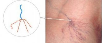
- abdominal growth with the development of varicose veins.
Symptoms of enlargement
Over a fairly long period of time, hypertrophy of the left cardiac ventricle may not manifest itself due to compensation for the proliferation of cardiomyocytes during normal blood circulation. After you have found out what it is - an enlargement of the left atrium, it is worth talking about its obvious signs.
The first signs of thickening of the heart tissue, which should alert the patient, are not recommended to be ignored:
- increased fatigue due to increased physical activity;
- shortness of breath, breathing problems;
- periodic painful sensations (burning, pressing or squeezing) in the heart, especially after exercise;
- frequent dizziness and fainting;
- pressure surges;
- atrial fibrillation;
- feeling of a sinking heart;
- angina pectoris;
- increased swelling of the legs;
- bad dream.
Signs of left atrium enlargement depend on the level of septal compaction, thickening of the heart and myocardium. This disease may indicate interruptions and inadequate functioning of the cardiac system. Thus, the patient may not suspect a developing disease for many years. Heart failure may occur due to fainting, which occurs due to insufficient oxygen in the myocardium. Such an increase is usually a concomitant symptom of a certain disease.
Among such diseases are myocardial infarction, pulmonary edema, kidney disease, heart disease, etc. If any abnormalities are detected, it is very important to immediately contact a doctor. If treatment is not timely, there is a risk of complications resulting in death or disability.
Varieties
The type of right atrium hypertrophy is directly related to the cause of the development of the pathology.
There are three types of GPP:
- Regeneration is a post-infarction condition in which a scar forms on the affected area. To restore the contractile functions of cells, a muscle layer grows around the scar.
- Replacement - the inclusion of a compensatory mechanism by increasing muscle mass for normal function in various diseases.
- Myofibrillar (or working) - develops as a result of constant physical overexertion in professional athletes or people in professions that involve heavy physical labor (miners, loaders, etc.).
How to solve a problem
When detecting enlargement of the left atrium, it is necessary to accurately determine the cause of this disorder. The main thing in treatment is to normalize myocardial function and prevent possible complications as much as possible. This can be achieved through surgical or medical methods. Complex treatment consists of:
- normalization of the rest regime, improvement of lifestyle;
- weight control;
- maintaining a balanced diet;
- monitoring hormonal balance;
- moderate loads;
- minimizing stressful situations.
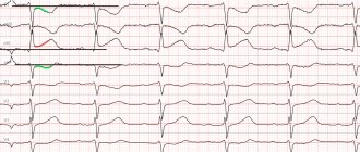
Signs on ECG
An electrocardiogram is the most informative research method for heart pathologies. What indicators may appear on the ECG with the development of right atrial hypertrophy:
| Indicators | Description |
| Increasing amplitude of the P wave with a sharpening (acute) | With the innervation of the atrium, a change in a flat tooth with a rounded apex is noted. |
| Increase in P wave height and width | Exceeding the norm (up to 2.5 mm) of amplitude, fluctuations in width within 0.12 seconds. |
| EO deviation | A sharp displacement of the electrical axis in different directions. |
The appearance of P-signs on the cardiogram can be caused by various reasons. Including overload of the atrium under the influence of pathology or increased load on the body.
In pregnant women, the risk of developing this pathology increases due to hormonal changes, changes in blood pressure, stress and difficulty breathing caused by increased body weight.
To assess the ability to bear a fetus normally, women are prescribed repeated procedures. This is especially true in the presence of congenital pathologies of the cardiovascular system or those discovered during gestation.
Deviation of the electrical axis is also not always considered a critical sign. A slight displacement is often observed in people of asthenic physique, for whom such a phenomenon is considered the norm.
To clarify the data, additional diagnostic methods are usually prescribed.
Treatment for enlargement
The goal of treating an enlarged left ventricle in the heart is to normalize the functioning of its muscles. First, the doctor must identify the cause of origin, as well as the existing features of the disease.
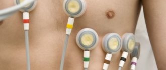
A variety of diagnostic procedures must be performed: the patient must have a complete blood test, blood pressure must be regularly monitored, an ultrasound scan, an echocardiogram and an electrocardiogram must be performed. Taking into account the data obtained, it is possible to accurately make a diagnosis, as well as determine the most optimal treatment regimen.
The basis of therapy are drugs with a negative effect, as well as those with an ionotropic effect - calcium blockers or antagonists (for example, Cardizem, Verapamil, Carvedilol, Procardia, Diltiazem). These drugs should be prescribed for life in optimal doses.
If disturbances in heart rhythm acquire serious consequences, antiarrhythmic drugs (Amiodarone, Ri, Disopyramide), as well as nitroglycein preparations, which allow dilation of the coronary heart vessels, can be prescribed.
When systolic dysfunction and dilatation of the cavities are detected, therapy for increasing the size of the left ventricle of the heart is performed according to generally accepted rules. For this, diuretics, ACE inhibitors, blockers, ongiotensin receptor antagonists, and spironolactones are used. Increased doses of ACE and saluretics usually increase obstruction.
Treatment of this condition using surgical methods is prescribed in case of ineffective drug treatment, especially in the presence of asymmetric hypertrophy of the septum between the cardiac ventricles, with clinical manifestations, severe obstruction and subaortic pressure.
Risk groups, diagnosis and treatment of cardiac arrhythmia
Arrhythmia is a violation of the frequency, rhythm and/or sequence of contractions of the heart. The term combines heartbeat disturbances that are different in their causes, clinical manifestations and consequences: increased heart rate (> 100 beats/min, tachycardia); deceleration (
- The main one is the sinus node in the upper part of the right atrium. It is called the “pacemaker of the first order”, since it is responsible for the rhythm frequency of the heart muscle.
- The impulses originating in it, like the rays of the sun, spread along the nerve and muscle fibers. Some of them stimulate the atrium to contract, others are sent to the next “power plant”: the atrioventricular node (AV node). At this point, the impulse slows down so that the chambers of the heart have time to contract and drive blood into the ventricles.
- After this, the nerve signals reach the bundle of His, which is branched into two “legs”: the right one conducts them to the right ventricle, and the left one to the left one, causing their excitation and, accordingly, contraction.
In an adult, the heart at rest contracts at a rate of 60–100 beats/min. Athletes resting heart rate Causes and risk factors
In a healthy heart, rhythm regulation occurs automatically. But if a failure occurs in one of the links of a well-functioning system (the formation of an electrical impulse or its conduction is disrupted), cardiac arrhythmia occurs.
Episodic arrhythmias also occur in completely healthy people: with a cold, overwork, from fear, joy, excitement; The heart rate increases after a cup of strong tea or coffee, excessive amounts of alcohol or cigarettes. In women, they can be caused by menopausal changes in the body. Such cardiac arrhythmias usually do not require treatment. However, most often they occur against the background of diseases of the cardiovascular system and their consequences.
Diseases of the heart and blood vessels
First on the list of diseases with the greatest risk of heart rhythm disturbances is coronary heart disease (CHD). It provokes many types of heart failure, but the most strongly associated with it are ventricular arrhythmias and sudden cardiac death. The occurrence of arrhythmias is also affected by atherosclerosis of the coronary arteries, myocardial infarction, inflammation of the heart muscle (myocarditis) or the outer lining of the heart (pericarditis).
- When blood flow to the heart is reduced, it affects the ability of cardiomyocytes to form and properly conduct electrical impulses.
- The death of a muscle section and scars on it after myocardial infarction, cicatricial changes after myocarditis, pericarditis change the path of propagation of impulses.
- Cardiomyopathy (stretching and thinning of the walls of the heart chambers or thickening of the walls of the left ventricle) also weakens the myocardium and impairs the contractility of the heart muscle.
Arrhythmia can be caused by diseases of the heart valves. Due to degenerative degeneration of myocardial tissue or infectious myocarditis, the valve openings narrow or their leaflets no longer close tightly. When the chambers of the heart become stretched and weakened due to valve insufficiency, blood flow in the organ is impaired and the risk of arrhythmias increases.
Other risk factors
Among the factors that increase the risk of developing arrhythmias are genetic predisposition, hormonal imbalance, persistent increase in blood pressure, and diabetes mellitus. Those who have been smoking for a long time, abusing alcohol, coffee, energy drinks, or taking drugs often complain about heart problems. Amphetamines and cocaine damage the heart muscle and can cause any of the arrhythmias and even lead to sudden cardiac death due to the development of ventricular fibrillation.
The likelihood of arrhythmias increases when the electrolyte balance in the body is disturbed, which happens, for example, with certain diseases of the gastrointestinal tract. Both excessively high and insufficient concentrations of electrolytes (potassium, sodium, magnesium, calcium) in the blood affect the electrical activity of the heart and can cause rhythm and conduction disturbances.
Symptoms
In some cases, arrhythmia is detected even before it makes itself felt, during a clinical examination or during examination for other diseases. But in most cases, heart rhythm disturbances give noticeable symptoms of cardiac arrhythmia: a feeling of strong heartbeat, interruptions and chest pain, shortness of breath, dizziness, a feeling of “lightheadedness” or fainting.
But you need to keep in mind that bright symptoms do not always indicate a serious problem and the need for treatment. Conversely, a person with a life-threatening arrhythmia who has not had any complaints may one day experience sudden cardiac arrest.
Tachycardia
In a healthy person, sinus tachycardia usually occurs in response to psychoemotional stress, physical activity, strong tea, coffee, and energy drinks. It is not accompanied by any unpleasant sensations, and the heart rate quickly returns to normal when the action of the provoking factors stops. You only need to worry if tachycardia occurs at rest.
Paroxysmal ventricular tachycardia is a more serious heart rhythm disorder. This is evidenced by:
- Atrial flutter is a sudden onset and just as suddenly stopped attack of rapid (up to 150–240 per minute) ventricular contractions, while maintaining the correct regular heart rhythm.
- Atrial fibrillation is a chaotic, very fast (250–480 per minute) contraction of myocardial fibers; ventricular contractions are not coordinated.
In the first case, there may be no symptoms, or the patient complains of “fluttering in the chest,” pain behind the sternum, or shortness of breath. In the case of atrial fibrillation, the first symptoms may be sudden cardiac arrest with loss of consciousness; It is difficult to feel the patient's pulse, breathing stops, the pupils are dilated, and convulsions are possible.
Bradycardia
In healthy, trained people, the heartbeat slows down at rest and during sleep. In pathological cases, there are two main types of bradycardia:
- atrioventricular block (decreased conductivity of myocardial cells), complete or incomplete;
- sick sinus syndrome.
With bradycardia, short-term fainting, lasting a few seconds, is possible: “entered the room, woke up on the floor.” Before an attack, some patients feel a rush of heat to the head. It must be remembered that loss of consciousness during bradycardia very rarely lasts > 5–10 minutes. Complete transverse blockade can cause heart failure and even sudden cardiac death.
Sinus bradycardia (sinus node weakness) often accompanies heart disease and hypotension. It is felt as pressure and heaviness in the heart, the patient often feels “broken”, gets tired quickly, and becomes dizzy.
Performing an operation
What it is - an enlargement of the left atrium, and how to treat it with medication is clear. Surgical methods are also worth considering. Typically, transaortic septal myectomy according to Morrow is used to treat this disease. Also quite effective is the operation performed according to K. Borisov and L. Bockeria, as a result of which the surgeon eliminates the enlarged area of the left septum. An excellent alternative is septal transcatheter ablation.
These surgical techniques reduce obstruction by reducing the septum between the ventricles of the heart. Surgeons often use dual-chamber pacing, which has atrioventricular delay.
Other diagnostic methods
An initial appointment with a cardiologist to identify signs of increased load on the right atrium includes preliminary diagnostic methods:
- interviewing the patient to describe a detailed medical history;
- percussion - tapping in the area of the heart;
- palpation - pressing on certain areas of the body to identify pathological abnormalities;
- Auscultation - listening to the heart rhythm.
To establish an accurate diagnosis, instrumental studies are prescribed:
- ECHO-CG (echocardiography) or ultrasound of the heart - examination of the anatomical structure of the organ (increase in atrium volume, thickening of the walls), as well as determination of the type of defect.
- Contrast radiography and CT (computed tomography) - identifying changes in the boundaries of the atrium and ventricle on the right, checking the condition of the arterial network.
- MRI - performed when it is difficult to evaluate echocardiography.
- Duplex scanning + Doppler sonography - to obtain hemodynamic characteristics.
Cardiomyopathy
This operation makes it possible to remove a more advanced hypertrophied area of the heart muscles. Often a complete or partial heart transplant is performed.
So, if the enlargement of the ventricles of the left atrium is caused by damage to the septum or heart valve, an organ transplant is performed first, which is simpler than whole heart surgery.
After performing cardiomyopathy, the patient should be monitored for life by a surgeon and cardiologist, and also take medications that will prevent thrombosis of the coronary vessels.