An objective assessment of the patient's condition, based on laboratory data, provides more accurate results than subjective data obtained from the patient. The results of laboratory tests allow not only to make a timely accurate diagnosis, but also to evaluate the quality of the therapy. That is why medical personnel need to ensure a high degree of reliability of the results.
Several factors can influence the degree of reliability:
- preliminary preparation of a person for blood collection;
- time of day at which the material was taken for laboratory research;
- the instruments used for sampling and the technique used to obtain the material;
- compliance with the sampling algorithm.
The main reason for the appearance of errors in the results of laboratory tests is non-compliance with the standards of the preanalytical stage of working with venous blood due to poor mastery of the technique for collecting biomaterial using vacuum systems.
Why is it important to use vacuum systems?
Laboratory diagnostics is carried out in three stages:
- Preanalytical.
- Analytical.
- Post-analytical.
The duration of the stages and the degree of their influence on the reliability of the data are different.
The longest is the first stage, occupying two-thirds of the duration of any study. Errors made at the preanalytical stage lead not only to an increase in time spent on making a diagnosis, but also to unnecessary waste of budget funds due to the appointment of a repeat procedure. They affect the entire process of correct diagnosis and evaluation of therapy.
The degree of reliability of the data obtained depends on a huge number of variables:
- personal characteristics of a person (gender, age, race, etc.);
- features of eating behavior before submitting laboratory material (fasting, abuse of a certain type of food, etc.);
- intensity of physical and emotional stress;
- natural changes in hormonal levels (phases of the menstrual cycle, pregnancy, menopause, etc.);
- weather and climatic conditions;
- medicines taken by humans;
- position of the patient at the time of material collection.
In addition to the above, the accuracy and correctness of the results depends on the technique of taking blood from a vein, the instruments used for this, the conditions of transportation and storage of the collected material.
When collecting blood from a vein using needles or syringes, it is impossible to standardize the technology for collecting the material. Using needles to collect venous blood can result in the collected material and pathogens of bloodborne infections getting into the hands of medical staff. This creates the danger of further transfer of pathogens to other patients. Taking biomaterial with a syringe practically eliminates this possibility, but when transferring it from a syringe to a test tube, hemolysis of red blood cells caused by mechanical action is possible.
Taking venous blood with a syringe does not exclude contact of medical staff with the patient’s blood and is therefore unsafe
Thus, vacuum systems have become the optimal tool for collecting venous blood.
Benefits of using negative pressure systems
All the benefits of negative pressure systems come from their design. Their use allows:
More article: Consequences of air entering a vein
- completely eliminate contact of medical personnel with the patient’s blood during sampling;
- standardize the process of blood collection and sample preparation, create a simple algorithm of actions;
- reduce the number of operations spent on preparing a sample for testing in the laboratory;
- Primary tubes included in negative pressure systems can be used directly in many automated analyzers. This saves money on purchasing secondary plastic tubes and time on transferring samples into them;
- make the transportation and centrifugation of biomaterials safer, since the tubes are sealed and made of unbreakable materials;
- facilitate the identification and labeling of samples by type of examination, thanks to color coding of the caps of negative pressure systems;
- reduce laboratory material costs for the purchase and processing of additional secondary tubes;
- simplify staff training methods;
- reduce the occupational risk of infection;
- reduce the time spent collecting venous blood using the method discussed in the article.
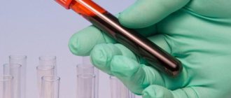
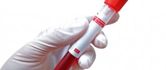
Multi-colored test tubes made from high-strength modern materials ensure the safety of medical staff working with blood
Test tube with red cap (with silica and separating gel).
These tubes are used to study blood serum.
Blood serum, unlike plasma, does not contain fibrinogen; therefore, in order to obtain a serum sample, the clotting process of the sample must be activated. In test tubes with a red cap, this process is activated due to silicon dioxide (SiO2) deposited on the inner walls in the form of microparticles. In addition to the reagent, tubes of this type may contain a separation gel. It creates a reliable barrier between the clot and the serum, which is important during sample storage and transportation. Test tubes with a coagulation activator are used for research in: biochemistry, immunology, microbiology, serology and others.
Sequence of obtaining venous blood using vacuum systems
The process of collecting venous blood consists of three stages:
- preparation for the procedure;
- performing a fence;
- end of material collection.
At the preparation stage in the procedure for taking biomaterial from a vein, medical personnel need to:
- Treat your hands using the scheme provided by WHO.
- When working with blood, each person is considered a potential carrier of a blood-contact infection. Therefore, before starting the blood collection procedure, it is necessary to change into protective clothing.
- Fill out a referral for a blood test in the registration journal. This is necessary for labeling instruments and filling out documents related to one person. The referral contains the patient’s passport details, the date and time of the blood draw, the registration data of the analysis in the laboratory, and the details of the doctor who ordered the analysis.
- Compare the information in the referral with the data of a specific patient.
- Check whether the patient has given informed consent to the procedure, explain in detail the purpose and sequence of its implementation.
- Clarify the patient’s compliance with the rules of food restrictions adopted before taking the tests.
- Conveniently accommodate the patient.
- Prepare the workplace: arrange all the devices necessary for drawing blood, having first ensured their integrity and suitability for use (safety of sterility seals, expiration date, etc.). Select test tubes with the desired color coding and the required volume. Take a needle of the appropriate size.
- Wear a mask, safety glasses, and rubber gloves.
Correct positioning of the patient is one of the important principles of the correct blood sampling procedure.
After completing all the steps in the first stage, you can proceed to blood sampling.
Algorithm for collecting biomaterial using a vacuum system
The second stage of the procedure is performed step by step:
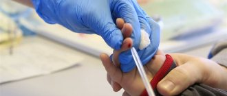
Correct placement of the needle when collecting venous blood
- Examine the proposed venipuncture sites, select a point for the procedure, and palpate the vein. The ulnar veins are most often used, but if necessary, blood can be taken from the veins of the wrist, the back of the hand, above the thumb, etc.
- Secure the tourniquet 10 centimeters above the venipuncture site. When applying a tourniquet, women should not use the hand on the side of the mastectomy. Prolonged compression of tissues and blood vessels (more than two minutes) can lead to shifts in coagulogram parameters and the concentration of certain substances.
- Take the needle and remove the protective cap from it.
- Connect the needle to the holder.
- Ask the patient to clench his hand into a fist. You should not make sudden movements, as this can lead to changes in blood counts. If the vein is poorly visible, you can apply a warm napkin to your arm, or massage your arm from the hand to the elbow. If there are no vessels suitable for venipuncture on one arm, the other should be checked.
- Treat the puncture site with a disinfectant using circular movements from the center to the edge.
- Wait for the antiseptic to evaporate, or remove excess with a sterile dry cloth.
- Remove the protective colored cap from the vacuum system.
- Secure the vein by clasping your forearm. Place your thumb 3˗5 centimeters below the injection site. Stretch the skin.
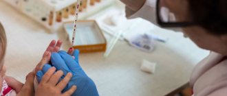
- Insert the needle with the holder into the vein at an angle of 15°. When inserted correctly, blood will appear in the indicator chamber of the holder.
- Secure the test tube in the holder with the cap facing up. Under the influence of negative pressure, blood will begin to flow into the test tube.
- As soon as blood begins to accumulate in the test tube, loosen the tourniquet or remove it.
- Tell the patient to relax his arm and unclench his fist.
- When the flow of blood into the test tube stops, remove it from the holder.
- Mix the biomaterial with the preservative. Do not shake! The test tube can only be gently inverted.
- If several samples are taken from the patient, the holder with the needle is left in the vein and steps 11-15 are repeated sequentially.
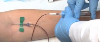
The vacuum collection system allows you to collect several tubes of material without removing the needle
After completing all of the above steps, you can proceed to the final stage of blood sampling.
Stage of completion of the procedure At the final stage of taking biomaterial from a vein, medical personnel need to:
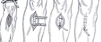
- Cover the venipuncture site with a dry sterile cloth.
- Remove the needle from the vein, cover it with a protective cap, and place it in a waste container.
- Apply a fixing bandage.
- Ask the patient how he is feeling. Provide assistance if necessary.
- Label the samples and sign each tube.
- Place samples in shipping containers and send to the laboratory.
Taking blood using vacuum tubes
Taking blood from a vein using a vacuum system is the safest and most effective method of collection. The use of vacuum tubes, so-called vacutainers, ensures the correct procedure for sample collection, transportation and qualitative analysis.
Features and advantages of vacutainers
The three-component system for venous blood collection consists of:
- sterile vacuum tube with preservative;
- double-sided automatic needle for intravenous injection;
- automatic needle holder.
The advantages of negative pressure systems are related to their design features:
- safety, sterility and guarantee of sample integrity;
- minimizing microclots and hemolysis;
- maintaining a constant time between collection and connection with the additive;
- exact ratio of sample and additive;
- minimizing the tourniquet effect.
Algorithm for taking blood using a vacuum system
The method of collecting venous blood with vacuum tubes is similar to using a syringe, but provides greater safety, efficiency and convenience. The collection is carried out quickly, which is important to guarantee an accurate research result.
When collecting blood from a peripheral vein using a vacuum system, you will need:
- vacuum tubes;
- tourniquet;
- cotton wool (cotton swabs) or napkins;
- antiseptic (medicinal alcohol);
- bactericidal patch;
- sterile medical tray;
- medical clothing (gown, goggles, mask and gloves).
Before the procedure, it is necessary to fill out a patient referral, treat your hands with a special solution, and put on protective medical clothing.
Technique for collecting blood from a vein
- Prepare test tubes that correspond to the stated tests or laboratory tests required by the patient, a needle, a holder, alcohol wipes or a cotton swab, and a patch.
- Place a tourniquet on the patient's shirt or diaper 7-10 cm above the venipuncture site. Ask the patient to make a fist.
- Select a venipuncture site. The middle ulnar and saphenous veins are the most commonly used, but smaller and more plethoric veins of the dorsum of the wrist and hand can also be punctured.
- Take the needle and remove the cap from the rubber membrane side. Insert the needle into the holder and screw it in until it stops.
- Disinfect the venipuncture site with a gauze pad. You must wait until the antiseptic solution has completely dried.
- Remove the protective cap on the other side. Insert the holder-needle vacuum system into the vein in accordance with the algorithm for conventional blood collection using a syringe. Make sure that the needle is cut upward at an angle of 15º relative to the surface of the skin. Since the other end is covered with a membrane, blood does not flow through the needle. Using smooth and quick movements, the skin and vein wall are punctured. Deep immersion of the needle should be avoided.
- Insert the test tube as far as it will go into the holder. As a result, the needle pierces the membrane and the plug, and a channel is formed between the vacuum tube and the vein. The needle should not be moved when blood begins to flow. The process continues until the vacuum in the test tube is compensated.
- The tourniquet should be removed or loosened as soon as blood begins to flow into the vacutainer. Make sure the patient unclenches their fist.
- After the blood flow stops, the tube is removed from the holder. The membrane returns to its original position, the blood flow through the needle is blocked. If necessary, other tubes can be connected to the holder to collect the required volume of blood. Immediately after filling, the test tube must be carefully inverted to mix the sample with the filler: test tube without anticoagulants - 5-6 times; test tube with citrate - 3-4 times; test tube with heparin, EDTA and other additives - 8-10 times.
- After filling the last tube, disconnect it from the holder and remove the holder-needle system from the vein. To ensure safety, remove the needle from the holder and place it in a special container for disposal.
- A sterile napkin/cotton ball moistened with an antiseptic is applied to the puncture site, or a bactericidal patch is applied.
- The tubes are labeled and placed in a special container for transportation to the laboratory.
Possible errors when using vacuum tubes
| Problem | Possible reasons | Solution |
| Blood does not flow into the tube after connecting to the holder | The needle did not enter the vein | In all of these cases, it is necessary to carefully adjust the position of the needle. There is no need to disconnect the tube from the holder if there is no need to remove the needle and under the skin. |
| The tip of the needle rests against the venous wall | ||
| The vein is pierced through | ||
| The blood in the test tube was received in less quantity than required for the analysis. | The venous vessel collapsed due to low pressure | It is necessary to disconnect the tube from the holder and wait a while until the vein is filled again |
| The system needs to be replaced and the procedure repeated | Air got into the test tube |
You can order high-quality laboratory consumables. When collecting blood using a vacuum system, follow the algorithm. This will ensure the safety of the procedure and the reliability of the research results.