Blood is the most important system in the human body, performing many different functions. Blood is a transport system through which vital substances are transported to the organs and waste substances, decay products and other elements that must be eliminated from the body are removed from the cells.
The blood also circulates substances and cells that provide protection to the body as a whole.
Blood consists of cells and a liquid part of serum, consisting of proteins, fats, sugars and trace elements.
There are three main types of cells in the blood:
- red blood cells,
- Leukocytes,
- Platelets.
Red blood cells are cells that transport oxygen to tissues
Red blood cells are highly specialized cells that do not have a nucleus (lost during maturation). Most of the cells are represented by biconcave disks, the average diameter of which is 7 µm, and the peripheral thickness is 2-2.5 µm. There are also spherical and dome-shaped red blood cells.
Due to the shape, the surface of the cell is significantly increased for gas diffusion. Also, this shape helps to increase the plasticity of the red blood cell, due to which it is deformed and moves freely through the capillaries.
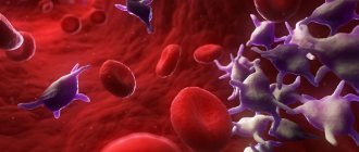
Human red blood cells and leukocytes
In pathological and old cells, plasticity is very low, and therefore they are retained and destroyed in the capillaries of the reticular tissue of the spleen.
The erythrocyte membrane and the anucleation of cells provide the main function of erythrocytes - the transport of oxygen and carbon dioxide. The membrane is absolutely impermeable to cations (except potassium) and highly permeable to anions. The membrane consists of 50% proteins that determine the blood group and provide a negative charge.
Red blood cells differ from each other in:
- size,
- Age,
- Resistance to adverse factors.
Video: Red blood cells
Red blood cells are the most numerous cells in human blood
Red blood cells are classified according to their degree of maturity into groups that have their own distinctive characteristics
maturation stage; features
| Erythroblast | diameter - 20-25 microns, nucleus occupying more than 2/3 of the cell with nucleoli (up to 4), cytoplasm is brightly basophilic, purple. |
| Pronormocyte | diameter - 10-20 microns, nucleus without nucleoli, chromatin is rough, cytoplasm becomes lighter. |
| Basophilic normoblast | diameter - 10-18 microns, chromatin is segmented, zones of basochromatin and oxychromatin are formed. |
| Polychromatophilic normoblast | diameter - 9-13 microns, destructive changes in the nucleus, oxyphilic cytoplasm due to the high hemoglobin content. |
| Oxyphilic normoblast | diameter - 7-10 microns, pink cytoplasm. |
| Reticulocyte | diameter - 9-12 microns, cytoplasm yellow-green. |
| Normocyte (mature red blood cell) | diameter - 7-8 microns, red cytoplasm. |
In peripheral blood there are both mature, young and old cells. Young red blood cells that contain remnants of nuclei are called reticulocytes.
The number of young red blood cells in the blood should not exceed 1% of the total mass of red cells. An increase in the content of reticulocytes indicates increased erythropoiesis.
The process of formation of red blood cells is called erythropoiesis.
Erythropoiesis occurs in:
- Bone marrow of the skull bones,
- Taza,
- Torso,
- Sternum and vertebral discs,
- Until the age of 30, erythropoiesis also occurs in the humerus and femur.
Every day, the bone marrow produces more than 200 million new cells.
After full maturation, the cells penetrate into the circulatory system through the capillary walls. The lifespan of red blood cells ranges from 60 to 120 days. Less than 20% of red blood cell hemolysis occurs intravascularly, the rest is destroyed in the liver and spleen.
Functions of red blood cells
- Perform a transport function. In addition to oxygen and carbon dioxide, cells transport lipids, proteins and amino acids,
- Helps remove toxins from the body, as well as poisons that are formed as a result of the metabolic and life processes of microorganisms,
- Actively participate in maintaining the balance of acid and alkali,
- Participate in the process of blood clotting.
Leukocytes: structure and functions
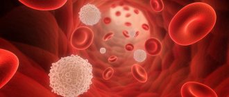
White blood cells - leukocytes
White blood cells (WBC) are a group of cells, each of which has a specialized protective function. Leukocytes contain nuclei; the cells contain hydrolytic enzymes, a protein synthesis system, biologically active compounds and other organelles. Leukocytes have the ability to migrate through the vascular wall, rushing towards foreign particles to capture and destroy them. The destruction of harmful cells is carried out by leukocytes using the process of phagocytosis - absorption and digestion. Leukocytes include 5 groups of protective cells.
1. Basophils (BAS). They make up only 1% of all leukocytes. These cells are round in shape, their diameter is approximately 12 - 15 microns. Basophils contain irregularly shaped granules that contain histamine, heparin, serotonin, prostaglandin and other substances. If necessary, basophilic leukocytes release the contents of their granules, participating in allergic reactions, blocking poisons, protecting blood vessels from the formation of blood clots, and attracting other helper cells to the site of inflammation.
2. Eosinophils (EOS). Their number in leukocytes is also small - from 1 to 4%. The cells have a round shape, the nucleus forms 2 segments connected by a bridge. The diameter is about 12 - 17 microns. Eosinophil granules contain collagenase, elastase, peroxidase, acid phosphatase, prostaglandins, alkaline protein, etc. Eosinophils are able to attach to parasites and introduce enzymes from their granules into the cytoplasm of harmful organisms, dissolving their membrane.
Agranulocytic leukocytes - lymphocytes
3. Lymphocytes (LYM). They make up about 30% of leukocytes and are the main immune cells. Lymphocytes are spherical formed elements, most of them are small cells with a dark nucleus, 5 - 7 microns in diameter. Large lymphocytes have a bean-shaped nucleus, their diameter exceeds 10 microns. These cells are functionally divided into types:
- B lymphocytes. They form antibodies against harmful agents.
- Killer T cells destroy pathogenic cells (parasitic, viral, tumor).
- T-helpers help in the processes of proliferation and differentiation of lymphocytes and promote the production of antibodies.
- Suppressor T cells stop helper T cells from working when needed.
- T-memories “record” information about microbes that have entered the body, so that when a new attack by harmful microorganisms occurs, the appropriate antibodies are directed against them.
- NK lymphocytes destroy abnormal cells.
Band neutrophil
4. Neutrophils (NEU). The most numerous group of leukocytes, makes up up to 75% of the number of protective cells. The diameter is approximately 12 - 15 microns; they circulate in the blood in the form of two subtypes:
- Rods. They are immature elements, their nuclei are similar to rods, which will then divide into segments, forming the next subspecies.
- Segmented. Their nuclei are segmented and usually contain 3 lobes connected by chromatin threads.
Neutrophils actively absorb bacteria, fungi and some viruses. They are the first to rush to the source of infection, capture pathogenic particles with their pseudopods and place them inside the cytoplasm, releasing the contents of their granules. Their granules contain collagenase, aminopeptidase, cationic proteins, acid hydrolases, and lactoferrin. Having digested harmful microorganisms, neutrophils usually die, at this moment releasing a number of substances that help suppress the remaining bacteria and fungi, and also enhance the process of inflammation, which becomes a signal for other immune cells. The mass of dead neutrophils, mixed with cellular detritus, represents pus.
5. Monocytes (MON). These leukocytes do not have granules; their nuclei can be presented in the form of an oval, horseshoe, bean, and the diameter is 12 - 20 microns. They make up about 4 - 10% of the number of immune cells. They are active phagocytes, capable of absorbing large microorganisms, and usually do not die after the digestion process. They remain at the site of inflammation and clean it, separating healthy tissue from damaged tissue. Monocytes destroy both pathogenic microbes and dead leukocytes, promoting the subsequent regeneration of damaged tissues.
Hemoglobin
The erythrocyte contains a complex iron-containing protein, hemoglobin, the main function of which is the transfer of oxygen between tissues and lungs, as well as partial transport of carbon dioxide.
Hemoglobin contains:
- Large protein molecule globin,
- The non-protein structure of heme embedded in globin. The core of heme contains an iron ion.
In the lungs, iron binds with oxygen, and it is this connection that contributes to the acquisition of a characteristic color by the blood.
Hemoglobin
HEMOPOIESIS OR BLOODPOISIS: HOW IS BLOOD ORIGINATED IN OUR BODY?
Blood is a unique liquid connective tissue, the structure of which contains a liquid medium - plasma, red and white blood cells. Its movement through a closed system is carried out by the heart. But where does blood come from in the body? How does this process happen?
Hematopoiesis or how blood is born
Blood cannot come from nowhere. This is a complex process that is controlled by many organs and systems and is called hematopoiesis. During this process, stem blood cells are converted into mature blood cells. When red blood cells are born, this process is called erythropoiesis, leukocytes - leukpoiesis, platelets - thrombocytopoiesis, etc.
Hematopoietic stem cells, that is, those from which the body can make blood, are concentrated in the red bone marrow, but they can circulate in organs not related to hematopoiesis.
The content of cells in the blood of a relatively healthy person is stable, but during certain adaptation processes, for example, in high altitude conditions, blood loss or infection, the differentiation of these cells accelerates, which is reflected in a blood test.
It is known that 2-5 billion cells are lost every day, but they are replaced by an equal number of new ones. Therefore, hematopoiesis does not stop throughout life. Scientists calculated the total weight of cells that are formed over a lifetime (approximately 70 years): 460 kg of red blood cells, 40 kg of platelets and 275 kg of lymphocytes.
The idea of hematopoiesis is not based on the theory of A.A. Maksimov about stem cells. According to this theory, there is one progenitor cell that can subsequently turn into any blood cell, be it an erythrocyte, platelet or lymphocyte. There are 2 main lines of hematopoiesis: lymphoid, during which various types of lymphocytes are formed, and myeloid, leading to the formation of all other blood cells.
Hematopoietic stem cells
Stem cells are unique in nature; they can transform not only into blood cells, but also into cells of other tissues, for example, reproduce all the tissues of the fetus during intrauterine development, build tissues of internal organs, blood, etc. after birth.
All stem cells have a number of common properties:
- their structure is unique, because there are no structural components. But the structure of a blood cell is significantly different; its components perform certain functions;
- capable of dividing into tens, hundreds and thousands of cells;
- can degenerate and turn into mature cells, and their structure corresponds to its type;
- capable of asymmetric division: if in this process one stem cell is formed, then the second turns into a specialized one;
- can move to areas of damage and literally patch up holes, and this is how tissue regeneration occurs, for example, skin when it is damaged.
Where does blood come from?
After birth, the main organ of hematopoiesis is the red bone marrow, which is concentrated in most bones, for example, ribs, sternum, and the epiphysis of long bones.
Regulation of the process of blood formation occurs in accordance with the needs of the body. To start the process of stem cell differentiation, a signal is needed that comes from cytokines, hormones that “tell” about the composition of the blood. And they are the ones who slow down or accelerate the process of hematopoiesis.
Vitamins, macro- and microelements and, of course, water take part in this process and play an important role.
Vitamin B12 and B9 (folic acid) are involved in the process of cell maturation and division. Iron and copper are necessary for the synthesis of hemoglobin, as well as for the maturation of erythroblasts, the precursors of red blood cells.
Erythropoiesis
Or the formation of red blood cells, which occurs in the bone marrow of the pelvic and other bones, and in children - in the epiphysis of the tubular bones. The lifespan of red blood cells is 3-4 months, and their utilization (apoptosis) occurs in the liver and spleen.
Before being released into the blood, future red blood cells go through successive stages of maturation, corresponding to the red germ of hematopoiesis.
The stem cell produces a progenitor cell, a unipotent cell that has receptors for erythropoietin, a hormone produced by the kidneys that controls the maturation of red blood cells.
The colony-forming unit of red blood cells gives rise to the erythroblast, and after several stages of development they give rise to “offspring” according to the following scheme:
- erythroblast;
- pronormocyte;
- several sequential forms of noocytes;
- reticulocyte;
- noocyte is a mature erythrocyte when it enters the bloodstream and becomes a full-fledged erythrocyte in a short time.
Leukopoiesis
Leukocytes can be formed during successive cellular transformations that occur in the hematopoietic organs; it begins in the red bone marrow.
There are 5 types of leukocytes: granulocytes - neutrophils, eosinophils, basophils, and agranulocytes, which include monocytes and lymphocytes. They make up the leukocyte formula.
From a class I stem cell, a precursor cell for myelopoiesis or lymphopoiesis is formed. And these cells, through a certain number of divisions and differentiation stages, turn into mature leukocytes, and for each type of leukocyte the number of these stages is not the same.
Regulation of the hematopoiesis process is a complex and genetically determined system. Any disturbances in this system, be it disturbances in the production of hormones or diseases, lead to disruption of the normal composition of the blood and the development of certain diseases.
Since blood circulates throughout the body, it can be said to be a carrier of information and can tell a lot about the state of health, the main thing is to be able to interpret the results obtained, but more on that in other materials.
Text: Yulia Lapushkina
Blood groups and Rh factor
On the surface of red blood cells there are antigens, of which there are many varieties. This is why one person's blood can be different from another's. Antigens form the Rh factor and blood group.
antigen; blood type
| 0 | I |
| 0A | II |
| 0B | III |
| AB | IV |
The presence/absence of Rh antigen on the surface of an erythrocyte is determined by the Rh factor (if Rh is present, Rh is positive, if Rh is not present, Rh is negative).
Determining the Rh factor and blood group of a person is of great importance when transfusing donor blood. Some antigens are incompatible with each other, causing destruction of blood cells, which can lead to the death of the patient. It is very important to receive a blood transfusion from a donor whose blood type and Rh factor match those of the recipient.
Organs of lymphopoiesis
In addition to red bone marrow, lymphopoiesis organs play a great role in the process of hematopoiesis. Their role is to continue the development of intermediate forms of lymphocytes. These cells are a type of leukocyte: they, like other types, are colorless (white), and their main function is immune defense. They recognize all proteins that are encoded in the genes of foreign chromosomes and strive to destroy these antigens. They also perform a transport function and participate in metabolism.
The thymus is responsible for the formation of T-lymphocytes (that’s why they are called that). It is here that their development from blast forms occurs. Different types of T-lymphocytes are formed in the thymus, which, depending on the function they perform, are called killers, helpers or suppressors. Killer T cells independently destroy foreign antigens, helper T cells contribute to the formation of humoral immunity against these antigens, and suppressors that suppress an excessive immune response are of great importance in maintaining the balance between protection and destruction.
In addition, the thymus destroys those cells that are hostile to their own tissues: without this, life would be impossible - the human body would destroy itself from the inside, perceiving its own tissues as antigens. The development of B lymphocytes occurs in the spleen. In addition, lymph nodes and Peyer's patches play a huge role in their maturation. Without these organs, it would not be possible to form an immune response and protect the body from carriers of a foreign set of chromosomes. The spleen is involved not only in the creation, but also in the destruction of obsolete blood cells. Another of its functions is to act as a depot of red blood cells, from where, if necessary, they enter the general bloodstream.
Leukocytes are blood cells that perform the function of phagocytosis
Leukocytes, or white blood cells, are blood cells that perform a protective function. White blood cells contain enzymes that destroy foreign proteins. Cells are able to detect harmful agents, attack them and destroy them (phagocytose). In addition to eliminating harmful microparticles, leukocytes take an active part in cleansing the blood of decay and metabolic products.
Thanks to antibodies produced by white blood cells, the human body becomes resistant to certain diseases.
Leukocytes have a beneficial effect on:
- Metabolic processes
- Providing organs and tissues with the necessary hormones,
- Enzymes and other necessary substances.
Leukocytes are divided into 2 groups: granular (granulocytes) and non-granular (agranulocytes).
Granular leukocytes include:
- Neutrophils,
- Basophils,
- Eosinophils.
The group of non-granular leukocytes includes:
- Lymphocytes,
- Monocytes.
Types of leukocytes
Neutrophils
The largest group of leukocytes, accounting for almost 70% of their total number. This type of leukocyte received its name due to the ability of the granularity of the cell to be stained with paints that have a neutral reaction.
Neutrophils are classified according to the shape of their nucleus into:
- Young , without a core,
- Rods , the core of which is represented by a stick,
- Segmented , the core of which consists of 4-5 segments connected to each other.
Neutrophils
When counting neutrophils in a blood test, the presence of no more than 1% of young, no more than 5% of band cells and no more than 70% of segmented cells is acceptable.
The main function of neutrophil leukocytes is protective, which is realized through phagocytosis, the process of detecting, capturing and destroying bacteria or viruses.
1 neutrophil can neutralize up to 7 microbes.
Neutrophils also take part in the development of inflammation.
Basophils
The smallest subtype of leukocytes, the volume of which is less than 1% of the number of all cells. Basophilic leukocytes are named due to the ability of the granular cells to be stained only with alkaline dyes (basic).
Basophils
The functions of basophilic leukocytes are determined by the presence of active biological substances in them. Basophils produce heparin, which prevents blood clotting at the site of the inflammatory reaction, and histamine, which dilates the capillaries, which leads to rapid resorption and healing. Basophils also contribute to the development of allergic reactions.
Eosinophils
A subtype of leukocytes, which got its name due to the fact that its granules are stained with acidic dyes, the main of which is eosin.
The number of eosinophils is 1-5% of the total number of leukocytes.
Cells have the ability of phagocytosis, but their main function is the neutralization and elimination of protein toxins and foreign proteins.
Eosinophils also participate in the self-regulation of body systems, produce neutralizing inflammatory mediators, and participate in blood purification.
Eosinophil
Monocytes
A subtype of leukocytes that does not have granularity. Monocytes are large cells resembling a triangle shape. Monocytes have a large nucleus of various shapes.
Monocyte formation occurs in the bone marrow. During the process of maturation, a cell goes through several stages of maturation and division.
Immediately after a young monocyte matures, it enters the circulatory system, where it lives for 2-5 days. After this, some of the cells die, and some go to mature to the stage of macrophages, the largest blood cells, whose lifespan is up to 3 months.
Monocytes perform the following functions:
- Produce enzymes and molecules that promote inflammation,
- Participate in phagocytosis,
- Promote tissue regeneration,
- Helps in the restoration of nerve fibers,
- Promotes the growth of bone tissue.
Monocytes
Macrophages phagocytose harmful agents found in tissues and suppress the proliferation of pathogenic microorganisms.
Lymphocytes
The central link of the defense system, which is responsible for the formation of a specific immune response and provides protection from everything foreign in the body.
The formation, maturation and division of cells occurs in the bone marrow, from where they are sent through the circulatory system to the thymus, lymph nodes and spleen for full maturation. Depending on where complete maturation occurs, T lymphocytes (matured in the thymus) and B lymphocytes (matured in the spleen or lymph nodes) are distinguished.
The main function of T lymphocytes is to protect the body by participating in immune reactions. T lymphocytes phagocytose pathogenic agents and destroy viruses. The reaction carried out by these cells is called nonspecific resistance.
B-lymphocytes are cells that are capable of producing antibodies - special protein compounds that prevent the proliferation of antigens and neutralize the toxins released by them during their life processes. For each type of pathogenic microorganism, B lymphocytes produce individual antibodies that eliminate the specific type.
Lymphocytes
T-lymphocytes phagocytose mainly viruses, while B-lymphocytes destroy bacteria.
What antibodies do lymphocytes produce?
B lymphocytes produce antibodies, which are found in cell membranes and in the serum portion of the blood. As an infection develops, antibodies begin to rapidly enter the bloodstream, where they recognize pathogenic agents and inform the immune system about this.
The following types of antibodies are distinguished:
- Immunoglobulin M accounts for up to 10% of the total amount of antibodies in the body. They are the largest antibodies and are formed immediately after the introduction of the antigen into the body,
- Immunoglobulin G is the main group of antibodies that plays a leading role in protecting the human body and forms immunity in the fetus. The cells are the smallest among antibodies and are able to cross the placental barrier. Together with this immunoglobulin, immunity from many pathologies is transmitted to the fetus from the mother to her unborn child,
- Immunoglobulin A protects the body from the influence of antigens entering the body from the external environment. The synthesis of immunoglobulin A is produced by B lymphocytes, but they are found in large quantities not in the blood, but on the mucous membranes, breast milk, saliva, tears, urine, bile and secretions of the bronchi and stomach,
- Immunoglobulin E antibodies secreted during allergic reactions.
Lymphocytes and immunity
After a microbe meets a B-lymphocyte, the latter is able to form memory cells in the body, which determines resistance to pathologies caused by this bacterium. To create memory cells, medicine has developed vaccines aimed at building immunity to particularly dangerous diseases.
Where are leukocytes destroyed?
The process of destruction of leukocytes is not fully understood. To date, it has been proven that of all the mechanisms of cell destruction, the spleen and lungs take part in the destruction of white blood cells.
Cells that are found in blood
Substances necessary for the full functioning of all our organs constantly circulate through the bloodstream. There are also elements in the blood that protect the human body from diseases and the effects of other negative factors.
Attention! Blood is divided into two components. These are the cellular part and plasma.
Plasma
In its pure form, plasma is a yellowish liquid. It makes up about 60% of the total blood mass. Plasma contains hundreds of chemicals that belong to different groups:
- protein molecules;
- ion-containing elements (chlorine, calcium, potassium, iron, iodine, etc.);
- all types of saccharides;
- hormones secreted by the endocrine system;
- all types of enzymes and vitamins.
All types of proteins that are in our body are also in plasma. For example, from blood test results we can remember immunoglobulins and albumins. These plasma proteins are responsible for defense mechanisms. There are about 500 of them. All other elements enter the blood due to its constant circulating movement. Enzymes are natural catalysts for many processes, and three types of blood cells make up the main part of plasma.
This is interesting! Blood plasma contains almost all the elements of D.I. Mendeleev’s periodic table.
About red blood cells and hemoglobin
Red blood cells are very small. Their maximum size is 8 microns, and their number is large - about 26 trillion. The following structural features are distinguished:
- absence of nuclei;
- lack of chromosomes and DNA;
- they do not have an endoplasmic reticulum.
Under a microscope, a red blood cell looks like a porous disk. The disc is slightly concave on both sides. He looks like a little sponge. Each pore of such a sponge contains a hemoglobin molecule. Hemoglobin is a unique protein. Its basis is iron. It actively contacts the oxygen and carbon environment, transporting valuable elements.
At the beginning of maturation, the red blood cell has a nucleus. Later it disappears. The unique shape of this cell allows it to participate in the exchange of gases, including the transport of oxygen. The red blood cell has amazing plasticity and mobility. While traveling through the vessels, it undergoes deformation, but this does not affect its operation. It moves freely even through small capillaries.
In simple school tests on medical topics, you can come across the question: “What are the names of the cells that transport oxygen to the tissues?” These are red blood cells. It’s easy to remember them if you imagine the characteristic shape of their disk with a hemoglobin molecule inside. And they are called red because iron gives our blood a bright color. When the blood binds with oxygen in the lungs, it turns bright scarlet.
On a note! Few people know that the precursors of red blood cells are stem cells.
The name of the hemoglobin protein reflects the essence of its structure. The large protein molecule that is part of it is called “globin”. The structure that does not contain protein is called heme. In its middle there is an iron ion.
The process of producing red blood cells is called erythropoiesis. Red blood cells are formed in flat bones:
- cranial;
- pelvic;
- sternum;
- intervertebral discs.
Until the age of 30, red blood cells are produced in the bones of the shoulders and hips.
Collecting oxygen in the alveoli of the lungs, red blood cells deliver it to all organs and systems. The process of gas exchange takes place. Red cells give oxygen to cells. In return, they collect carbon dioxide and carry it back to the lungs. The lungs remove carbon dioxide from the body, and everything repeats all over again.
At different ages, a person experiences different degrees of red blood cell activity. The fetus in the womb produces hemoglobin, which is called fetal hemoglobin. Fetal hemoglobin transports gases much faster than in adults.
If the bone marrow produces few red blood cells, a person becomes anemic or anemic. Oxygen starvation occurs throughout the body. It is accompanied by severe weakness and fatigue.
The lifespan of one red blood cell can be from 90 to 100 days.
There are also red blood cells in the blood that have not had time to mature. They are called reticulocytes. With large blood loss, the bone marrow releases immature cells into the blood, since there are not enough “adult” red blood cells. Despite the immaturity of reticulocytes, they can already be carriers of oxygen and carbon dioxide. In many cases this saves human life.
Antigens, blood groups and Rh factor
In addition to hemoglobin, red blood cells contain another special antigen protein. There are several antigens. For this reason, the composition of blood in different people cannot be the same.
Important! Blood group and Rh factor depend on the type of antigen.
If there is an antigen on the surface of the red blood cell, the Rh factor of the blood will be positive. If there is no antigen, then the rhesus is negative. These indicators are critical if blood transfusion is necessary. The donor's group and Rh must match those of the recipient (the person receiving the blood transfusion).
Leukocytes and their varieties
If red blood cells are carrier cells, then leukocytes are called protectors. They contain enzymes that fight foreign protein structures, destroying them. White blood cells detect harmful viruses and bacteria and begin to attack them. By destroying harmful substances, they cleanse the blood of harmful decay products.
Leukocytes provide the production of antibodies. Antibodies are responsible for the body's immune resistance to a number of diseases. White blood cells take part in metabolic processes. They provide tissues and organs with the necessary composition of hormones and enzymes. Based on their structure, they are divided into two groups:
- granulocytes (granular);
- agranulocytes (non-granular).
Among granular leukocytes, neutrophils, basophils and eosinophils are distinguished.
Leukocytes are divided into 2 groups: granular (granulocytes) and non-granular (agranulocytes). Non-granular bodies include monocytes and lymphocytes.
Neutrophils
They make up about 70% of all white blood cells. The prefix “neutro” means that a neutrophil has a special property. Due to its grainy structure, it can only be painted with neutral paint. Based on the shape of the nucleus, neutrophils are:
- young;
- stab;
- segmented.
Young neutrophils do not have nuclei. In rod cells, the nucleus looks rod-shaped under a microscope. In segmented neutrophils, the nuclei consist of several segments. There can be from 4 to 5. When conducting a blood test, the laboratory technician calculates the number of these cells as a percentage. Normally, young neutrophils should be no more than 1%. The norm for the content of rod cells is up to 5%. The permissible number of segmented neutrophils should not exceed 70%.
Neutrophils carry out phagocytosis - they detect, capture and neutralize harmful viruses and microorganisms.
This is interesting! One neutrophil can destroy about 7 microorganisms.
Eosinophils
This is a type of leukocyte, the granules of which are stained with dyes that have an acidic reaction. Basically, eosinophils stain with eosin. The number of these cells in the blood ranges from 1 to 5% of the total number of leukocytes. Their main task is to neutralize and destroy foreign protein structures and toxins. They also take part in the mechanisms of self-regulation and purification of the bloodstream from harmful substances.
Basophils
Few cells among leukocytes. Their percentage of the total is less than 1%. Cells can only be stained with alkali-based dyes (“bases”).
Basophils are producers of heparin. It slows down blood clotting in areas of inflammation. They also produce histamine, a substance that expands the capillary network. The expansion of capillaries ensures resorption and healing of wounds.
Monocytes
Monocytes are the largest human blood cells. They look like triangles. This is a type of immature white blood cell. Their kernels are large and of different shapes. The cells are formed in the bone marrow and mature through several stages.
The lifespan of a monocyte is from 2 to 5 days. After this time, the cells partially die. Those that survive continue to mature into macrophages.
Fun fact! A macrophage can live in the human bloodstream for about 3 months.
The role of monocytes in our body is as follows:
- participation in the process of phagocytosis;
- restoration of damaged tissues;
- regeneration of nervous tissue;
- bone growth.
Lymphocytes
They are responsible for the body’s immune response, protecting it from foreign invasions. The place of their formation and development is the bone marrow. Lymphocytes that have matured to a certain stage are sent through the bloodstream to the lymph nodes, thymus and spleen. There they ripen to the end. Cells that mature in the thymus are called T lymphocytes. B lymphocytes mature in the lymph nodes and spleen.
T lymphocytes protect the body by participating in immune responses. They destroy harmful microorganisms and viruses. With such a reaction, doctors talk about nonspecific resistance - that is, resistance to pathogenic factors.
The main task of B lymphocytes is to produce antibodies. Antibodies are special proteins. They prevent the spread of antigens and neutralize toxins.
Important! B lymphocytes produce antibodies to each type of harmful virus or microbe.
In medicine, antibodies are called immunoglobulins. There are several types:
- M-immunoglobulins are large proteins. Their formation occurs immediately after antigens enter the blood;
- G-immunoglobulins are responsible for the formation of the fetal immune system. Their small size makes it easy to cross the placental barrier. The cells transmit immunity from mother to child;
- A-immunoglobulins - include defense mechanisms in the event of a harmful substance entering from the outside. Type A immunoglobulins are synthesized by B lymphocytes. They enter the blood in small quantities. These proteins accumulate on mucous membranes and in women's breast milk. They are also contained in saliva, urine and bile;
- E-immunoglobulins - released during allergies.
In the human bloodstream, a microorganism or virus can encounter a B lymphocyte on its way. The response of the B lymphocyte is to create so-called “memory cells”. “Memory cells” determine a person’s resistance (persistence) to diseases caused by specific bacteria or viruses.
We can obtain “memory cells” artificially. Vaccines have been developed for this purpose. They provide reliable immune protection against those diseases that are considered especially dangerous.
Platelets
Their main function is to protect the body from critical blood loss. Platelets ensure stable hemostasis. Hemostasis is the optimal state of the blood, which allows it to fully supply the body with the elements necessary for life. Under a microscope, platelets appear as cells that are convex on both sides. They lack nuclei and can range from 2 to 10 microns in diameter.
Platelets can take on a round or oval shape. When they are activated, growths appear on them. Because of the growths, the cells look like small stars. The formation of platelets occurs in the bone marrow and has its own characteristics. First, megakaryocytes arise from megakaryoblasts. These are cells with huge cytoplasm. Several separation membranes are formed inside the cytoplasm and division occurs. After division, parts of magekaryocytes “bud off” from the mother cell. These are already full-fledged platelets that are released into the blood. Their lifespan ranges from 8 to 11 days.
Platelets are divided by their diameter (in microns):
- microforms – up to 1.5;
- standard forms – from 2 to 4;
- macroforms – 5;
- megaloforms – 6-10.
The site of platelet formation is the red bone marrow. They mature in six cycles.
This is interesting! The growths that appear on platelets during the period of their activity are called pseudopodia. This is how cells stick together. They close the damaged vessel and stop bleeding.
Stem cells and their features
Stem cells are called immature structures. Many living beings have them and are capable of self-renewal. They serve as the initial material for the formation of organs and tissues. Blood cells also appear from them. There are more than 200 types of stem cells in the human body. They have the ability to renew (regenerate), but the older a person gets, the fewer stem cells his bone marrow produces.
Medicine has long practiced successful transplantation of certain types of stem cells. Among them, hematopoietic structures are distinguished. As already mentioned, hematopoiesis is a complete process of hematopoiesis. If it is normal, the composition of a person’s blood does not cause concern among doctors.
When treating leukemia or lymphoma, donor stem cells responsible for hematopoietic functions are transplanted. In systemic blood diseases, hematopoiesis is impaired, and bone marrow transplantation helps restore it.
Fun fact! Stem structures can turn into any type of cell, including blood cells.
Platelets are cells that protect the body from fatal blood loss.
Platelets are formed blood elements that participate in hemostasis. They are represented by small biconvex cells that do not have a nucleus. The diameter of the platelet varies between 2-10 microns.
Platelets are produced by red bone marrow, where they undergo 6 cycles of maturation, after which they enter the bloodstream and remain there for 5 to 12 days. Platelet destruction occurs in the liver, spleen and bone marrow.
Platelets
While in the bloodstream, platelets have the shape of a disk, but when activated, the platelet takes on the shape of a sphere, on which pseudopodia are formed - special outgrowths with the help of which platelets connect to each other and adhere to the damaged surface of the vessel.
In the human body, platelets perform 3 main functions:
- Create plugs on the surface of a damaged blood vessel, helping to stop bleeding (primary thrombus),
- Participate in blood clotting, which is also important for stopping bleeding,
- Platelets provide nutrition to vascular cells.
Platelets are classified into:
- Microforms – platelet with a diameter of up to 1.5 microns,
- Normal forms of platelets with a diameter of 2 to 4 µm,
- Macroforms of a platelet with a diameter of 5 microns,
- Megaloform platelets with a diameter of up to 6-10 microns.
Platelets
These blood cells are small, anucleate plates and can be round or oval in shape. During activation, when they are near the damaged vessel wall, they form outgrowths, so they look like stars. Platelets contain microtubules, mitochondria, ribosomes, and specific granules containing substances necessary for blood clotting. These cells are equipped with a three-layer membrane.
Platelets are produced in the bone marrow, but in a completely different way than other cells. Blood plates are formed from the largest cells of the brain - megakaryocytes, which, in turn, were formed from megakaryoblasts. Megakaryocytes have a very large cytoplasm. After the cell matures, membranes appear in it, dividing it into fragments that begin to separate, and thus platelets appear. They leave the bone marrow into the blood, stay in it for 8-10 days, then die in the spleen, lungs, and liver.
Blood plates can have different sizes:
- the smallest are microforms, their diameter does not exceed 1.5 microns;
- normoforms reach 2-4 microns;
- macroforms – 5 microns;
- megaloforms – 6-10 microns.
Platelets perform a very important function - they participate in the formation of a blood clot, which closes the damage in the vessel, thereby preventing blood from leaking out. In addition, they maintain the integrity of the vessel wall and promote its rapid recovery after damage. When bleeding begins, platelets adhere to the edge of the injury until the hole is completely closed. The adhered plates begin to break down and release enzymes that affect the blood plasma. As a result, insoluble fibrin threads are formed, tightly covering the injury site.
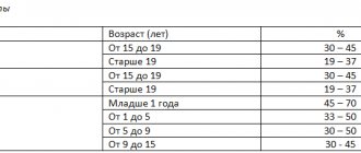
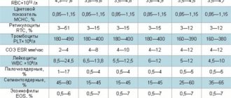
Norm of red blood cells, leukocytes and platelets in the blood (table)
age; polyerythrocytes (x 10 12 / l); leukocytes (x 10 9 /l); platelets (x 10 9 /l)
| 1-3 months | husband | 3,5 — 5,1 | 6,0 — 17,5 | 180 — 490 |
| wives | ||||
| 3-6 months | husband | 3,9 — 5,5 | ||
| wives | ||||
| 6-12 months | husband | 4,0 — 5,3 | 180 — 400 | |
| wives | ||||
| 1-3 years | husband | 3,7 — 5,0 | 6,0 — 17,0 | 160 — 390 |
| wives | ||||
| 3-6 years | husband | 5,5 — 17,5 | ||
| wives | ||||
| 6-12 years | husband | 4,5 — 14,0 | 160 — 380 | |
| wives | ||||
| 12-15 years | husband | 4,1 — 5,5 | 4,5 — 13,5 | 160 — 360 |
| wives | 3,5 — 5,0 | |||
| 16 years | husband | 4,0 — 5,5 | 4,5 — 12,0 | 180 — 380 |
| wives | 3,5 — 5,0 | 150 — 380 | ||
| 16-65 years old | husband | 4,0 — 5,6 | 4,5 — 11,0 | 180 — 400 |
| wives | 3,9 — 5,0 | 150 — 340 | ||
| over 65 years old | husband | 3,5 — 5,7 | 180 — 320 | |
| wives | 3,5 — 5,2 | 150 — 320 |
BLOOD
Blood is a viscous red liquid that flows through the circulatory system: it consists of a special substance - plasma, which carries various types of formed blood elements and many other substances throughout the body.
FUNCTIONS OF BLOOD:
•;Supply oxygen and nutrients to the entire body. •;Transfer metabolic products and toxic substances to the organs responsible for their neutralization. •;Transfer hormones produced by endocrine glands to the tissues for which they are intended. •;Take part in the thermoregulation of the body. •;Interact with the immune system.
MAIN BLOOD COMPONENTS:
— Blood plasma. It is a liquid consisting of 90% water that transports all the elements present in the blood throughout the cardiovascular system: in addition to transporting blood cells, it also supplies the organs with nutrients, minerals, vitamins, hormones and other products involved in biological processes, and carries away metabolic products.
Some of these substances themselves are freely transported by the plasma, but many of them are insoluble and are transported only together with the proteins to which they are attached, and are separated only in the corresponding organ. - Blood cells. When looking at the composition of blood, you will see three types of blood cells: red blood cells, the same color as blood, the main elements that give it its red color; white blood cells responsible for many functions; and platelets, the smallest blood cells.
RED BLOOD CELLS
Red blood cells , also called erythrocytes or red blood platelets, are fairly large blood cells. They are shaped like a biconcave disc and have a diameter of about 7.5 microns; they are not actually cells as such because they lack a nucleus; Red blood cells live for about 120 days. Red blood cells contain hemoglobin, a pigment made of iron that gives blood its red color;
It is hemoglobin that is responsible for the main function of the blood - the transfer of oxygen from the lungs to the tissues and the metabolic product - carbon dioxide - from the tissues to the lungs. Red blood cells under a microscope. If you lined up all the red blood cells of an adult, you would get over two trillion cells (4.5 million per mm3 times 5 liters of blood), which could be placed 5.3 times around the equator.
WHITE BLOOD CELLS
White blood cells , also called leukocytes , play an important role in the immune system, protecting the body from infections. There are several types of white blood cells ;
All of them have a nucleus, including some multinucleated leukocytes, and are characterized by segmented, oddly shaped nuclei that are visible under the microscope, so leukocytes are divided into two groups: polynuclear and mononuclear. Polynuclear leukocytes are also called granulocytes, because under a microscope you can see several granules in them, which contain substances necessary to perform certain functions. There are three main types of granulocytes:
— Neutrophils , which absorb (phagocytose) and process pathogenic bacteria; — Eosinophils , which have antihistamine properties, their numbers increase during allergies and parasitic reactions; — Basophils , which secrete a special secretion during allergic reactions.
Let us dwell in more detail on each of the three types of granulocytes. You can consider granulocytes and cells, which will be described later in the article, in Scheme 1 below.
Scheme 1. Blood cells: white and red blood cells, platelets.
Neutrophil granulocytes (Gn/n) are motile spherical cells with a diameter of 10-12 µm. The nucleus is segmented, the segments are connected by thin heterochromatic bridges. In women, a small, elongated appendage called the rod tympani (Barr's body) may be visible; it corresponds to the inactive long arm of one of the two X chromosomes. On the concave surface of the nucleus there is a large Golgi complex; other organelles are less developed. Characteristic of this group of leukocytes is the presence of cell granules. Azurophilic or primary granules (AG) are considered primary lysosomes from the moment when they already contain acid phosphatase, aryle sulfatase, B-galactosidase, B-glucuronidase, 5-nucleotidase d-aminooxidase and peroxidase. Specific secondary, or neutrophil, granules (NG) contain the bactericidal substances lysozyme and phagocytin, as well as the enzyme alkaline phosphatase. Neutrophil granulocytes are microphages, i.e. they absorb small particles such as bacteria, viruses, and small parts of decaying cells. These particles enter the cell body by being captured by short cell processes and are then destroyed in phagolysosomes, into which azurophilic and specific granules release their contents. The life cycle of neutrophil granulocytes is about 8 days.
Eosinophilic granulocytes (Gr/e) are cells reaching a diameter of 12 microns. The nucleus is bilobed; the Golgi complex is located near the concave surface of the nucleus. Cellular organelles are well developed. In addition to azurophilic granules (AG), the cytoplasm includes eosinophilic granules (EG). They have an elliptical shape and consist of a fine-grained osmiophilic matrix and single or multiple dense lamellar crystalloids (Cr). Lysosomal enzymes, lactoferrin and myeloperoxidase, are concentrated in the matrix, while a large basic protein, toxic to some helminths, is located in the crystalloids.
Basophilic granulocytes (Gr/b) have a diameter of about 10-12 microns. The nucleus is kidney-shaped or divided into two segments. Cellular organelles are poorly developed. The cytoplasm includes small, sparse peroxidase-positive lysosomes, which correspond to azurophilic granules (AG), and large basophilic granules (BG). The latter contain histamine, heparin and leukotrienes. Histamine is a vasodilator, heparin acts as an anticoagulant (a substance that inhibits the activity of the blood coagulation system and prevents the formation of blood clots), and leukotrienes cause constriction of the bronchi. Eosinophilic chemotactic factor is also present in granules; it stimulates the accumulation of eosinophilic granules at sites of allergic reactions. Under the influence of substances that cause the release of histamine or IgE, basophil degranulation can occur in most allergic and inflammatory reactions. In this regard, some authors believe that basophilic granulocytes are identical to mast cells of connective tissues, although the latter do not have peroxidase-positive granules.
There are two types of mononuclear leukocytes : - Monocytes , which phagocytose bacteria, detritus and other harmful elements; — Lymphocytes that produce antibodies (B-lymphocytes) and attack aggressive substances (T-lymphocytes).
Monocytes (Mc) are the largest of all blood cells, measuring about 17-20 microns. A large kidney-shaped eccentric nucleus with 2-3 nucleoli is located in the voluminous cytoplasm of the cell. The Golgi complex is localized near the concave surface of the nucleus. Cellular organelles are poorly developed. Azurophilic granules (AG), i.e. lysosomes, are scattered throughout the cytoplasm.
Monocytes are very motile cells with high phagocytic activity. Since the absorption of large particles such as whole cells or large parts of broken cells, they are called macrophages. Monocytes regularly leave the bloodstream and enter the connective tissue. The surface of monocytes can be either smooth or contain, depending on the cellular activity, pseudopodia, filopodia, and microvilli. Monocytes are involved in immunological reactions: they participate in the processing of absorbed antigens, the activation of T lymphocytes, the synthesis of interleukin and the production of interferon. The lifespan of monocytes is 60-90 days.
White blood cells , in addition to monocytes, exist as two functionally distinct classes called T and B lymphocytes , which cannot be distinguished morphologically based on conventional histological examination methods. From a morphological point of view, young and mature lymphocytes are distinguished. Large young B- and T-lymphocytes (CL), 10-12 µm in size, contain, in addition to a round nucleus, several cellular organelles, among which there are small azurophilic granules (AG), located in a relatively wide cytoplasmic rim. Large lymphocytes are considered a class of so-called natural killer cells.
Mature B and T lymphocytes (L) with a diameter of 8-9 microns, have a massive spherical nucleus surrounded by a thin rim of cytoplasm, in which rare organelles can be observed, including azurophilic granules (AG). The surface of lymphocytes can be smooth or dotted with many microvilli (MV). Lymphocytes are amoeboid cells that freely migrate through the epithelium of blood capillaries from the blood and penetrate into the connective tissue. Depending on the type of lymphocytes, their lifespan varies from several days to several years (memory cells).
Colored leukocytes under an electron microscope.
PLATELETS
Platelets are corpuscular elements that are the smallest particles of blood. Platelets are incomplete cells; their life cycle is only up to 10 days. Platelets concentrate at bleeding sites and take part in blood clotting.
Platelets (T) are spindle-shaped or disc-shaped biconvex fragments of the cytoplasm of a megakaryocyte with a diameter of about 3-5 microns. Platelets have few organelles and two types of granules: a-granules (a), containing several lysosomal enzymes, thromboplastin, fibrinogen, and dense granules (DG), which have a highly condensed interior containing adenosine diphosphate, calcium ions, and several types of serotonin.
Platelets under an electron microscope.
| BLOOD DISEASES / SPLEN / BLOOD PRESSURE AND PULSE / BLOOD CLOTTING / BLOOD TYPES / RH FACTOR / ARTERIES / VEINS / NOSELEED / STRUCTURE OF THE CONDUCTION SYSTEM OF THE HEART |
Human blood smear (Romanovsky-Giemsa method)
Self-preparation task for diagnostic lesson No. 1
List of study drugs:
Human blood smear (Romanovsky-Giemsa method)
Be able to find all the shaped elements!
Blood is a kind of connective tissue (refers to the tissues of the internal environment of a person) with a liquid intercellular substance (plasma), which contains various cells (leukocytes) and postcellular structures (erythrocytes, platelets).
Originates from mesenchyme.
Blood functions:
Transport - a universal function of the blood associated with ensuring the transfer of various substances.
Includes:
respiratory function - transfer of gases in a dissolved and chemically bound state
trophic function - transfer of nutrients from areas of their absorption and accumulation to tissues
excretory function - removal of metabolic products from tissues and their release from the body
regulatory function - transfer of hormones and other biologically active substances to cells of different tissues, thermoregulatory function
homeostatic - maintaining a constant internal environment of the body
· protective - neutralization of foreign antigens, neutralization of microorganisms
The composition of plasma includes: water, proteins (albumin, globulins, fibrinogen), lipids, low molecular weight organic compounds, inorganic ions.
Shaped elements:
1. Red blood cells
Morphology: devoid of nuclei, stained pink with eosin, round in shape and clear in the center (biconcave disc).
Functions: respiratory, regulatory, protective.
2. Leukocytes
Basophils
Morphology: the presence of large basophilic granules of violet-cherry color, filling almost the entire cytoplasm, the nuclei are lobulated, difficult to distinguish behind the granules.
Functions: regulatory, homeostatic (through accumulated or synthesized biologically active substances), protective.
Eosinophils
Morphology: the nucleus consists of 2 segments, the cytoplasm contains many oxyphilic granules.
Functions: protective, immunoregulatory.
Neutrophils
There are: segmented, rod, young.
Morphology: the nucleus consists of 3-4 connected segments; in the cytoplasm, fine granularity is difficult to distinguish.
Functions: destruction of microorganisms, destruction and digestion of damaged cells and tissues, participation in the regulation of the activity of other cells.
Lymphocytes
Morphology: round, highly stained nucleus, narrow rim of basophilic cytoplasm without granules.
Functions: ensuring immune reactions, regulating the activity of other cells.
Monocytes
Morphology: large bean-shaped or horseshoe-shaped light nucleus, weakly basophilic cytoplasm.
Functions: providing nonspecific defense reactions, participation in immune reactions, capture and digestion of aging and dead cells, secretion of regulatory substances.
3. Platelets
Morphology: nuclear-free fragments of cytoplasm, small in size.
Functions: stopping bleeding when the vascular wall is damaged, hemocoagulation, participation in wound healing reactions, ensuring normal vascular function.
2. Red bone marrow smear (environmental azur2+eosin).
Be able to find the stages of development of erythrocytes, granulocytes, megakaryocytes.
https://www.histol.chuvashia.com/atlas/bon-mar.htm
Loose fibrous connective tissue. Film preparation (environmental hematoxylin).
Be able to find fibroblasts and macrophages.
Dense formed connective tissue (tendon on a longitudinal section) (surrounding hematoxylin and eosin).
Be able to find parallel bundles of collagen fibers, fibrocytes and layers of loose connective tissue - endotenonium (separates bundles of 2 orders from each other).
Dense formed connective tissue (tendon on a cross section) (surrounding hematoxylin and eosin).
Be able to find the connective tissue membrane on the outside of the tendon - peritenonium, identify tendon bundles of the 1st and 2nd order.
6. Dense, unformed connective tissue of the skin of a human finger (environment orcein + picrofuchsin + hematoxylin).
Pigment fabric
Self-preparation task for diagnostic lesson No. 1
List of study drugs:
Human blood smear (Romanovsky-Giemsa method)
Be able to find all the shaped elements!
Blood is a kind of connective tissue (refers to the tissues of the internal environment of a person) with a liquid intercellular substance (plasma), which contains various cells (leukocytes) and postcellular structures (erythrocytes, platelets).
Originates from mesenchyme.
Blood functions:
Transport - a universal function of the blood associated with ensuring the transfer of various substances.
Includes:
respiratory function - transfer of gases in a dissolved and chemically bound state
trophic function - transfer of nutrients from areas of their absorption and accumulation to tissues
excretory function - removal of metabolic products from tissues and their release from the body
regulatory function - transfer of hormones and other biologically active substances to cells of different tissues, thermoregulatory function
homeostatic - maintaining a constant internal environment of the body
· protective - neutralization of foreign antigens, neutralization of microorganisms
The composition of plasma includes: water, proteins (albumin, globulins, fibrinogen), lipids, low molecular weight organic compounds, inorganic ions.
Shaped elements:
1. Red blood cells
Morphology: devoid of nuclei, stained pink with eosin, round in shape and clear in the center (biconcave disc).
Functions: respiratory, regulatory, protective.
2. Leukocytes
Basophils
Morphology: the presence of large basophilic granules of violet-cherry color, filling almost the entire cytoplasm, the nuclei are lobulated, difficult to distinguish behind the granules.
Functions: regulatory, homeostatic (through accumulated or synthesized biologically active substances), protective.
Eosinophils
Morphology: the nucleus consists of 2 segments, the cytoplasm contains many oxyphilic granules.
Functions: protective, immunoregulatory.
Neutrophils
There are: segmented, rod, young.
Morphology: the nucleus consists of 3-4 connected segments; in the cytoplasm, fine granularity is difficult to distinguish.
Functions: destruction of microorganisms, destruction and digestion of damaged cells and tissues, participation in the regulation of the activity of other cells.
Lymphocytes
Morphology: round, highly stained nucleus, narrow rim of basophilic cytoplasm without granules.
Functions: ensuring immune reactions, regulating the activity of other cells.
Monocytes
Morphology: large bean-shaped or horseshoe-shaped light nucleus, weakly basophilic cytoplasm.
Functions: providing nonspecific defense reactions, participation in immune reactions, capture and digestion of aging and dead cells, secretion of regulatory substances.
3. Platelets
Morphology: nuclear-free fragments of cytoplasm, small in size.
Functions: stopping bleeding when the vascular wall is damaged, hemocoagulation, participation in wound healing reactions, ensuring normal vascular function.
2. Red bone marrow smear (environmental azur2+eosin).
Be able to find the stages of development of erythrocytes, granulocytes, megakaryocytes.
https://www.histol.chuvashia.com/atlas/bon-mar.htm
Didn't find what you were looking for? Use Google search on the site:
Leukocytes - protectors of the body
Blood cells, leukocytes, are the protectors of the body. They contain enzymes that attack and destroy foreign protein compounds. Leukocytes fight viruses and bacteria, clearing the blood of their waste products. Leukocytes look like a ball.
White blood cells produce antibodies that provide the body with resistance to certain diseases. White blood cells are involved in metabolism, responsible for delivering hormones and enzymes to organ tissues. Structurally they are divided into two parts:
- Grainy. These include neutrophils, eosinophils, and basophils.
- Non-grainy. Represented by monocytes and lymphocytes.
Types of neutrophils
They occupy 70% of leukocytes. Since the neutrophil has a granular structure, it can be stained with a substance with a neutral reaction. Neutrophils differ in the shape of their nucleus, so they are:
- Young. Such neutrophils are anucleate.
- Band-nuclear. The cell nuclei are rod-shaped.
- Segmented. They differ in segments in the core, there are 4-5 of them.
During a blood test, the laboratory technician displays the percentage of cell content. The norm for young people is up to 1%. Bands should be present up to 5%. Segmented ones should not be more than 70% of the mark. Neutrophils are needed to neutralize microorganisms that harm the body.
Eosinophils and basophils
The granules of this variety of white bodies are painted with an acidic paint. In the blood, eosinophils represent up to 5%, including all leukocytes. Their tasks include the destruction of parasites and their toxins, and cleansing the blood of harmful structures.
Of the total number of leukocytes, basophils make up only 1%. Cells are stained with alkaline reaction dye. Basophils produce heparin, which inhibits the blood clotting process in areas of inflammation. Plus, histamine is produced, which expands the capillary so that the wounds resolve and heal faster.
Monocytes and lymphocytes
Monocytes are the largest, triangular blood cells. They belong to a type of leukocyte, they have large nuclei of various shapes. Formed in the bone marrow. Monocytes live only five days, the survivors mature and turn into macrophages, which remain in the blood for about three months. They regenerate damaged tissue, restore nerve fibers, and affect bone growth.
Lymphocytes form the immune system, which is a barrier to infections. Formed in the bone marrow. Almost mature lymphocytes are sent through the bloodstream to the thymus, spleen and lymph nodes, where they mature completely. Those that enter the thymus are called T-lymphocytes, and those that enter the lymph nodes and spleen are called B-lymphocytes.
T-lymphocytes have a protective function; their tasks are to neutralize viruses and bacteria. They are resistant to pathogenic reactions. B lymphocytes produce antibodies (special proteins) that inhibit the spread of antigens and neutralize toxins. For each mutation of viruses, B lymphocytes are forced to produce antibodies, the latter are called immunoglobulins.
Once in the bloodstream, a virus or foreign bacterium can come across a B-lymphocyte, which instantly “photographs” it and creates a “memory cell.” It will help the body resist diseases caused by a specific pathogen.
We can form such protection with the help of vaccines, which help the body form an immune response to dangerous diseases.