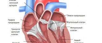
The heart, which is responsible for creating the optimal pressure for transporting blood in the vascular system, itself needs high-quality and very intensive blood supply. The arteries of the heart are responsible for it - short vessels with a very strong and elastic wall that deliver blood to the heart muscle, valves and septa of the heart. The main thing that distinguishes coronary arteries from other types of vessels is complete self-regulation, which allows this limited system to uninterruptedly supply the heart with oxygen and nutrients.
Anatomical features
According to the diagram of the vessels of the heart, there are two main trunks of the coronary vessels:
- right coronary artery – comes from the right aortic sinus, is responsible for blood supply to the right and posteroinferior wall of the left ventricle and some part of the interventricular septum;
- left – comes from the left aortic sinus, then is divided into 2-3 small arteries (less often four); The most significant are the anterior descending (anterior interventricular) and circumflex branches .
In each individual case, the anatomical structure of the heart vessels may vary, therefore, for a full study, cardiography of the heart vessels (coronary angiography) using an iodine-containing contrast agent is indicated.
Anatomy of the coronary arteries
The main branches of the right coronary artery are: branch of the sinus node, branch of the conus, right ventricular branch, branch of the acute margin, posterior interventricular artery and posterolateral artery.
The left coronary artery begins with a trunk that divides into the anterior interventricular and circumflex arteries. an intermediate artery (a.intermedia) arises between them . The anterior interventricular artery (anterior descending) gives off diagonal and septal branches. The main branches of the circumflex artery are the branches of the obtuse margin.
3rd or cardiac circle of blood circulation
Heart blood flow
Every adult knows that there are 2 circles of blood circulation in the body: large and small. But anatomists say that there are three of them! So is the basic anatomy course misleading? Not at all! The third circle, named figuratively, refers to the vessels that fill the blood and “serve” the heart itself. It deserves personal vessels, doesn't it? So, the 3rd or cardiac circle begins with the coronary arteries, which are formed from the main vessel of the human body - Her Majesty the aorta, and ends with the cardiac veins, merging into the coronary sinus.
It in turn opens into the right atrium. And the smallest venules open into the atrial cavity on their own. It was noticed very figuratively that the vessels of the heart entwine and envelop it like a real crown, a crown. Therefore, arteries and veins are called coronary or coronary. Remember: these are synonymous terms. So what are the most important arteries and veins the heart has at its disposal? What is the classification of coronary arteries?
Types of myocardial blood circulation
Based on the blood supply to the posterior wall of the heart, balanced, left and right types of blood circulation are distinguished. Determination of the predominant type depends on whether one of the arteries reaches the avascular area, which was formed as a result of the intersection of two grooves - the coronary and interventricular. One of the arteries reaching this area gives off a branch that passes to the apex of the organ.
Consequently, the predominant right type of blood circulation of the organ is provided by the right artery, which has a structure in the form of a large trunk, while the circumflex artery to this area is poorly developed.
The predominance of the left type, accordingly, implies the predominant development of the left artery, which goes around the root of the heart and provides blood supply to the organ. In this case, the diameter of the right artery is quite small, and the vessel itself reaches only the middle of the right ventricle.
The balanced type involves uniform blood flow to the above-mentioned area of the heart through both arteries.
Main veins
Cardiac veins
Arteries branch into arterioles and capillaries, which, having completed cellular exchange and taking decay products and carbon dioxide from cardiomyocytes, are organized into venules, and then larger veins. Venous blood can flow into the venous sinus (from which blood then flows into the right atrium), or into the atrial cavity. The most significant cardiac veins that drain blood into the sinus are:
- Big. It takes venous blood from the anterior surface of the two lower chambers and lies in the interventricular anterior groove. The vein begins at the apex.
- Average. It also originates at the apex, but runs along the posterior groove.
- Small. It can flow into the middle one and is located in the coronal sulcus.
The veins that drain directly into the atria are the anterior and smallest cardiac veins. The smallest veins are not named so by chance, because the diameter of their trunks is very small; these veins do not appear on the surface, but lie in the deep cardiac tissues and open mainly into the upper chambers, but can also flow into the ventricles. The anterior cardiac veins supply blood to the right upper chamber. In this way, you can imagine in the most simplified way how the blood supply to the heart occurs and the anatomy of the coronary vessels.
Once again I would like to emphasize that the heart has its own, personal, coronary circle of blood circulation, thanks to which separate blood circulation can be maintained. The most important cardiac arteries are the right and left coronary, and the veins are large, middle, small, and anterior.
Atherosclerotic lesion of heart vessels
Atherosclerotic disease of the heart and blood vessels is a dangerous lesion of the vascular walls, characterized by the formation of cholesterol plaques, which cause stenosis and prevent the normal supply of oxygen and nutrients to the heart. Symptoms of atherosclerosis of the heart vessels often manifest themselves in the form of angina attacks, leading to myocardial infarction, cardiosclerosis, as well as thinning of the vascular walls, which threatens their rupture and, without timely treatment, leads to disability or death.
Types of coronary arteries of the heart
Coronary arteries are located differently in the tissues of the heart. They are epicardial and subendocardial. Each type has features in structure, location and functional features.
Characteristic:
- Epicardial coronary arteries. Located on the surface of the myocardium. Their peculiarity is their smaller diameter compared to the second type of arteries. Their narrowness often contributes to the development of stenosis (blockage by a blood clot or blockage by an atherosclerotic plaque).
- Subendocardial coronary arteries. Located deeper in the tissues of the heart.
How does IHD manifest itself?
Coronary heart disease develops against the background of damage to the internal walls of blood vessels, which provokes a decrease in their lumen and deterioration of blood circulation in the heart muscle. Insufficient supply of oxygen and nutrients leads to myocardial ischemia with the subsequent development of acute or chronic processes, most often in the form of a heart attack and angina attacks.
To provide timely medical care, it is important to recognize the early symptoms of an impending vascular accident and call an ambulance.
Clinical manifestations of myocardial infarction:
- the main symptom is severe pain in the chest, which can be reduced only after taking narcotic analgesics;
- in patients with diabetes mellitus there may be no pain;
- in some cases, patients feel discomfort in the chest area, accompanied by pain in the abdomen and shoulder blade;
- sticky sweat appears;
- some patients develop symptoms of heart failure (the frequency and depth of breathing is impaired, which complicates respiratory function, coughing attacks occur that do not bring relief);
- heart rate is disturbed.
Symptomatic complex of angina attacks:
- a feeling of discomfort or painful sensations of a pressing nature appears in the chest area;
- pain occurs after physical exertion, nervous strain, stressful situations and after eating;
- pain radiates to the left shoulder area, between the shoulder blades and neck;
- the duration of attacks does not exceed 15 minutes;
- the sensation of pain and discomfort is easily eliminated after taking nitroglycerin.
As a rule, people with coronary circulatory insufficiency suffer from ascites, enlarged liver and paroxysmal cough. For timely diagnosis of coronary artery disease, a coronary examination of the heart vessels is performed - selective coronary angiography, which makes it possible to accurately identify the nature, degree and location of the narrowing. When the disease is advanced, post-infarction cardiosclerosis develops and is diagnosed as a complication after a heart attack or as an independent form of coronary artery disease. According to medical reviews, with the help of coronary angiography of the heart vessels in case of cardiosclerosis, it is possible to determine the location of stenoses or occlusions, vascular aneurysms, and identify possible arterial thrombosis; Such consequences of coronary vessel pathologies are often incompatible with life.
Another serious condition is sudden coronary (cardiac) death , characterized by sudden cardiac arrest.
The exact causes of acute pathology have not been identified; according to some medical hypotheses, cardiac arrest is associated with electrical conduction disorders.
Pathologies
Unlike other large vessels, coronary vessels are practically not subject to excessive stretching. However, they have a much more dangerous problem - short-term or chronic narrowing of the lumen of the tube, which leads to a deterioration in the blood supply to the myocardium and heart structures.
Doctors call short-term spasms of the coronary arteries the least dangerous. They always occur against the background of acute symptoms:
- intense pain in the heart;
- sudden dizziness;
- breathing problems;
- nausea.
The condition is relieved fairly quickly by taking vasodilator drugs. With a minor spasm, symptoms also subside with proper rest.
Cardiologists call long-lasting narrowing of a vessel coronary artery occlusion. This condition most often provokes chronic cardiac symptoms:
- general loss of strength;
- decreased physical endurance;
- severe fatigue and shortness of breath with increasing load;
- sleep problems;
- periodic dull but tolerable pain in the heart.
With prolonged progression, a significant decrease in body weight, deterioration of cognitive functions, etc. are possible.
Such conditions are most often provoked by atherosclerosis of the coronary arteries, that is, blocking of their lumen with cholesterol plaque. The constantly present insufficient blood supply to tissues leads to organ ischemia: part of the myocardium experiences acute oxygen deficiency and gradually “dies.” As a result, the patient sooner or later experiences a “catastrophe” of the heart—myocardial infarction.
On MSCT of the coronary artery, areas of ischemia are clearly visible, so this study is prescribed to identify atherosclerosis and its consequences. Coronary artery CT with contrast has similar performance, but it is more often used to identify atherosclerotic areas.
Causes of coronary circulatory disorders
The process of development of atherosclerosis of the coronary artery
The main cause of the development of coronary artery disease is atherosclerotic deposits on the vascular walls. Other causes of poor circulation include:
- unhealthy diet (predominance of animal fats, fried and fatty foods);
- age-related changes;
- men are several times more likely to suffer from vascular diseases;
- diabetes;
- excess weight;
- genetic predisposition;
- persistent increase in blood pressure;
- disturbed ratio of lipids in the blood (fat-like substances);
- bad habits (smoking, drinking alcohol and drugs);
- sedentary lifestyle.
Examination of the coronary sinuses
In order to take timely measures to treat various pathologies of the coronary veins, it is necessary to undergo regular examinations. It takes place in several stages:
- Clinical examination. It is performed to study the rhythm of the coronary sinus and evaluate symptoms such as shortness of breath and rapid heartbeat.
- Medical checkup. Cardiac or Doppler ultrasound may be performed to establish or confirm the diagnosis. They can be supplemented with coronary angiography, CT and MRI.
- Electrocardiogram. This examination allows you to analyze the electrical activity of the organ.
- Electrocardiogram of stress. Allows you to analyze the electrical activity of the heart during physical activity.
Diagnostics of heart vessels
The most informative method for checking the vessels of the heart is angiography. To study the coronary arteries, selective coronary angiography of the heart vessels - a procedure that allows one to assess the condition of the vascular system and determine the need for surgical intervention, but it has contraindications and in rare cases leads to negative consequences.
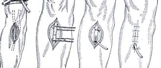
During the diagnostic study, a puncture is made in the femoral artery, through which a catheter is inserted into the vessels of the heart muscle to supply a contrast agent, resulting in an image being displayed on the monitor. Next, the area of narrowing of the artery walls is identified and its degree is calculated. This allows the specialist to predict the further development of the disease.
In Moscow, prices for coronary angiography of the heart vessels on average vary from 20,000 to 50,000 rubles, for example, the Bakulev Center for Cardiovascular Surgery provides high-quality examination of coronary vessels, the cost of the procedure starts from 30,000 rubles.
Sick sinus syndrome
The term was coined in 1962 by American cardiologist Bernard Lown. The diagnosis can be made if at least one of the typical findings on the electrocardiogram has been demonstrated:
- inadequate bradycardia of the coronary sinus;
- freezing of the sinus node;
- sinoatrial block;
- atrial fibrillation;
- atrial flutter;
- supraventricular tachycardia.
The most common cause of sick sinus syndrome is hypertension, which leads to chronic strain on the atrium and then overstretching of the muscle fibers. The key examination method is long-term ECG.
General methods of treating heart vessels
To treat and strengthen blood vessels, complex methods are used, consisting of dietary and lifestyle adjustments, drug therapy and surgical intervention.
General recommendations:
- adherence to dietary nutrition, with increased consumption of fresh vegetables, fruits and berries, which is useful for strengthening the heart and blood vessels;
- light gymnastic exercises for the heart and blood vessels are prescribed at home, swimming, jogging and daily walks in the fresh air are recommended;
- vitamin complexes are prescribed for the vessels of the brain and heart with a high content of retinol, ascorbic acid, tocopherol and thiamine;
- droppers are used to support the heart and blood vessels, nourishing and restoring the structure of tissues and walls in the shortest possible time;
- medications are used for the heart and blood vessels, reducing pain, removing cholesterol, lowering blood pressure;
- a new technique for improving the activity of the heart and blood vessels is listening to therapeutic music: American scientists have proven a positive effect on the contractile function of the myocardium while listening to classical and instrumental music;
- good results are observed after using traditional medicine: some medicinal plants have a strengthening and vitamin effect for the heart and blood vessels, the most popular are a decoction of hawthorn and motherwort.
How to reduce the risk of complications
To reduce the risk of complications after acute coronary syndrome, it is necessary to lead a healthy lifestyle. The same recommendations will help reduce the risk of recurrence of this syndrome:
- Constant monitoring of blood pressure using a tonometer. To carry it with you, it is better to purchase a more compact version. Normal blood pressure is considered to be 120/80, but there may be individual differences. The frequency of blood pressure measurements will be determined by your attending physician. Any deviation from the norm is a reason to seek advice.
- Regular monitoring of blood cholesterol levels. If the norm is exceeded, the doctor will prescribe a strict diet.
- Smoking is prohibited. This has an extremely negative effect on the functioning of the heart.
- It is prohibited to consume alcoholic beverages. In some situations, doctors allow limited consumption of high-quality alcohol, but caution must be exercised, because it helps increase blood pressure.
- Body weight control. Excess weight contributes to the development of hypertension, increases cholesterol levels in the blood, and increases the load on the heart and the entire body.
- Proper nutrition. It is widely known that fatty, fried, salty and sweet foods have a negative impact on health. It increases blood cholesterol levels and increases the risk of obesity. Maintaining a proper diet should come to the fore.
- Avoidance of stress and/or ability to cope with it. If you cannot control your emotions on your own, then you should seek help from a professional psychologist. You shouldn’t immerse yourself entirely in work, because the main thing in such a state is to be in balance.
Moderate physical activity is important. It is important to maintain balance when playing sports. If you have suffered an ACS, you should not over-stress your heart with physical exercise.
On a note! Slow walking and light exercise at a comfortable pace are suitable.
It is important to understand that all loads must be increased gradually, and if discomfort occurs, stop immediately.
Surgical methods for treating heart vessels
X-ray surgeons at work performing angioplasty and cardiac stenting
To improve blood circulation in the coronary arteries, balloon angioplasty and stenting are performed.
The balloon angioplasty method involves inserting a specialized instrument into the affected artery to inflate the vessel walls at the site of narrowing. The effect after the procedure remains temporary, since the operation does not involve eliminating the main cause of the stenosis.
For the most effective treatment of stenosis of the vascular walls, stents are installed in the vessels of the heart. A specialized frame is inserted into the affected area and expands the narrowed walls of the vessel, accordingly the blood supply to the myocardium improves. According to reviews of leading cardiac surgeons, after stenting of cardiac vessels, life expectancy increases, provided that all medical recommendations are followed.
The average cost of cardiac stenting in Moscow ranges from 25,000 to 55,000 rubles, excluding the cost of instruments; prices depend on many factors: the severity of the pathology, the number of stents and balloons required, the rehabilitation period, etc.
Stent deployed in coronary artery
In terms of open cardiac surgery, everyone knows the operation of coronary artery bypass grafting. Previously, cardiac arrest, cardioplegia, heart-lung machine, etc. were required. Today, such operations are possible in some cases on a beating heart. There is also an option - mammary coronary bypass surgery. Moreover, the latter is also possible from a mini-access - through a mini-thoracotomy.
Coronary artery bypass grafting with coronary artery bypass grafting
Mammarocoronary bypass surgery
The best help for coronary artery diseases is timely seeking qualified help for further diagnosis and treatment of vascular diseases.
Causes of lesions
Damage to the coronary arteries in most cases is associated with insufficient attention to one’s own health.
Every year, such violations lead to the deaths of millions of people around the world. Moreover, most people are residents of developed countries and are quite wealthy.
Provoking factors contributing to violations are:
- Tobacco use. Not only is smoking harmful, but also inhaling cigarette smoke.
- Abuse of foods containing large amounts of cholesterol.
- Weight problems. Obesity creates additional stress on blood vessels.
- Insufficiently active lifestyle.
- Excessive blood sugar.
- Constant emotional stress.
- Constant fluctuations in pressure in the arteries.
No less important influence is exerted by age-related changes, hereditary predisposition, and gender. Such diseases in acute form affect men, so they die from them much more often. Women are more protected due to the influence of estrogen, so they are more likely to have a chronic course.
Coronary angiography
Coronary angiography (coronary angiography) is x-ray visualization of the coronary vessels after the injection of a radiopaque contrast agent. The X-ray image is simultaneously recorded on 35 mm film or digital media for subsequent analysis. At the moment, coronary angiography is the “gold standard” for determining the presence or absence of stenoses in coronary disease. The purpose of coronary angiography is to determine the coronary anatomy and the degree of narrowing of the lumen of the coronary arteries. Information obtained during the procedure includes determination of the location, extent, diameter and contours of the coronary arteries, the presence and degree of coronary obstruction, characterization of the nature of the obstruction (including the presence of atherosclerotic plaque, thrombus, dissection, spasm or myocardial bridge). The data obtained determine the further tactics of treating the patient: coronary bypass surgery, intervention, drug therapy. To conduct high-quality angiography, selective catheterization of the right and left coronary arteries is necessary, for which a large variety of diagnostic catheters of various modifications have been created. The examination is carried out under local anesthesia and NLA through arterial access. The following arterial approaches are generally accepted: femoral arteries, brachial arteries, radial arteries. Transradial access has recently gained a strong position and has become widely used due to its low morbidity and convenience. After puncture of the artery, diagnostic catheters are inserted through the introducer, followed by selective catheterization of the coronary vessels. The contrast agent is administered in doses using an automatic injector. Filming is performed in standard projections, the catheters and intravener are removed, and a compression bandage is applied.