Cardiovascular diseases are a very common pathology for people over 40 years of age. And among these diseases, the most common are associated with imperfection of the vascular bed and limited nutrition of the heart muscle.
To clarify the causes of heart disease, there are many diagnostic methods. One of the most informative checks is coronary angiography of the heart vessels - what is it, is it dangerous to do it, and how is the examination performed?
general information
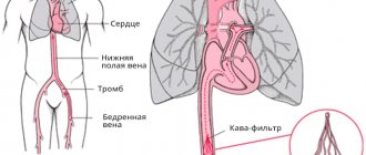
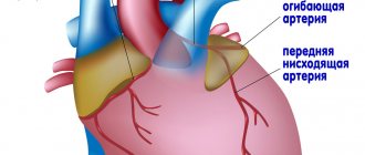
This is an invasive manipulation that serves to determine the condition of the vessels that carry blood and oxygen to the heart. They are called coronary. The left and right coronary arteries normally provide nutrition to the muscle and maintain the functionality of the entire organ.
In case of unfavorable development of events, these arteries, for various reasons, narrow (stenosis) or become blocked (occlusion) . The blood supply to the heart is significantly limited or stops altogether in a certain area, which causes coronary artery disease and heart attack.
To exclude such a defect or, if present, to determine its extent, coronary angiography is performed.
This is an X-ray examination of the lumen of the coronary vessels using an angiograph and a contrast agent introduced through a catheter precisely into the vestibule of the cardiac arteries. The survey is carried out from different angles, which allows you to create the most detailed picture of the condition of the object being examined.
Types of diagnostics
Examination of the heart vessels may include:
- General. Coronary angiography of all heart vessels is performed.
- Selective. A study of some (not all) vessels is carried out. The contrast is introduced so as to fill only the required vessels.
Depending on the type of equipment and technique for examining a vital organ of the cardiovascular system, several subtypes of angiography are used.
Invasive coronary angiography
A contrast agent is injected into the vessel, images are taken (the shooting frequency is several frames per second), recorded on film or digital media. The survey is characterized by high spatial resolution.
Tomographic examination (CT or MSCT)
After injection of a contrast agent, the patient is placed in a multislice tomograph. The doctor, studying the three-dimensional image, receives detailed data about the lumens and morphological pathologies. The disadvantages of tomographic coronary angiography include the high dose of radiation received by the patient.
Magnetic resonance coronary angiography
After intravenous administration of a special radiopharmaceutical, the patient is placed on a table that can change its position relative to the source of magnetic waves. Some tomographs provide highly accurate results without contrast.
Advantages of MTP coronary angiography: no need for large-scale vascular interventions, excellent visualization of the arterial bed, detection of vascular narrowing, spasms, thromboembolism, atherosclerosis, hypoxia and myocardial ischemia.
Positron emission coronary angiography
The technique involves injecting glucose or another substance into a vein along with a radionuclide that emits positrons (positively charged particles). The tomograph records changes in the intensity of ionizing radiation in various areas of the myocardium and blood vessels, creating clear images. The disadvantages of PET coronary angiography include the high cost of the procedure and significant radiation doses.
Single photon emission coronary angiography
The method is based on the generation of images by a tomograph that records radiation (gamma rays) of the administered radiopharmaceutical. The procedure for conducting an examination, which allows a comprehensive assessment of the nature of vascular damage, is similar to PET diagnostics.
Indications for the procedure
Routinely, coronary angiography is performed for:
- confirmation or refutation of the diagnosis of IHD;
- clarifying the diagnosis when other methods of determining the disease are ineffective;
- determining the nature and method of eliminating the defect during the upcoming operation;
- audit of the condition of the organ in preparation for open-heart surgery, for example, in case of a defect.
In emergency cases, the procedure is performed in the presence of the first signs and symptoms of a heart attack or in a pre-infarction condition that requires immediate intervention for life-saving reasons.
Let's look at how to prepare for coronary angiography of the heart, as well as how this procedure is done.
Indications for testing
Key indications for coronary angiography are suspicion of exacerbation of coronary heart disease, increased risk of complications, lack of effect from therapy for coronary artery disease, difficulties in choosing a treatment technique due to the inaccuracy of the clinical picture.
Let's look at the detailed readings. Coronary angiography is performed when:
- Severe (acute) symptoms of a heart attack. The study is carried out as soon as possible - no later than 10-12 hours after the onset of the attack.
- Post-infarction shock.
- Angina pectoris (primary, progressive, post-infarction, aggravated after stenting, bypass surgery).
- Insufficiency of blood supply to the myocardium, confirmed by the results of an electrocardiogram or daily ECG monitoring.
- Identification of ischemia after exercise tests.
- Ischemic pulmonary edema, pulmonary congestion.
- Prolonged hypotension.
- Severe forms of arrhythmia.
- The presence of prerequisites for differential diagnosis with non-ischemic heart pathologies.
- Chest injury.
- Painful sensations behind the sternum.
- Endocarditis.
- Cardiomyopathies.
- Kawasaki diseases.
- Cardiopulmonary resuscitation.
- Preparation for internal organ transplantation and cardiac intervention.
- Conducting professional medical examinations (pilots, astronauts, representatives of other extreme professions).
Preparation
Before prescribing coronary angiography, it is necessary to undergo a series of examinations in order to exclude or confirm the presence of factors that do not allow the use of this diagnostic method. Training program:
- blood tests (general, sugar, hepatitis B and C, bilirubin and other liver parameters, HIV, RW, group and Rh factor);
- urine test for the presence of renal pathology;
- ECG in 12 leads;
- examination and conclusion of specialists on existing chronic diseases.
Upon admission to manipulation, immediate preparation is carried out before the procedure :
- the doctor stops some medications in advance, for example, those that reduce blood clotting;
- exclude food intake on the day of diagnosis - to avoid complications such as vomiting, the study is carried out on an empty stomach;
- The doctor collects an allergic history and conducts a test with a contrast agent.
Immediately before coronary angiography, it is recommended to take a shower, shave the hair in the groin, remove jewelry from the body (earrings, rings, piercings), glasses, removable dentures, lenses, and use the toilet.
Risk factors
What is coronary angiography and what are the consequences of coronary angiography of the heart vessels? This is, first of all, an invasive procedure that allows you to assess the condition of the heart vessels by introducing into the body a special contrast agent that colors the arteries a special color at the time of the examination. When it comes to “penetration” through a person’s protective shell (in this case through the skin), one brief but important rule needs to be mentioned: “such intervention is always associated with a risk, both insignificant to health and posing a potential danger to life.”
In some cases, the likelihood of complications increases significantly. A special group of risk factors includes such ailments as:
- allergic reaction to injected contrast;
- serious condition associated with the psyche or somatics;
- pregnancy;
- atrial fibrillation (heartbeat disturbance with frequent contraction and excitation of the atria);
- hypokalemia;
- frequent extrasystole;
- renal and heart failure;
- fever;
- hemophilia, anemia and other forms of bleeding disorders;
- poisoning with special cardiac glycosides;
- advanced age of the patient;
- diseases of the cardiovascular system;
- diabetes mellitus and stroke;
- exhaustion or significant excess weight;
- severe lung disease, such as pulmonary failure;
- heart ailments;
- calcification of the coronary vessels (deposition of calcium salts in the valve leaflets and near the walls of the arteries).
If a patient at risk needs to urgently undergo coronography, the procedure is carried out under the close attention of a team of doctors. Within one day after diagnosis, special monitoring of ECG (electrocardiogram) and hemodynamics (blood movement through the vessels) is carried out.
In case of emergency intervention, you should try to inform the doctor about allergic reactions and possible concomitant diseases
It is worth noting that the probability of complications is approximately 0.05–0.2%. And death occurs in less than 0.08% of cases. More detailed information about risk factors and other indicators of coronography is presented in this article.
How it's made
The patient lies down on a special table. Heart monitors are attached to his chest. In the area where the catheter is inserted, local anesthesia and skin disinfection . A micro-incision is made in the vein through which a catheter is inserted.
The catheter is passed through the vessels under the control of an angiograph to the mouth of the coronary arteries. A contrast agent is injected into each of them one by one, which outlines the internal space of these vessels. are carried out from different positions . The location of stenosis or occlusion is determined.
During the operation, the patient is conscious and his condition is monitored by observing his appearance, heart rate and verbal behavior.
Once monitoring is complete, the catheter is carefully removed from the vein. The wound is carefully sutured. The patient remains lying down for some time, and the doctor writes a conclusion . It indicates the size of the smallest lumens in the vessels, the degree of narrowing and the recommended method of correcting the situation - stenting or bypass surgery of the heart vessels. If there are no problem areas, a general description of the coronary arteries is given.
Video about how outpatient coronary angiography of the heart vessels is performed:
How is coronary angiography done?
It is worth distinguishing an emergency and a planned procedure from each other: the second type involves a longer preparation stage, and the emergency, in turn, does not allow the main part of the check before diagnosis, since every second is worth its weight in gold. The initial stage of a routine inspection is a comprehensive examination:
- a comprehensive blood test, including general and biochemical analysis, establishment of the Rh factor and blood group, testing for hepatitis B and C, HIV and syphilis;
- ultrasound examination of the heart (ultrasound);
- electrocardiogram (ECG) in 12 leads;
- examination by a cardiologist.
If a patient has any concomitant chronic illnesses, he must definitely visit specialists in the relevant areas. The next stage represents the following sequence:
- If necessary, the person is given anti-allergenic and sedatives;
- electrodes are being connected, with the help of which doctors will be able to monitor heart parameters during an ECG study;
- The catheterization site is prepared; as a rule, the puncture is carried out in the area of the femoral artery, and in case of contraindications, in the area of the brachial, radial or axillary artery;
- the patient is anesthetized with local anesthesia and then sedatives are administered to promote relaxation and drowsiness;
- the corresponding place is treated with an antiseptic, and the body is covered with a sterile cloth;
- after catheterization, an introducer (plastic tube) is inserted into the vessel and diagnostic catheters are passed through it into the bloodstream;
- the arteries are filled with the required amount of contrast agent, and at the same time angiography is performed using X-rays;
- at the end of the medical procedure, the doctor removes the catheter and stops the bleeding;
- a special pressure bandage is applied to the puncture site;
- the patient is taken to the ward.
Some people want to know how long the research process takes. The answer to their question is quite simple: on average, the procedure takes 10–20 minutes, but special cases require an extension of the time period. The specifics of the procedure are briefly outlined in this video.
Conditions
Most often, coronary angiography is performed in a hospital setting as part of a routine examination for coronary artery disease. In this case, all tests are taken here, a few days before the intervention.
Diagnostics can also be carried out on an outpatient basis. But the patient must first independently undergo all the examinations on the list, obtain a cardiologist’s opinion on the possibility of coronary angiography and a referral for it, indicating the purpose of the study.
In an outpatient setting, the insertion of a catheter for coronary angiography is most often carried out through the wrist vein and in the arm - in the postoperative period, the load on it, in contrast to invasion through the femoral vessel, can be minimized in order to avoid dangerous bleeding.
Cardiography
The stages of the event themselves will depend on exactly how the heart vessels are examined.
Coronary angiography is performed as follows:
- First, the area to be examined is numbed. For these purposes, Novocaine or Lidocaine is used.
- After this, a catheter is passed to the mouth of the coronary vessels. This is done through the femoral artery.
- Then a contrast agent is injected.
- After all of the above, an angiographer conducts an examination and records the results of the study.
To perform electrocardiography, the patient is undressed to the waist and placed on a couch. Then an electrically conductive gel is applied to the area being examined and electrodes are installed. Phonocardiography is performed in almost the same way. But instead of electrodes, five special microphones are used.
Contraindications
A number of conditions do not allow the use of this diagnostic method, so they resort to alternative ones. Preliminary examination may reveal these conditions:
- uncontrolled arterial hypertension - intervention can provoke stress, resulting in a hypertensive crisis;
- post-stroke condition - anxiety can cause a re-attack of the disease;
- internal bleeding in any organ - with invasion, blood loss may increase;
- infectious diseases - the virus can contribute to the formation of blood clots at the incision site, as well as peeling of areas on the walls of blood vessels;
- diabetes mellitus in the stage of decompensation is a state of significant kidney damage, high blood sugar levels, and the possibility of a heart attack;
- elevated temperature of any origin - accompanying high blood pressure and rapid heartbeat can lead to heart problems during and after the procedure;
- severe kidney disease - the contrast agent can cause organ damage or worsen the disease;
- intolerance to contrast agent - a test is carried out on the eve of diagnosis;
- increased or decreased blood clotting - can cause thrombosis or blood loss.
With preliminary preparation, all these conditions are identified and treatment is prescribed to compensate for them. There are no absolute contraindications to the procedure. Once stabilization has been achieved, the procedure can be performed in a hospital setting.
Possible complications after the procedure
Like any other surgical intervention and violation of the integrity of the vessel, coronary angiography can have a number of complications, even if it is performed by an experienced specialist. Possible complications include:
- heart attack;
- rupture of an artery or heart;
- heart attack or stroke due to a blood clot breaking off from the wall of a vessel;
- arrhythmia;
- bleeding;
- allergies.
Of course, complications do not always arise and are often limited to small hematomas or swelling at the puncture site. However, the possibility of serious consequences cannot be excluded, so before the procedure you need to carefully study the contraindications to it.
Contraindications for the study
Contraindications for cardiac vascular examination with contrast are:
- various respiratory diseases that are accompanied by elevated body temperature;
- anemia;
- open bleeding of various locations;
- low potassium levels in the blood;
- poor blood clotting;
- diabetes;
- obesity or underweight;
- renal, heart failure;
- lung damage.
Risks, complications and consequences
Coronary angiography, like any invasion, can have side effects caused by the body’s improper response to the intervention and the patient’s stress. Rarely, but the following events occur:
- bleeding at the insertion gate;
- arrhythmia;
- allergy;
- detachment of the inner layer of the artery;
- development of myocardial infarction.
Pre-procedural testing is intended to prevent these conditions, but sometimes they happen. The doctors involved in the examination cope with the situation, the procedure is stopped at the first unfavorable signs , the patient is removed from the dangerous state and transferred to a hospital for observation.
Recommendations during the recovery period after the procedure
The patient can remain in the hospital after angiography from 5 to 24 hours. During this period, bed rest is recommended, you can drink water and fruit juices. If the heart function is stable, the patient is discharged.
At home, you need to follow a gentle regimen for at least a week, avoid physical activity, drinking alcohol and smoking. There is no need to take a bath for 2-3 days; the puncture site should remain dry when taking a shower. The car can be driven in 3 - 5 days.
You should immediately consult a doctor if you have the following symptoms:
- bleeding from the puncture site of the artery;
- pain, swelling and redness of the skin;
- there is hardening near the catheterization area;
- body temperature has increased;
- the skin has changed color and the limb that was used to insert the catheter becomes numb and feels cold or hot to the touch;
- excessive weakness, chest pain and shortness of breath occurred.
Recommendations after completion
Based on the conclusion of the doctor who conducted the study, the cardiologist determines the path of treatment for the patient . If there are indications, the time for stent installation is scheduled (in the same way as coronary angiography - using a catheter).
Sometimes this procedure is performed directly during diagnosis, if there is the prior consent of the patient. The cardiologist may also prescribe outpatient treatment or coronary artery bypass surgery.
How is outpatient coronary angiography performed?
The algorithm for performing coronary angiography on an outpatient basis includes three stages. At the first stage, patients are selected for a diagnostic procedure, and the necessary additional examinations are carried out. Indications for coronary angiography : - identified or suspected ischemic heart disease; - pain in the chest, suspicious for angina pectoris; - myocardial infarction; - planned surgery for heart defects; - heart failure, ventricular arrhythmias. Indications for coronary angiography are determined by the attending physician in accordance with accepted criteria. In preparing the patient for coronary angiography , the necessary tests and studies are performed. In addition to these, additional studies may be prescribed. The second stage of outpatient CAG angiography procedure itself . The patient is admitted to the day hospital ward. After assessing the stability of his condition, premedication is administered and he is transported to the cath lab, where the coronary angiography . After anesthetizing the access area, research begins - a special catheter is passed through the artery of the forearm into the lumen of the coronary arteries . Using a catheter, a radiopaque substance is injected into the blood, thanks to which the lumen of the vessels becomes visible on a special device - an angiograph. During coronary angiography, the degree and size of damage to the coronary vessels is determined, which determines further treatment tactics. This procedure is low-traumatic, which allows it to be performed under local anesthesia without the use of general anesthesia. The duration of the procedure, as a rule, does not exceed 20 minutes. From the operating room, the patient, accompanied by medical personnel, is taken to the day hospital ward. The third stage of outpatient coronary angiography is observation of the patient in a day hospital ward for 4-5 hours after the examination. In the ward, the patient can drink water or juices without restrictions and have lunch. If there are no complications, the patient is sent home. On the day of the outpatient coronary angiography, the patient receives a conclusion with recommendations on further treatment tactics and a disk with the results of coronary angiography . If complications arise during coronary angiography or during the control period, patients are hospitalized in the intensive observation unit of the hospital.
Diagnostic cost
If you have a compulsory medical insurance policy, coronary angiography according to indications is performed free of charge. But the equipment of most hospitals does not allow covering everyone with this diagnostic method in a short time. Usually the queue lasts for months, because... Limited quotas for examination are provided. It is possible to undergo this study on a commercial basis.
The cost in Russia is in a wide range - from 10 to 45 thousand rubles. Abroad, this intervention is also not always covered by insurance and is also not cheap - from 300 dollars to 2500 euros.
Coronary angiography is included in the mandatory list of diagnostic procedures to determine the degree of damage to the heart vessels. The procedure has been worked out and standardized for a long time - this serves as a guarantee of patient safety. The level of cardiology in the country makes it possible to identify pathology at an early stage and take measures to eliminate it or prevent its development.
How to avoid consequences
Although the incidence of complications is not very high, there are recommendations that, if followed, can reduce the risk of their development.
It should be remembered that the main way to prevent the development of complications is to choose experienced medical personnel. According to foreign colleagues, a doctor who performs more than 100 coronary angiographies per year can be considered experienced.
Preoperative preparation
In some cases, coronary angiography is performed very urgently - in the first hours of myocardial infarction. In these conditions, preparation takes a minimum of time and boils down to the fact that the medical staff quickly asks the patient about complaints and medical history, conducts the minimum necessary examination, takes an ECG and draws blood for tests. In addition, the patient receives the necessary medications for the treatment of acute coronary syndrome, and he undergoes catheterization of a peripheral vein. After this, the patient is transported to the operating room. This urgency is due to the fact that the time before surgery for acute myocardial infarction plays a huge role - the sooner it is performed, the better the result.
In most cases, coronary angiography is performed routinely. In order to prepare for it, the patient undergoes a detailed examination by a doctor, who conducts a survey and examination of the patient, evaluates laboratory and instrumental data. The patient should inform the doctor about his diseases that may affect the performance and complications of coronary angiography (for example, diabetes mellitus and kidney disease); allergies to medications and food products; the medications he is taking. A laboratory (complete blood count, urinalysis, coagulogram, biochemical blood test) and instrumental (ECG, echocardiography) examination is carried out, which allows diagnosing concomitant pathologies.
Typically, before the procedure, the patient must:
- Follow the doctor's recommendations; You cannot independently use medications that were not prescribed to the patient.
- Do not eat or drink after midnight the day before your coronary angiography; Take prescribed tablets with a small sip of water.
- Shave the groin area and/or forearm through which the intervention will be performed. It is better to carry out this procedure using an electric razor so as not to damage the skin - this will reduce the risk of developing infectious complications.
- Take a hygienic shower the day before coronary angiography.
- Ask your doctor about the possibility of performing diagnostic surgery through the radial artery.
Carrying out coronary angiography through the radial artery can reduce the incidence of severe complications and mortality after the procedure.
Most often, the patient is prescribed sedatives before surgery, which will allow him to relax and unwind a little.
Postoperative period
After the procedure, the patient remains in the hospital for at least one more day. At this time, his blood pressure and pulse are monitored, and medication correction is carried out.
Immediately after coronary angiography, the patient must strictly follow the doctor's recommendations about bed rest. The duration of the supine position depends on the site of surgical access (femoral or radial artery), whether the sheath was removed, and the method of hemostasis.
If hemostasis was carried out by pressing the femoral artery, you need to lie down for 6-8 hours; if a special device was used to stop bleeding, the patient can sit down after 1-2 hours.
Since the contrast agent is excreted in the urine, the patient should drink enough water, unless he has contraindications to this, and monitor diuresis (count the amount of urine).
You should immediately report any complaints or complications to medical personnel.
The intravenous catheter is removed a few hours after surgery, and the bandage over the arterial puncture site is removed the next day.