No one will argue that ultrasound diagnostics to this day remains one of the most effective methods for identifying and preventing pathologies of vital organs. In turn, ultrasound of the heart or echocardiography is an incredibly effective diagnostic method that allows you to detect certain diseases of the cardiovascular system and treat them in a timely manner.
Everyone knows that echocardiography is prescribed not only for adults, but also for children, and therefore many young parents still have many questions regarding this procedure. Is it safe to perform an ultrasound of the heart on a child, how does this manipulation work, can it be done on a newborn - we will answer all these and other questions in our article.
Indications for echocardiography
Heart ultrasound is prescribed in cases where:
- the doctor, while auscultating the baby’s heart, will hear any noises;
- the baby complains of pain in the chest area on the left side;
- during crying and sucking, the baby's nasolabial triangle turns blue;
- periodically the child experiences loss of consciousness;
- the child’s body weight is clearly insufficient for his age, he gets tired quickly and has increased sweating;
- the baby suffers from frequent pneumonia (read more about the symptoms of pneumonia in a child);
- The baby's family has relatives with serious cardiovascular pathologies.
However, even without any clear indications, this study is recommended for all children without exception before they reach one year of age to exclude possible pathologies. At the same time, other ultrasound examinations must also be carried out: the brain through the fontanelle, the kidneys, the pelvis and abdominal cavity, as well as the hip joints (read more about hip dysplasia).
However, it is advisable to carry out such studies at the age of 1.5 – 2 months.
Decoding the results
Based on the results of the study, the patient receives a conclusion where the parameters are presented in numerical values. They cannot be considered a final diagnosis. After echocardiography, a consultation with a pediatric cardiologist is needed, who will interpret them. This is due to the fact that the obtained figures may differ depending on the severity of the pathology and the age of the patient.
Standards for children of different ages: table
Table of normal ultrasound parameters of the heart of an infant weighing up to 3.5 kg.
| Index | Girls (in mm) | Boys (in mm) |
| LV EDC | 15-20 | 16-21 |
| LV ESR | 10-16 | 10-16 |
| Left atrium diameter | 10-15 | 11-16 |
| Left ventricular diameter | 5-12 | 6-15 |
| TZSLZH | 2-4 | 3-4 |
| MZhP | 2-5 | 3-6 |
| Right ventricular free wall | 2-3 | 2-3 |
| Ejection fraction | 64-74% | 64-74% |
| Pulmonary valve blood flow velocity | 1.4-1.5 m/s | 1.4-1.5 m/s |
Table of normal ultrasound parameters of the heart of a newborn weighing up to 4.5 kg.
| Index | Girls (in mm) | Boys (in mm) |
| LV EDC | 18-25 | 20-26 |
| LV ESR | 11-18 | 11-18 |
| Left atrium diameter | 11-18 | 14-19 |
| Left ventricular diameter | 6-14 | 7-15 |
| TZSLZH | 3-6 | 3-6 |
| MZhP | 3-6 | 3-6 |
| Right ventricular free wall | 2-3 | 2-3 |
| Pulmonary valve blood flow velocity | 1.4 m/s | 1.4 m/s |
Attention! The results of the study in adolescents (over 14-15 years old) are assessed according to adult age standards.
Pathologies shown by ultrasound of the heart
Usually, an ultrasound examination of the heart is enough for a pediatric cardiologist to make a diagnosis. Using this method, the following diseases are detected:
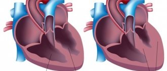
- Incomplete fusion of the interventricular septum. There remains a hole between the ventricles. The defect is characterized by enlargement of the walls of the heart and its cavities.
- Ventricular septal defects. An ultrasound visualizes an opening and an increase in the thickness of the atrium walls.
- Mitral valve disease. A violation of the hole diameter is often detected.
- Aortic stenosis. The study noted a narrowing of the aortic lumen, an increase in the thickness of the walls of the atria and ventricles.
- In the case of inflammatory changes in cardiac tissue, the ejection function is impaired. It decreases with a parallel increase in the cavities of the heart.
- Tumor-like formations.
EchoCG results are interpreted by a pediatric cardiologist or pediatrician. The ultrasound diagnostic doctor only describes the abnormalities he sees, he does not make a diagnosis. The pediatrician makes a final conclusion based on a comprehensive examination of the child.
What does an ultrasound of the heart show?
Echocardiography makes it possible to examine:
- anatomical features of the heart structure;
- “serviceability” of its valves;
- pressure inside the heart muscle;
- blood flow speed.
It can show the presence of such pathologies in a child:
- congenital and acquired heart defects;
- enlargement of the heart chambers;
- hypertrophy or hypotrophy of the heart;
- blood clots and other unnatural neoplasms;
- heart tumors;
- endocarditis (inflammation of the inner lining of the heart muscle);
- pericarditis (inflammation of the serous membrane of the heart muscle);
- aortic stenosis, which interferes with normal blood flow, etc.
When is an ultrasound performed for a child?
Doctor's suspicions during auscultation
There are a number of indications when cardiac echocardiography is desirable or necessary for children:
- Noises heard during auscultation. If the doctor does not like the nature of the noise, and in order to determine its cause, an ultrasound of the heart may be needed.
- A feeling of trembling in the area of the child’s heart, which is noticed by the parents and confirmed by the doctor during the examination.
- Child's complaints about unpleasant or painful sensations in the heart area (pulling, aching, stabbing, pressing, etc.)
- Unusual behavior of infants - refusal of the breast, sluggish sucking, blueness of the nasolabial triangle while sucking the breast.
- Periodic coldness of the arms and legs of children at a constant temperature. In this case, it is necessary to consult a doctor so that, if necessary, he can refer the child for an ultrasound of the heart.
- Loss of consciousness during intense physical activity.
- The child gets tired quickly, sweats a lot and gains weight poorly compared to his peers.
- When a child cries, the area of the nasolabial triangle begins to turn blue.
- Frequent colds in a child (up to 6-8 times a year).
- Family history (there are relatives suffering from cardiovascular diseases).
Preparation for this procedure
As such, no preparation is required for echocardiography. However, due to the fact that during this study the baby’s pulse and breathing should not be rapid , and he should be as calm as possible, parents still need:
- reassure the child, explain to him that it is absolutely not painful and why such a procedure is needed;
- exclude physical activity before echocardiography;
- do not drink tonic drinks such as coffee or strong tea;
- The newborn should also be fed before the procedure so that he is fed and calm.
How is cardiac ultrasound performed?
Echocardiography, like any other ultrasound examination, should be carried out by a specialist competent in this field - an ultrasound diagnostician. This procedure does not take much time, about 15 minutes (if serious diseases are detected, the duration of the ultrasound, of course, can increase to about 30 - 40 minutes), and takes place in this order: the child is undressed from the waist up and placed on the couch on the left side or on the back , and the doctor applies a special water-based gel to the area of his chest and begins to move the sensor over it, pausing at one point or another to record the data.
Complete immobilization of the baby is not required , since small movements of the arms or legs will not affect the diagnostic results in any way. At the end of this manipulation, the child’s chest must be wiped of any remaining gel. And then you should contact a pediatric cardiologist to decipher the indicators and make a conclusion based on the ultrasound results.
What pathologies can be identified?
Based on the results of the ultrasound, the following can be determined:
- Septal defects - this type of pathology is characterized by thickening of the walls of the atria.
- Mitral valve defects.
- Narrowing of the aorta - there is a reduced size of the aorta, while the ventricles and atria are thickened.
- The presence of inflammation in the tissues of the heart - in this case, the cardiac cavities are enlarged, and the function of blood ejection is reduced compared to the norm.
- Violation of the structure and location of the chambers of the heart.
- Presence of blood clots.
- Increased pressure in the pericardial cavity.
- Presence of fluid in the pericardial cavity.
- Scars on the surface of the heart muscle.
- Aneurysms of the great vessels.
How safe is echocardiography?
The most common question about this study is: is ultrasound harmful and is it safe for the child’s health? It is safe to say that, unlike the unsafe X-ray examination, echocardiography does not use radiation, but only mechanical wave vibrations.
This procedure is absolutely harmless, and therefore has no age restrictions: ultrasound of the heart can be performed already in the first hours of a baby’s life and even in premature babies.
As you can see, the doctor does not wear any protective equipment during this procedure, unlike, again, an x-ray examination.
What to take with you to the examination
To conduct an ultrasound of the heart, a newborn baby does not require any special preparation, but we still recommend that you put the necessary things in your bag in advance:
- referral for examination and the child’s medical record. It would be useful to record the height and weight of the subject;
- disposable diaper and wet wipes. You will need the first one in order to place the baby on it before the ultrasound, and the second ones in order to remove the remnants of the inert gel;
- pacifier or bottle of milk. The effectiveness of the study depends on the child’s behavior. For more accurate results, the patient must be calm. Therefore, worry in advance about what will distract your baby from the procedure;
- if cardiac ultrasound is not performed for the first time, prepare the results of previous procedures.
Before the event, we advise you to dress your child in things that can be quickly and easily removed, since ultrasound examination suggests this. Clothes with buttons and ties will only spoil the baby’s mood, which will definitely affect the quality of the procedure.
How often can you do an ultrasound of the heart?
As already mentioned, this manipulation does not cause any harm and is absolutely safe, and therefore it can be carried out as often as circumstances require. If necessary, echocardiography can even be performed several times in one day . There is absolutely no need to maintain any intervals between procedures.
Video about how an ultrasound of a child’s heart is performed
This video clearly shows how this procedure is carried out, and also provides answers to the most common questions about echocardiography: for what purpose it is performed, at what age and what it reveals.
From all of the above, we can conclude that echocardiography is an incredibly important procedure and all children, without exception, should undergo it, even without obvious indications. How do you feel about this manipulation, do you consider it safe? Has your baby ever had a heart ultrasound? How did he behave during this and how did it all go? Leave your comments on this matter.
Advantages and disadvantages of the method
This method of studying the myocardium has a number of advantages, which include:
- Does not require the use of anesthesia.
- Not harmful.
- Has a relatively low cost.
- Allows you to visualize soft tissues and identify the disease in the early stages.
The disadvantages of the procedure include low image quality compared to MRI and CT.
Ultrasound of the myocardium in newborns is performed according to certain indications. The study is completely safe, requires virtually no special preparation and allows you to identify defects and disorders of the heart muscle.