How does the embryo develop in the period from 3 to 5 weeks?
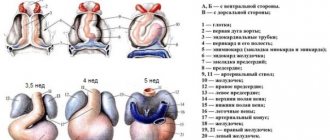
The period of pregnancy from the 3rd to the 5th week is considered very important for the development of the fetus. This is due to the fact that it is at this time that embryogenesis occurs. The embryo has the appearance of the auricle, as it is curved in the shape of the letter C. At this time, when performing an ultrasound, specialists can already examine the rudiments of the head, back, upper and lower extremities.
In the period from 3 to 5 weeks of development, the spinal cord is released in the embryo (the development of the spinal cord and spine begins). The correct formation of the brain can be indicated by the process of flattening (at the wide end of the embryo) of the neural tube, which should occur gradually. During an ultrasound examination, specialists can see how tissue segments called somites are formed and subsequently rapidly increase, which are responsible not only for the development of muscles, but also of all tissue structures present in the human body.
Gynecologists pay close attention to this period also because the fetus begins to develop vascular and cardiac systems, which develop very quickly. Using a highly sensitive ultrasound machine, it is possible to examine the process of formation of large blood vessels. They are located in the central part of the embryo and are closely connected with a glomerulus of tissue, which at this time is only a prototype of the future heart. This tangle is a very important substance that takes an active part in the process of organogenesis.
If pregnancy is successful, it is from these tissues that the respiratory tract (upper) will begin to develop. In this case we are talking about the trachea and larynx. They also take an active part in the process of laying the pancreas, liver, gonoblasts (germ cells), which determine the sex of the child.
A photo of an embryo at the 3rd – 4th week is not informative, since the size of the embryo does not reach 0.2 mm
In the period from 3 to 5 weeks of pregnancy, gynecologists and uzologists are unable to hear the baby’s heartbeat (even when using ultra-sensitive equipment), but they confidently claim that the embryos already have all the necessary rudiments of the vascular and cardiac systems.
Control at home
Situations arise when the fetal heartbeat is monitored every day at home. After all, the mother is worried about the baby’s condition; it is important for her to know that his heart is beating and developing.
Listen to the fetal heartbeat at home in several ways. The simplest one is to put your ear to your stomach. With this diagnostic method, the baby’s heartbeat can be heard only after 30 weeks of pregnancy. This method is ineffective because it depends on the woman’s fatty tissue, the thickness of which distorts the conduction of the sound of the beating heart.
A more reliable method is to calculate the fetal heart rate using an obstetric stethoscope, which is applied to the pregnant abdomen with the widest part. However, this requires daily training and a patient assistant. In addition to knowledge of auscultation points, it is necessary to distinguish between extraneous noises (pulse of the expectant mother, intestinal peristalsis). By correctly applying the diaphragm of the device to the woman’s anterior abdominal wall, using a phonendoscope, the typical sound of a fetal heartbeat is heard.
Today, the expectant mother evaluates the baby’s heart function without leaving home, with minimal error. For this purpose, they purchase a device for listening to the fetal heartbeat at home - a fetal doppler. The work is based on the use of ultrasound, the level of which is eight times lower than when performing ultrasound. Therefore, its use is considered safe for children. Such a device for listening to the heartbeat is sold in a pharmacy or medical equipment store.
Advantages of such a device:
- convenience and ease of use;
- the ability to quickly independently assess the fetal heart rate;
- use from 12 weeks.
The disadvantages include:
- limited application time;
- high price.
How do the heart and blood vessels develop in a human embryo?
Two weeks after fertilization, the embryo begins to develop heart rudiments. This occurs in the cervical region, and the visceral layer of mesoderm is directly involved in this process. Even with hardware diagnostics, doctors are unlikely to be able to consider this point. This is due to the fact that the embryo is very small in size (length does not exceed 2 mm).
Many pregnant women are interested in the question of the formation of the hearts of their future children, since their health and life expectancy directly depend on this. This organ is formed in the form of two rudiments (paired and identical), which are located in front of the foregut. In the 3rd week of pregnancy, the process of the embryos bringing each other closer together occurs, due to which the embryo begins to form a heart tube (single).
Ultrasound in the early stages of pregnancy will help you find out whether the heart is developing normally in the embryo:
Experts gave such a rudimentary organ a name - a simple heart, the location of which is the center of the embryo. If at this time doctors conduct an ultrasound examination of a pregnant woman, they will be able to detect one interesting point - the heart bud will be located slightly below the cervical region of the child. A thorough ultrasound will show that the cardiac rudiment has not only an arterial trunk and a venous sinus, but also a single ventricle, as well as a single atrium.
If a three-week embryo does not stop developing, then at approximately the 5th week specialists will be able to detect significant changes. When performing an ultrasound examination, you can notice a changed heart primordium, which by this time has acquired an S-curve. During this period, septa (internal and transverse) develop in the sigmoid heart of the embryo, due to which the organ becomes two-chambered. With a successful pregnancy, the baby's heart and longitudinal septa begin to develop.
Starting from the fifth week of pregnancy, the doctor performing ultrasound diagnostics can clearly hear the fetal heartbeat
If you perform an ultrasound at the 5th week, you will see that the unborn child’s heart becomes three-chambered. This is due to the completion of the process of formation of longitudinal and transverse partitions. At this time, specialists can already determine the heartbeat, since thanks to the developed interatrial septa (secondary and primary), blood is pumped from the right atrium to the left.
During this period, the embryo's arterial trunk finally divides into the pulmonary trunk and aorta. A septum (dividing septum) also grows into the ventricular cavity, which connects to the longitudinal septum (it grows in the direction of the interventricular septum, which is the main one). When the embryo reaches eight weeks of age, its heart becomes four-chambered.
At what age does a baby's heart begin to beat?
Read the article: At what period is fetal CTG done?
Even during the process of heart formation, a small part of the tissue begins to contract (later it is transformed into full-fledged cardiac ventricles). It is worth noting that such contractile movements are in no way connected with the nervous system of the embryo. Modern medicine has proven that nervous tissue does not regulate the heartbeat process.
If, during an ultrasound examination of the patient (using heavy-duty devices), specialists are able to determine heart contractions in the 5th week of fetal development, then pregnant women will experience these amazing sensations between the 6th and 8th weeks.
How do doctors monitor a baby's heart activity using ultrasound?
Thanks to modern diagnostic techniques, specialists are able to determine the heartbeat of the embryo. Pregnant women today undergo transvaginal ultrasound examination, thanks to which it is possible to detect heartbeats earlier. Unlike a transabdominal study (detects heart rate only at 6–7 weeks), performed on the abdomen, a vaginal sensor allows you to determine the heartbeat at 5–6 weeks.
A very important point is heart rate (HR). Modern medicine has established the following standards that are used by gynecologists when examining every pregnant woman:
| Gestational age (in weeks) | Heart rate (beats per minute) | Deviations from the norm |
| 6-8 | Heart rate appears, 110-130 | Reduced shocks to 85-100 |
| 9-10 | 170-190 | Increased heart rate, exceeding 200 beats |
| 11-40 | 140-160 | Can't hear the heartbeat |
The digital difference can easily be explained by the formation of the nervous system (vegetative) at different stages of pregnancy. The work of all internal organs and systems of the baby will depend on it. If, during an examination of a fetus whose size exceeds 8 mm, the specialist does not detect heartbeats, he will make a diagnosis of a non-developing pregnancy.
In such a situation, the woman is prescribed a repeat ultrasound examination, the purpose of which is to confirm or refute the primary diagnosis. This diagnosis is prescribed 5-7 days after the last ultrasound, and during the examination of a pregnant woman, the doctor pays attention to the following indicators:
- heart rate (at this stage the child can hear 140-160 beats per minute);
- location of the heart (in an embryo at this age, the organ should occupy 1/3 of the chest and be located on its left side);
- the nature of heart contractions (a specialist must determine irregularity or rhythmicity), etc.
The baby’s heartbeat is one of the most important points that people pay attention to when examining an expectant mother.
In later stages of pregnancy, the child’s heart rate may change, since this process can be affected by various factors: the mother’s physical activity, the child’s mobility, the presence of diseases, etc.
Why is the fetal heartbeat determined?
Establishing the fact of developing pregnancy
After the first delay of menstruation and the appearance of two cherished stripes, the expectant mother is usually sent for an ultrasound. With the help of modern devices, already in the third week of pregnancy you can hear the rapid beating of the heart of a small embryo. If during the first ultrasound the fetal heartbeat is not heard when there is a fertilized egg in the uterus, then this is not a reason to panic. Usually, when re-examined a week later, the grown embryo allows you to hear its heartbeat. But in some cases, a heartbeat does not appear, and the fertilized egg is deformed. This condition is called frozen pregnancy. In this case, a medical abortion (complications) is performed with the help of hormonal drugs, and a new attempt to become pregnant is recommended after 3-6 months.
Fetal assessment
The baby's heart reacts to the slightest changes in the world around him. Stress, illness or physical activity of the mother, the state of sleep or activity of the fetus, the concentration of oxygen in the surrounding air are instantly reflected in the heart rate. But these changes are temporary. If the heart beats too fast for a long time, this indicates a violation of the blood supply to the fetus, the so-called fetoplacental insufficiency. Most often it is chronic. In rare cases, the baby’s compensatory capabilities are depleted, the heart begins to beat slower than normal, which indicates a deterioration in his condition. In such cases, emergency delivery is often required. The choice of treatment largely depends on the week at which the fetal heartbeat became pathological.
Monitoring the condition of the fetus during labor
At the time of birth, the child experiences enormous stress, compression and oxygen deficiency. In most cases, his cardiovascular system successfully copes with these difficulties. But sometimes umbilical cord compression, placental abruption, or other emergencies occur that require immediate medical attention. Therefore, during childbirth, the baby’s heart rate is checked after each contraction so as not to miss signs of an acute lack of oxygen.