At an early stage of pregnancy, during a repeated ultrasound examination, the expectant mother may be puzzled by the news of the presence of such a diagnosis as a hyperechoic focus in the left ventricle of the fetal heart.
Such an incomprehensible combination of words, undoubtedly, can plunge one into a state of shock and cause the most sincere worry and anxiety in any woman who is not experienced in the subtleties and specifics of medical terminology.
A similar diagnosis is currently found in almost every fourth fetus. It is worth highlighting that there is no unanimous opinion among scientists and doctors regarding this formation in the heart of the fetus.
Most are inclined to argue that the hyperechoic focus is a unique, slightly modified version of the norm. The exception is those cases when the focus of increased echogenicity is detected against the background of symptoms and signs indicating pathological abnormalities at the genetic level.
If all of the above is translated into simple language, the conclusion will be quite optimistic. Diagnosing a hyperechoic focus in the left ventricle of the heart in a fetus does not pose any danger to the child’s health; the only exceptions are those cases when there are abnormalities in the chromosomes.
What is the GEF?
We also recommend: When does the fetal heartbeat begin?
To refer to a hyperechoic focus in the left ventricle of the fetal heart, doctors use the characteristic term “golf ball.”
This phenomenon was first described in medical literature at the end of the 19th century in Foggy Albion, when this term was first used.
Most often, such an inclusion is found in women over 35 years of age, predominantly of the Asian race.
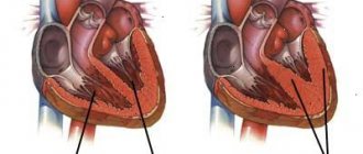
During ultrasound examination, the focus of echogenicity on the monitor appears as a small, white dot located in the area of the heart, most often in the left ventricle.
With a more detailed study, if the ultrasound image is enlarged several times, you can see a clearly visible formation, slightly oval in shape, which seems to jump with each contraction of the heart. This is what explains the use of the term “golf ball”.
In the place where the hyperechoic focus of the left ventricle is found, the elastic structures of the heart muscle become denser.
In most cases, such compaction is caused by a number of related factors:
- deposition of excess calcium salts;
- pathologies at the genetic level;
- the formation of an additional string (a kind of thread stretched from the heart valves to the ventricles), the presence of which in most cases does not affect the normal functioning of the fetal heart.
If the formation of a compaction in the left ventricle of the fetus is caused by one of the above factors, then there is no reason for concern.
In cases caused by the deposition of excess calcium salts, the formation occurs on its own. In most cases, the string disappears even before birth and does not pose a danger to the health of the unborn child. Of course, it can cause small murmurs heard in the left ventricle, which also in most cases disappear by the third year of life.
The only recommendation when detecting a heart murmur is to monitor the dynamics of this formation by a cardiologist
A hyperechoic focus in the left ventricle of the fetal heart can be a dangerous pathology only if its diagnosis is accompanied by the presence of a genetic abnormality or chromosomal defect (the appearance of an additional chromosome section). In 20%, the presence of a compacted inclusion in the cavity of the fetal heart may be a sign of the presence of Down syndrome.
Hyperechoic focus in the fetal heart - Understanding ultrasound
Most doctors claim that a hyperechoic focus in the fetal heart is one of the normal variants.
However, it can also pose a danger in cases where the deviation is detected in the presence of other symptoms indicating genetic disorders.
In general, this deviation does not pose any threat to the life and health of the baby in any case. It may be one of the “markers” (signs) of chromosomal abnormalities or just an age-related change that will pass before birth.
What is the GEF of the fetal heart?
This conclusion was first found in medical literature at the end of the 19th century in Great Britain, and was described at the same time. Most often, the disorder is diagnosed in women after 35 years of age, and predominantly in Asian women.
On ultrasound, the GEF appears on the monitor as a small white dot in the area of the heart, usually in the left ventricle. If the image is enlarged several times, an oval-round formation will become clearly visible, moving slightly with each heartbeat.
That is why this violation is often called a “golf ball.”
The formation occurs due to compaction of the elastic structures of the myocardium (heart muscle). Typically, ultrasound specialists find a similar area in women at 18-22 weeks of pregnancy during a repeat (second) screening ultrasound. The lump may remain until the third trimester, and in some cases until the birth of the baby.
Causes
One of the common causes of this deviation is excessive saturation of a particular area of the myocardium with calcium salts. This phenomenon is called calcification.
The most susceptible to calcification are the papillary (papillary) muscles, whose function is to open/close the valves between the atrium and the ventricle.
The discovery of GEF in this case is explained by the active mineralization of the child’s bones and the peculiarities of mineral metabolism precisely at 18-22 weeks. That is why by the end of pregnancy this phenomenon goes away, and the tricks themselves disappear on their own.
In some cases, the cause of GEF may be the presence of certain structures in the heart. Usually they are additional or false chords. These are congenital developmental abnormalities. In a healthy person, the notochord is a thin thread of connective tissue that connects the papillary muscle to the heart valve.
There is one chord for each muscle. In some people, additional chordae are identified. They do not have any useful function, but they do not harm health, and therefore are not considered a pathology.
Sometimes, when there are a large number of chords in the ventricles, the doctor also listens to noise.
It occurs due to the vibration of the blood as it passes through the plexus of these threads. In most cases, even such situations do not affect a person’s health in any way and do not require treatment.
In the case of a hyperechoic focus, even these thin filaments can cause deviations and provoke excessive deposition of calcium salts.
For a long time, there was a misconception that GEF in the left ventricle is one of the symptoms of chromosomal pathologies of the fetus, including Down syndrome.
Recently, scientists have abandoned such a conclusion, since they have recorded that the presence of such a deviation in the absence of other signs of a genetic disease is not at all a reliable sign.
GEF in a fetus without additional symptoms no longer requires consultation with a geneticist; a woman with a similar finding is followed up as standard.
But there are also sadder cases. Sometimes a thickened cervical fold and anomalies of new bones are observed in a fetus at 11-14 weeks, and at other times gross anatomical abnormalities in the heart, pathologies of the bone structure of the skull, legs and arms, and the formation of the gastrointestinal tract are diagnosed.
In such cases, GEF is considered an additional symptom of a chromosomal abnormality and must be monitored by a geneticist.
In the presence of Down syndrome, a hyperechoic focus is detected in approximately 16% of cases, so if there are other reasons to worry, additional examination is necessary.
What to do if GEF is detected?
As already mentioned, this deviation does not require specific treatment. In approximately 30-35% of cases, the lump disappears on its own by the end of pregnancy. In approximately 80% of newborns, it turns into an additional trabecula in the internal tissue of the ventricles of the heart.
Such formations also do not affect health and do not require treatment.
Based on the above, you should only worry about the presence of such a disorder after repeated screening if there are other syndromes that may indicate genetic diseases.
If the doctor detects several markers (signs) of such an anomaly at once, he prescribes an additional medical genetic consultation, as well as other studies to determine the diagnosis. After this, if necessary, treatment is prescribed or other further actions for the management of pregnancy and childbirth are discussed with the mother.
Source:
What does a hyperechoic focus in the left ventricle of the fetal heart mean?
Pregnant women in our country undergo three screening ultrasounds, and during the second of them, approximately in the middle of gestation, a hyperechoic focus is sometimes detected in the left ventricle of the fetal heart (most often in the left).
According to statistics, this phrase appears in approximately 7% of survey reports. This inclusion looks like a dot. In medicine, this acoustic phenomenon is called “Golf Ball Syndrome.”
This pathology does not pose a threat to the baby’s health if it is not accompanied by chromosomal abnormalities.
Description
Where the dot is located on the ultrasound machine screen, the child has a thickening of the heart tissue.
The cause of the pathology may be:
- Salt deposits - if this is the cause of the disorder, then by the third trimester of pregnancy there will be no trace left of the compaction, and this will not negatively affect the formation of the child.
- Chromosomal disorders - in combination with them, compaction of the heart muscle is the most dangerous option. A hyperechoic focus in a baby can be one of the signals for a detailed examination of the baby for the presence of Down syndrome.
- Formation of an additional chord (which does not prevent the heart from functioning fully). This feature may disappear without a trace even before the birth of the child, or it will persist until 2-3 years of age, and then it is a likely cause of murmurs in the baby’s heart.
If your daughter or son is diagnosed with an additional chord, this is a reason for regular visits to a pediatric cardiologist.
When is the examination carried out?
A hyperechoic focus can be detected by ultrasound, which may result in a 3D ultrasound or fetal echocardioscopy. The reason for ordering an examination may be:
- age of the expectant mother;
- infectious diseases suffered by a pregnant woman in the first trimester;
- diabetes mellitus or cardiac pathology in the mother or close relatives;
- previously identified heart problems in the fetus;
- developmental delay of the child (assessed by ultrasound);
- detection of markers of chromosomal diseases.
“Golf ball syndrome” can be easily detected by echocardioscopy if the gestational age exceeds 18 weeks, but has not reached 28 weeks. Later, it will not be possible to fully examine the baby’s heart, because the older child partially blocks the view.
Decoding and norms
When examining the fetus, all cavities of its heart are measured and the doctor compares the results with special standards:
- length of the right ventricle – from 0.5 to 1.75 cm;
- width of the right ventricle – from 0.4 to 1.1 cm;
- length of the left ventricle – from 0.9 to 1.8 cm;
- left ventricular width – from 0.44 to 0.89 cm;
- ratio of the width of the left ventricle to the right – from 0.45 to 0.9 cm;
- aortic mouth – from 0.3 to 0.52 cm;
- the mouth of the pulmonary artery – from 0.3 to 0.5 cm;
- mitral orifice – from 0.35 to 0.6 cm;
- tricuspid foramen – from 0.3 to 0.63 cm;
- the mouth of the pulmonary artery – from 0.3 to 0.5 cm;
- mitral orifice – from 0.35 to 0.6 cm;
- tricuspid foramen – from 0.3 to 0.63 cm;
- the number of heart contractions is from 140 to 160 beats/minute.
It should be taken into account that the size of a child’s heart and its individual components differs significantly from the corresponding sizes in an adult, because the size of the organs depends on the size of the body. Ultrasound in this case is very informative, and if there is a pathology, it will always be detected .
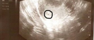
Photo 1. Hyperechoic focus in the left ventricle of the fetal heart.
What to do if a hyperechoic focus is identified?
If the doctor discovers the presence of a hyperechoic focus in the child’s heart, then the woman will need to undergo additional examinations as planned, which may include:
- Ultrasound with Doppler aimed at examining the fetal heart;
- cardiotocographic study;
- three- or four-dimensional ultrasound.
If the diagnosis is confirmed, it is worth consulting a geneticist to rule out possible chromosomal pathologies.
Important! The doctor may order a test of cord blood or amniotic fluid, but keep in mind that this test should only be performed in special cases, as it carries a significant risk of miscarriage or miscarriage of the baby.
But do not be afraid ahead of time - if the cardiac pathology is insignificant, then the doctor will not offer potentially dangerous diagnostic methods, but will limit himself to a consultation.
Possible complications and consequences
Most often, the accessory chord in the left ventricle of a baby disappears by the time of birth, but can be present in the heart until 2-3 years of age, and can persist for life.
Reference! This condition does not pose a threat to the baby’s well-being, but you need to be aware of it and not ignore preventive visits to the cardiologist.
The local pediatrician should also be warned about the child’s health characteristics in order to exclude erroneous diagnoses when listening with a stethoscope.
Has your child been diagnosed with GEF?
Usually, by the age of three, all signs of a hyperechoic focus disappear in the child, and all the worries associated with this diagnosis disappear in the parents.
Important! Once again, it is worth clarifying that this condition does not pose any danger to the life and health of the baby, because mothers are very scared when they discover any pathologies associated with the child’s heart.
If GEF is accompanied by the presence of an additional chord, then murmurs are possible in the heart and this is where the manifestations of the hyperechoic focus end.
If GEF is detected in the fetus during gestation after its birth, it is worth waiting until 2-3 months of age and undergoing an additional ultrasound of the heart, detailing the size of all cavities, the number of chords and the characteristics of the valves .
The study protocol will assess the general condition of the child’s heart and blood vessels and their suitability for age. For example, up to a year old, babies may have an open oval window; this is not considered a pathology.
Moms and dads are interested in: how do children live with a hyperechoic focus? Before the time, you shouldn’t worry too much and imagine a negative situation.
1. GEF.
According to statistics, only a small percentage of children with GEF become patients of a cardiologist, because in them this pathology leads to heart disease or other complications. But for such a development of events, one hyperechoic focus is not enough - usually a whole complex of disorders leads to serious consequences , one of which may be GEF.
“Golf ball syndrome” in the right ventricle
The acoustic shadow found in the right ventricle is no different from the GEF of the left ventricle. This finding requires only observation and correction and does not require treatment. Such a child is considered healthy, and even more so there is no question of a risk to his life. It is extremely rare that additional inclusions in the myocardium lead to the development of other diseases, such as heart defects.
Conclusion
An ultrasound sign, which is “golf ball syndrome,” may indicate microcalcifications in the heart muscle, the presence of an additional chord or more serious chromosomal pathologies.
To eliminate unnecessary worries, you need to undergo all additional examinations recommended by your doctor.
Remember that this condition extremely rarely becomes the cause of illness or a sign of severe pathologies, but requires attention to the health of the baby’s cardiac system.
Source:
Hyperechoic focus in the left ventricle of the fetal heart
Pregnant women often encounter unfamiliar terms during ultrasound examinations, which become causes of concern. Hyperechoic focus in the left ventricle of the fetal heart is one such term that requires explanation. What is a hyperechoic focus? How dangerous is this?
Source: https://uzi-tolyatti.ru/drugoe/giperehogennyj-fokus-v-serdtse-ploda.html
What to do if you have the syndrome?
This diagnosis is usually made in the 4th week of pregnancy, when the fetus's heart begins to form, using ultrasound.
If, during a routine ultrasound, a compaction is detected in the left cavity of the ventricle of the heart, doctors are strongly recommended to conduct a detailed examination to determine the possibility of detecting factors indicating the presence of a hidden pathology in the fetus at the chromosomal level:
- the presence of deviations in the anatomical structure of the fetus;
- congenital heart defects.
The isolated detection of a left ventricular hyperechogenicity factor, in the absence of signs of chromosomal pathology, is not a cause for concern. In 70% of cases, hyperechoic formation in newborns does not require correction and, in most cases, does not pose a threat to the health of the unborn child. In 30% of cases, hyperechoic inclusions disappear unnoticed by the end of pregnancy, leaving no trace of their presence.
In the early 90s, American specialists developed a special system for assessing signs of genetic pathologies in points using ultrasound. After some time, Russian scientists improved this system by initiating the recording of hyperechoic inclusions as a sign of chromosomal pathology with 1 point.
How to do an ultrasound of the fetal heart: details of the manipulation
Ultrasound of the fetal organs is a safe procedure that does not entail any complications.
The manipulation does not require special preparation, but it is better to come to the procedure in advance and catch your breath in order to normalize your heart rhythm.
An ultrasound examination of the fetus is carried out like a regular ultrasound: the woman is placed on her back, a special water-based gel is applied to her stomach and the condition of the organs is assessed with a special sensor.
The study is performed in various operating modes of the device:
- M-mode – simultaneously reflects the rhythm of contractions of the atria and ventricles, impulse conduction;
- pulsed wave Dopplerography – evaluates blood flow in an artery and vein simultaneously;
- color Doppler scanning - shows the flow of blood through the atria and ventricles, blood vessels.
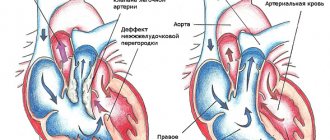
Obtaining an image of 4 chambers of the heart at once is called a four-chamber slice. It is very informative, but insufficient for some defects:
- tetralogy of Fallot;
- transposition of the great arteries;
- anomalies of the aortic arch;
- common arterial duct.
To identify them, special directions of the sensor are used.
What does an ultrasound of the fetal heart show: decoding the data
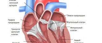
Ultrasound of the fetal heart has its own norms. The following indicators are determined:
- organ location;
- condition of the cameras;
- location of the heart axis;
- sizes of the ventricles and atria;
- are there any defects in the interventricular septum;
- changes in the tissues of the heart - enrocardium, pericardium, myocardium.
Heart rate and rhythm also matter. Normally, the fetal heart rate after 11 weeks is 140-160 beats per minute.
Echo diagnostics determines various types of defects.
A common finding is fetal cardiac rhabdomyoma.
. Ultrasound can determine its location, extent and size. Rhabdomyoma is a single or multiple benign tumor that develops in utero and after childbirth. The neoplasm is localized in the interatrial septum. The cause of rhabdomyoma is considered to be a violation of tissue embryogenesis. A small tumor does not manifest itself in any way. Increased attention is paid to her after the birth of a child.
Sometimes a hyperechoic inclusion in the fetal heart is detected on ultrasound
. This is not a defect - more distinct lesions simply appear on the image. They may be caused, for example, by increased calcium levels. In most cases, this phenomenon goes away on its own and does not require special treatment.
As we have already said, a hyperechoic inclusion can also occur in an absolutely healthy child. However, a hyperechoic focus is sometimes a sign of a chromosomal abnormality, Down syndrome. This is why a complete diagnosis is necessary.
Usually by 33-34 weeks this phenomenon goes away on its own, if not combined with other pathological changes. Sometimes it remains, but in the future does not affect the contractility of the heart, the management of pregnancy and childbirth.
Ultrasound is a highly informative diagnostic method that detects pathologies in 60-80% of cases.
Yulia Shevchenko, obstetrician-gynecologist, especially for Mirmam.pro
Useful video
Additional examination methods
When diagnosing “golf ball” syndrome in a fetus using ultrasound, there are additional examination methods that allow you to maximally calculate the risk of possible complications.
Hyperechoic focus in the left ventricle of the fetal heart
The most effective are:
- A blood test for the presence of chromosomal abnormalities of the fetus, showing the likelihood of having a child with chromosomal abnormalities;
- Fetal fetoscopy is an examination using a thin tube inserted into the cavity of the amniotic sac through the abdominal wall or through the wall of the uterus. This method allows you to visually detect or confirm assumptions about the presence of pathology and hereditary diseases in the fetus. The method is relevant in cases where the use of other diagnostic tools does not lead to the desired results. The procedure itself is dangerous. Therefore, many doctors prescribe it in extreme cases. Due to the risk of premature termination of pregnancy;
- Placentocentesis – This procedure is very similar to a biopsy. Its essence boils down to obtaining, for subsequent research, placental cells in order to study the genetic structure of the fetus. This method allows you to obtain confirmation or refutation of suspicions of the presence of hereditary abnormalities in the shortest possible time;
- Amniocentesis is a study of the composition of amniotic fluid by penetration through the abdominal wall. A summary analysis of hormonal, immune and biochemical indicators helps determine the degree of risk of having a child with chromosomal abnormalities.
An effective and widely used method of additional examination is echocardioscopy.
Hyperechoic focus in the fetal heart
Ultrasound today has become an integral part of pregnancy management. Using this technique, you can detect pathological processes in the heart and throughout the baby’s body, as well as the occurrence of any deviations in its development.
Fetal examination helps doctors guide a woman in position and promptly respond to the slightest disturbances in the formation of the child in the womb. However, many expectant mothers find it difficult to decipher the results of such diagnostics, which causes great fear and anxiety.
A hyperechoic focus in the fetal heart is often detected, but what does this phrase mean and what is the risk for the baby?
Description of diagnosis
A hyperechoic focus (HEF), shown on an ultrasound machine during examination of the fetal heart, is usually an incidental finding.
Diagnosis of the main organ is included in the list of mandatory tasks for every pregnant woman; it is this area that deserves special attention from doctors.
The patient attends all procedures as planned, and at a certain time the doctor talks about such inclusion when examining the child’s organ.
All parents can be reassured by saying that a hyperechoic focus in the fetal heart is not a dangerous pathology. This feature does not have a negative impact on the functioning of the organ, which is confirmed by the study of a similar condition in a baby. Usually the disorder is detected at 18-22 weeks, this period is considered suitable for a second screening using ultrasound.
Further, the deviation is observed unchanged and does not disappear until childbirth. Statistical data show that an echogenic focus most often appears in the left ventricle of the fetus; relatively rarely, pathology is found in the right ventricle, as well as in the area of other cardiac chambers. On average, approximately 7%-8% of all pregnant women may experience this problem.
When performing an ultrasound, the doctor notices a white spot measuring about 2-3 mm, round in shape, located in the area of the left, right ventricle or the cavity of the organ chambers.
Another name for such a deviation, which is often used by diagnosticians, is qolf ball or golf ball.
This comparison is indeed appropriate, since the round ball on the heart, which bounces during each contraction of the organ, resembles a ball.
A similar picture on the monitor of an ultrasound machine usually occurs as a result of the appearance of a dense formation consisting of bone tissue, so many doctors are amazed to see it in this particular place. No one knows how such a seal appears on the heart and where it disappears after childbirth.
What the GEF shows:
- The chorda accessory, which are fibers passing from the ventricles and attached to the valves.
- Salt deposits (usually calcium salts).
- Pathological process of a chromosomal nature.
- Another type of anomaly that does not pose a danger to the activity of the heart muscle.
The accessory chord will not go anywhere after childbirth, but it does not interfere with the functioning of the organ. It should be borne in mind that in this state, when the heart beats, it can make noises that will be audible to the doctor when listening. The reason may lie in any of these factors, but there is no need to identify it.
Features of the functioning of the fetal heart on ultrasound
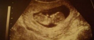
The formation of the heart muscle occurs in the 4th week of gestation. At this stage of development, the organ looks more like a hollow tube. In the middle of the 5th week, the activity of the heart begins and its first contractions occur. By the 9th week, this muscle already has 4 chambers and can be called a full-fledged organ, since ventricles and atria are observed.
If an ultrasound is performed correctly, the baby’s heartbeat can be heard already at 5-6 weeks of gestation. When using a transabdominal type sensor, it allows you to recognize the organ’s beating a little later, by about 1 week. A hyperechoic focus in the area of the left ventricle of the fetal heart (LV) has not yet appeared, so there is no need to worry about its possible formation.
At this stage of pregnancy, the doctor records the intensity of the organ’s work, the speed of its beating and other parameters. If heart activity is not observed, then one can suspect that pregnancy has been interrupted. In this case, the specialist recommends repeating the diagnosis after a few days to refute fetal freezing or confirm it.
The first ultrasound examination is carried out at three months to identify any pathologies. Down syndrome occurs frequently and it is necessary to undergo diagnostics during this period in order to exclude such a deviation. The work of the heart is also being studied for the first time at this stage of the baby’s formation. There are certain standards by which you can understand whether the fetus is healthy or not.
Heart rate indicators:
Duration Number of organ beats in 1 minute
| 6-8 weeks of fetal formation | 110-131 beats/min |
| 9-10 weeks of child development | 170-191 beats/min |
| From 11 weeks until birth | 140-162 beats/min |
Such changes in the rate of organ contractions are due to the formation and further development of the autonomic system. It is this area that is responsible for the correct and coordinated functioning of any of the internal organs, as well as systems. There are certain indicators that the doctor needs to pay attention to. Due to such factors, the baby in the womb experiences some disturbances in the form of a hyperechoic, hypogenic focus.67
What you need to pay attention to:
- in the anamnesis of children born earlier, there are records of a similar condition;
- the presence of congenital heart defects in either parent;
- pregnancy in a woman over 38 years of age;
- diseases of an infectious nature in the expectant mother;
- delayed fetal formation;
- diabetes mellitus in a pregnant woman;
- the presence of defects of other organs in the child in the womb.
Despite the fact that hyperechoic inclusion in the left ventricle of the fetal heart is not considered a dangerous pathology, there is a certain risk. In simple terms, the presence of hyperechogenicity in an organ can be a consequence of a disease, but only if the baby has problems at the genetic level, chromosome mutations and others.
During the examination, the doctor examines the structure of the child. When such deviations from the norm appear as shortening of the neck and changes in the area of the nasolabial triangle, it means that we can talk about Down syndrome. Such a diagnosis is established only in 20% of children with increased echogenicity in the heart.
Additionally, geneticists identify a chromosomal defect that confirms this pathology.
What to do?
When a routine ultrasound reveals a hyperechoic focus in the left ventricle of the heart, it is necessary to carry out a number of additional diagnostic measures. These examinations will help to study the organ in detail and find hidden disorders in this area, including at the chromosomal level: congenital heart defects, deviations in the anatomical features of the child in the womb.
If it becomes clear that only an echogenic focus is present in the baby, then there is no reason to worry.
Approximately 70% of all cases of such ultrasound results do not indicate a threat to the life and health of the child, but represent a minor defect that will disappear on its own.
In any case, if this organ condition is detected in a baby, a consultation with the attending physician is necessary; he will determine further actions and draw up a plan for additional diagnostic methods.
Additional examination methods:
- A genetic blood test to determine fetal chromosomal abnormalities, which shows the possibility of having a baby with abnormalities of a similar nature.
- Placentocentesis. The procedure is similar to the technique of taking a biopsy. The essence of the diagnosis is to obtain placental cells in order to further examine them to determine the genetic characteristics of the fetus. This method allows you to get a refutation or confirmation of a dangerous diagnosis in the shortest possible time.
- Fetoscopy. When carrying out this diagnostic method, doctors insert a thin catheter into the cavity of the amniotic sac, it enters through the wall of the peritoneum or through the wall of the uterus. This technique helps to detect the child’s pathologies and hereditary ailments. This method is used only in extreme cases, as it is dangerous and can lead to termination of pregnancy.
- Amniocentesis is a study of the composition of amniotic fluid, which is achieved by inserting a tube through the peritoneal wall. By analyzing the resulting fluid and studying hormonal levels, biochemical indicators and the immune system, it is possible to determine whether there is a risk of having a baby with chromosomal abnormalities.
The most effective diagnostic method in this regard is echocardioscopy. If a child has a hyperechoic heart focus in the womb, then detailed monitoring of the activity of the heart muscle or echocardioscopy is necessary.
This method, in combination with ultrasound, helps to monitor the main parameters of the organ, contraction of the ventricles, the volume of filling of the atria and much more. It should be borne in mind that this technique can only be used until the pregnancy reaches 25 weeks.
After this period, the procedure will turn out to be uninformative due to the large size of the baby in the womb and the small volume of amniotic fluid.
What are the consequences?
When a child is diagnosed with such a deviation, the doctor must take the pregnant woman under constant supervision.
This is done due to the fact that such a violation can lead to a malfunction of the myocardium, and the result of this change is often a circulatory disorder in the baby in the womb.
It is known that the main function of blood is to deliver nutrients to all organs and systems of the body, so if this process fails, serious problems can arise, including miscarriage.
Experts believe that identifying hyperechogenicity in the left ventricle of the heart is not as dangerous as a disorder in the right side of the organ. If such a compaction appears on the right, then this indicates an inevitable pathological process that can even lead to the death of the fetus. Therefore, a child with such a deviation must be observed by a cardiologist.
If a baby was born with impaired blood circulation and myocardial function, then his health may deteriorate greatly.
Symptoms:
- difficulty breathing;
- weakness, lethargy;
- pain in the heart area;
- swelling of the arms and legs;
- pallor of the skin, even blueness;
- disturbance of heart rhythm, increase in the rate of contraction of the organ;
- loss of appetite.
The consequences of the disease can be very different, so parents are obliged to regularly examine the baby and apply any treatment prescribed by the doctor. Sometimes drug therapy is powerless, in which case doctors decide on surgery.
The expectant mother should monitor her health, since the correct development of the child depends on her condition. You need to protect yourself from any infectious diseases, because such diseases most often cause severe complications in the fetus. If in the early stages of pregnancy the expectant mother has suffered from a disease of viral origin, then there is a high risk of pathology developing in the child.
Specialists should monitor fetal development regularly; a lot depends on such diagnostics. A woman is also obliged to pay close attention to the health of her child and attend all the required examination methods as planned.
A hyperechoic focus in the fetal left ventricle is not uncommon. This condition has been well studied, so there is no need to be alarmed if a similar deviation is detected in your baby.
Medicine today can eliminate almost any defect of the main organ; it is important to identify it in time so that doctors can immediately apply all treatment methods.
Source: https://MirKardio.ru/bolezni/tyazhelye/giperehkhogennyj-fokus-v-serdce-ploda.html
What is a hyperechoic focus?
A hyperechoic focus or HEF is an incidental finding during ultrasound examination of a pregnant woman. Ultrasound of the fetal heart is a mandatory component of any perinatal screening, so the doctor always pays attention to the structure and functioning of the child’s heart.
The woman regularly, without any complaints or worries, comes for an ultrasound scan, where the doctor discovers such an interesting feature of the structure of the fetal heart.
We hasten to reassure expectant mothers that GEF is only an “interesting feature” and not an anomaly or pathology.
The hyperechoic focus itself does not in any way affect the functioning of the heart of the fetus or an already born child, which has been confirmed in numerous studies.
As a rule, such an area is found during the second screening ultrasound - at 18-22 weeks of pregnancy and is observed until the third trimester, sometimes until childbirth. According to statistics from different countries, such a formation is more often found in the left ventricle of the fetus, less often in the right ventricle or other chambers of the heart.
On average, in 7% of pregnancies a hyperechoic focus of various localization is detected . In developed countries, there is even evidence that approximately a fifth of such findings are erroneous due to the poor quality of the ultrasound machine or incorrect conduct of the research procedure.
Below we will talk in a little more detail about what is associated with the appearance of such heart focuses.
On an ultrasound examination, the doctor sees a bright white spot within 2-3 mm of a rounded shape in the cavity of one of the chambers of the fetal heart.
Ultrasound diagnosticians are very fond of all kinds of artistic comparisons, so the hyperechoic focus is also called golf ball or golf ball symptom.
In fact, the small round structure that bounces with every heartbeat actually resembles a golf ball.
Only a very dense formation akin to bone tissue can give such a picture on an ultrasound, which causes some surprise to all scientists and doctors. How such a dense formation forms in the heart muscle and then disappears without a trace after birth remains an unsolved mystery.
Possible causes of the formation of a hyperechoic focus of the heart
In fact, GEF in the left ventricle remains an extremely controversial diagnosis to this day. Scientists have identified several of the most likely reasons for the formation of such inclusions:
- Impregnation of a limited internal area of the heart muscle with calcium salts. Typically, the papillary or papillary muscles that open and close the heart valves between the atrium and ventricle become calcified or saturated with calcium. The detection of such foci is associated with the active process of mineralization of fetal bones precisely in the period of 18-22 weeks, the peculiarities of calcium metabolism during this period, as well as with the start of taking calcium supplements by the expectant mother, which most often occurs precisely at the beginning of the second trimester. By the time of birth and the end of ossification of the child’s skeleton, such tricks resolve completely independently.
- The presence of any inclusions in the structure of the heart itself. Most often, the reasons for the appearance of a bright white inclusion are the so-called additional or false chords - congenital minor anomalies of the heart. Normally, the notochord is the thinnest connective tissue thread that connects the papillary muscle to the heart valve, just like slings connect a parachutist’s shoulder straps to the canopy of a parachute. There is one notochord for each papillary muscle. A huge number of people develop so-called additional or accessory chords, which do not carry a functional load, but are not a pathology. Occasionally, if there are a large number of such filaments in the ventricles of the heart, the doctor may listen to the noise produced by the vibration of the blood as it passes through the plexuses of the chords. Most often, additional chords are not subject to any treatment and do not cause any harm to the patient.
In the case of GEF, the chordae themselves can produce the golf ball sign, as well as provoke calcium deposits and increase the appearance of a hyperechoic focus.
- Genetic abnormalities. It has long been believed that a hyperechoic focus in the left ventricle of the fetal heart is one of the markers or signs of fetal genetic abnormalities, including Down syndrome. In recent years, doctors and scientists around the world have abandoned such conclusions, since numerous studies have found that isolated GEF without other markers of genetic abnormalities is not a criterion for suggesting such diagnoses . Isolated GEF is no longer subject to genetic counseling, and the woman is monitored according to standard pregnancy management protocols.
A completely different situation will be if GEF is combined with other markers of genetic abnormalities: with a thickened cervical fold and abnormalities of new bones on the first screening ultrasound at 11-14 weeks, other gross anomalies of the heart, pathology of the structure of the bones of the skull, limbs or gastrointestinal tract. In this case, a hyperechoic focus can be considered as an additional marker of genetic abnormalities and must be consulted with a geneticist.
Echocardioscopy
When diagnosing a focus of increased echogenicity of the left ventricle in a fetus, the use of echocardioscopy for detailed monitoring of the functioning of the heart muscle will be an effective step. This procedure allows you to monitor the main parameters of the fetal heart (atrial filling, ventricular contraction, etc.) in real time using ultrasound and detect possible deviations from the norm.
Cardiac echocardioscopy is the study of the structure of the heart and its functioning using ultrasound.
The main indications for examination using echocardioscopy are:
- the age of the pregnant woman exceeds 36 years;
- the presence of diabetes mellitus in the expectant mother or immediate relatives;
- recent infectious or viral disease in the early stages of pregnancy;
- the woman has a congenital heart defect, taking into account immediate relatives;
- the presence of markers of hereditary pathology in the blood of the expectant mother.
Echocardioscopy is an appropriate and effective method only up to the 25th week of pregnancy. At later stages, the procedure will be ineffective due to the large size of the fetus and the small amount of amniotic fluid.
Why is ultrasound of the fetal heart prescribed during pregnancy?
The heart is a vital organ. Its pathology can already affect itself in the prenatal period, but after birth all defects will manifest themselves in full.
At what period is an ultrasound of the fetal heart performed? Routine ultrasound of the fetal heart is performed during screening studies. If there are indications, which will be discussed below, the procedure is carried out additionally.
During the first screening at 11-12 weeks, not all heart structures are yet visible. At this time, the doctor has enough information that it is four-chambered and contracts in a normal rhythm. At the same time, at the 11th week of pregnancy, ultrasound already detects such fetal heart defects as:
- defects of the left atrium or ventricle;
- tricuspid valve atresia;
- hypoplasia of the valve or left trunk of the pulmonary artery;
Most of the detected early defects are inoperable and incompatible with life. In this case, the woman is offered to terminate the pregnancy.
In the second trimester, ultrasound is most informative from 18 to 28 weeks, just when the second screening is carried out, but optimally at 24. By this time, you can examine the structures as fully as possible and notice rhythm disturbances. The heart on the monitor is large and occupies a third of the chest.
At a later stage, ultrasound of the heart becomes uninformative. The large size of the organ does not always allow us to examine it completely.
Why is it so important to know about the condition of the baby’s heart before he is born? The presence of heart defects in the fetus determines the tactics of delivery. In most cases, a caesarean section is performed to avoid complications. A team of doctors prepares equipment in advance that will help the child during the first time after birth.
Indications for additional diagnostics of the fetal heart
Additional research is carried out if there is evidence from the mother or child. In the first case they are as follows:
- congenital heart defects in one of the parents;
- the previous child was born with anomalies;
- the pregnant woman has diabetes, phenylketonuria, or has thyroid disease;
- the expectant mother took antiepileptic drugs, antidepressants, and NSAIDs at the beginning of pregnancy;
- alcohol abuse, smoking;
- maternal autoimmune diseases;
- rubella, toxoplasma, cytomegalovirus infection detected during pregnancy;
- pregnancy using IVF.
Indications from the child:
- heart rhythm disturbances;
- change in the thickness of the collar zone;
- established hereditary chromosomal diseases - Down syndrome, Edward syndrome, Patau syndrome, Shereshevsky-Turner syndrome;
- blood flow abnormalities in the ductus venosus;
- feto-fetal transfusion syndrome in twins;
- the only artery of the umbilical cord.
Why register with a doctor?
Most scientific experiments conducted to study in more detail the hyperechoic focus in the left ventricle of the fetal heart have shown that the appearance of an additional string is a consequence (in most cases) of chromosomal pathology. This pathology is mainly inherited through the female line.
The need for a child with golf ball syndrome to be under constant medical supervision is primarily due to the fact that such structural compaction in the heart cavity often leads to disruption of the rhythm of the myocardium (heart muscle). Due to the fact that the string is short, the volume of the ventricular cavity decreases, and this leads to its incomplete relaxation and, accordingly, poor filling with blood. The result of this anatomical pathology is a violation of the blood circulation of the fetus itself.
The main function of blood in the body is to deliver oxygen and various nutrients to cells, tissues and organs. Weakening of blood flow can then cause oxygen starvation of the fetus or damage to the inner lining of the heart muscle, with a high probability of subsequent miscarriage.
But still, according to most experts, the presence of a compacted string in the left ventricle of the fetus is not such a serious pathology compared to the compaction that can occur in the right ventricle of the heart.
Diagnosing a focus of increased echogenicity of the right ventricle of the fetal heart inevitably leads to complications and often incompatible with life. In such circumstances, the best option is surgical correction of the defect.
All of the above is clear evidence that in order to timely identify problems with blood circulation, the child will definitely need to be registered with a cardiologist.
Reasons for education
During a second routine ultrasound, a pregnant woman may be informed that a hyperechoic focus has been detected in the left ventricle of the fetal heart. Of course, any woman will be scared after hearing such a long diagnosis, but is it really that dangerous?
We have found out what a hyperechoic focus is, but what are the reasons for its appearance? This:
- salt deposits;
- pathology in the chromosome set of the fetus;
- the presence of an additional chord in the heart, which is considered a normal variant and does not in any way affect the activity of the heart muscle.
If the appearance of compaction of myocardial structures was caused by salt deposition, then a pregnant woman has no reason to worry. Most often, such minor pathology goes away by the third trimester of pregnancy.
An additional chord in the heart is a structural feature that can create certain heart murmurs. At the age of 2-3 years, noises most often stop, but even if they remain, they do not pose a danger to the life and health of the child.
It’s another matter if the focus is caused by an incorrect set of chromosomes in the child. There is a risk of detecting Down syndrome in the fetus or other pathologies. If there is such a suspicion, additional tests are prescribed to clarify this information. However, most often the alarm turns out to be false.
The causes of a hyperechoic focus in the left ventricle of the fetal heart are individual and require examination to clarify them.
Consequences and complications
At the initial stage, dysfunction of the left ventricle does not have clearly defined symptoms.
The first signs of cardiac dysfunction appear closer to childbirth and are due to the fact that the fetal heart is anatomically formed and begins to operate independently.
The presence of a seal (a string stretched between the right atrium and the right ventricle) leads to increased pressure in the cavity of the left atrium. This condition occurs due to insufficient blood flow from the left atrium to the right ventricle.
The atrium is not completely emptied, and the ventricle does not receive blood in the required amount. A natural consequence of such dysfunction is that the fetal organs receive insufficient oxygen and nutrients. This state of affairs will negatively affect the state of all life systems of the unborn child, and in advanced cases (if diagnosis is delayed) can lead to the death of the fetus.
The occurrence of golf ball syndrome in utero often has negative consequences for both the newborn and the child during the first years of his life.
In this case, the most striking symptoms characteristic of this pathology are:
- increased fatigue;
- apathy;
- attacks of pain in the heart area;
- dyspnea;
- cardiopalmus;
- swelling of the limbs;
- characteristic pallor of the skin with a tendency to subsequent blue discoloration.
Attacks of tachycardia (due to rapid contraction of the heart muscle) are often observed; as a result, the pulse can exceed 200 beats per minute. Timely symptomatic therapy in most cases helps to cope with the consequences of intrauterine additional education.
In most cases, such compaction, either independently or with the help of complex treatment, disappears by the age of 4. In severe cases, when conservative treatment methods do not lead to the desired result, doctors recommend surgery.
Ultrasound of the fetal heart during pregnancy: interpretation of the results
Contents
hide
1 Ultrasound of the fetal heart during pregnancy: interpretation of the results
2 Why is ultrasound of the fetal heart prescribed during pregnancy? 2.1 Indications for additional diagnostics of the fetal heart
3 How an ultrasound of the fetal heart is done: details of the manipulation 3.1 What an ultrasound of the fetal heart shows: decoding the data
3.2 Useful video
4 Hyperechoic focus in the fetal heart
5 Echogenic foci in the heart of the fetus 5.1 Materials used in the article:
GEF prevention
A set of measures aimed at ensuring that pregnancy and subsequent births occur without complications (if the above syndrome is detected in the fetus) should include the need for treatment and monitoring of the expectant mother.
The primary task of doctors is the timely implementation of preventive measures aimed at preventing complications in women
It is extremely important to protect the expectant mother from contact with a possible source of pathogenic viruses or microorganisms. It is women who have suffered from various diseases of an infectious nature who form the main risk group in the early stages of pregnancy.
What does hyperechoic bowel mean?
To understand what this phrase means, you need to refer to ultrasound terminology. Echogenicity is a concept denoting the level of density of tissue examined by an ultrasound device. On the ultrasound screen this is manifested in the brightness of the display of certain organs.
Normally, the intestines of the unborn child should be higher in echogenicity than the liver, kidneys or lungs, but lower than the bones. If the brightness of the intestine is equal to the brightness of the fetal bone tissue, it is said to be hyperechogenic.
Examinations to identify such pathology are carried out only in the second trimester. Until the sixteenth week, any data will be inaccurate and uninformative. Although already at this time the doctor should pay attention to the level of echogenicity of the organs so as not to miss the problem in the future. The longer the pregnancy at which hyperechogenicity was detected, the more likely the presence of abnormalities in fetal development.