Human heart
The human heart is a hollow muscle. It is formed by four chambers: the right and left atrium, the right and left ventricle. The atria and ventricles are connected by leaflet valves. The heart contracts rhythmically, and blood flows in portions from the atria to the ventricles. The semilunar valves connect the ventricles to the blood vessels, through which blood is pushed out of the heart into the aorta and pulmonary artery.
Thus, blood with a high content of carbon dioxide passes through the right chambers and enters the lungs to be enriched with oxygen. And from the lungs, the blood is sent back into the bloodstream through the left side of the heart. Ensuring constant pumping of blood through the vessels is the main function performed by the heart.
Features of the functioning of the SSS
Over the course of 1 minute, the heart pumps approximately 5–6 liters of blood.
With increasing physical or emotional stress, this blood volume increases, and at rest it decreases. The heart acts as a muscular pump, the main role of which is to pump blood flow through the veins, vessels and arteries.
The cardiovascular system is presented in the form of two circles of blood circulation: large and small. The aorta carries it from the left side of the heart. From the aorta, flow passes through arteries, capillaries and arterioles.
In the process of movement, the blood gives oxygen to tissues and internal organs, taking from them carbon dioxide and products of the metabolic process. The blood that has given up oxygen turns from arterial to venous, heading to the heart. Through the vena cava it enters the right atrium of the heart, forming a systemic circulation .
From the right half of the heart it approaches the lungs, where it is enriched with oxygen. The circle repeats itself again.
Between the left and right ventricles there is a septum separating them. The cardiac atria and ventricles have different purposes.
Blood accumulates in the atria, and during cardiac systole, the flow is pushed under pressure into the ventricles. From there, the blood is distributed through the arteries throughout the body.
The healthy state of the cardiovascular system directly depends on how well the heart valves function, as well as on the specific direction of blood flow.
Heart valves
The valve apparatus is necessary in the process of pumping blood. Heart valves ensure blood flows in the right direction and in the right amount. Valves are folds of the inner lining of the heart muscle. These are kind of “doors” that allow blood to flow in one direction and prevent it from moving back. The valves open at the moment of rhythmic contraction of the heart muscle. There are four valves in the human heart: two cuspid and two semilunar:
- Bicuspid mitral valve.
- Tricuspid tricuspid valve.
- Semilunar valve of the pulmonary trunk. Its other name is pulmonary.
- Aortic semilunar valve or aortic valve.
Heart valves open and close according to the sequential contraction of the atria and ventricles. The blood flow of blood vessels, and therefore the oxygen saturation of all cells of the human body, depends on their synchronous work.
Mechanism and features of operation
The main function of the bicuspid atrioventricular valve is to prevent backflow (regurgitation) into the left atrium and direct its flow into the ventricle.
MK functions
All valve leaflets are mobile and pliable structures that move during contraction phases under the influence of directed blood flow. At the moment of diastole, the muscles of the cardiac cavities relax and fill with blood, the large anterior valvular valve closes the aortic cone, thereby preventing reflux into the aorta.
During systole, when the atrium and ventricles contract, the elements of the left atrioventricular orifice are compressed along thickened valve lines, which are held by the chordae. This prevents regurgitation and maintains normal hemodynamics in the systemic circulation.
Normal valve performance
Echocardiographic examination (ultrasound of the heart) identifies average normal values:
- the diameter of the fibromuscular ring is 2.0-2.6 cm, the valve is up to 3 cm;
- MK area up to 6.5 cm2;
- the thickness of the valves is 1-2 mm;
- the movement of all doors is active and smooth;
- the surface is uniform and smooth;
- in the systole phase, the deflection of the elements into the ventricular cavity is no more than 1.5-2 mm;
- chords in the form of long, linear and thin fibers.
Functions of the valve apparatus
Blood flowing through the vessels into the heart accumulates in the right atrium. Its further advancement is delayed by the tricuspid valve. When it opens, blood enters the right ventricle, from where it is expelled by the pulmonary valve.
Next, the blood flow enters the lungs to be oxygenated, and from there it is sent to the left atrium through the aortic valve. The heart's mitral valve connects the left chambers and restricts blood flow between them, allowing blood to accumulate. After blood enters the left ventricle and accumulates in the required amount, the blood is pushed into the aorta through the aortic valve. From the aorta, renewed blood continues its movement through the vessels, enriching the body with oxygen.
Anatomy and structure of the heart
The structure of the heart muscle is simple - the hollow muscular organ has 4 chambers: 2 ventricles and 2 atria, separated by valves and septa. Venous blood circulates in the right half of the heart muscle, and arterial blood circulates in the left half. The upper parts of the heart (atria) are separated from the lower parts (ventricles) by an incomplete septum.
In the left half of the muscle, the opening between the atrium and the ventricle is closed by a bicuspid heart valve, which doctors call the mitral valve. On the right side of the muscle is the tricuspid heart valve, which closes the atrioventricular orifice. The second name for the tricuspid valve is tricuspid.
The length of the heart muscle in adults varies between 10–15 centimeters. To imagine what kind of heart you have, clench your palm into a fist - this size will be your individual size. The mass of the organ differs slightly for women and men: the male myocardium weighs from 33 to 350 grams, the female myocardium is slightly less - from 250 to 300.
The main blood vessel of the body, the aorta, originates in the left ventricle and then branches into the coronary arteries. It provides blood circulation in a large circle. The pulmonary artery, which is responsible for blood circulation in the pulmonary circle, begins from the right ventricle. At the beginning of the pulmonary artery and aorta there are semilunar valves that work in a special way, allowing blood to flow in only one direction - from the heart.
People often have a question: how many valves are there in the human heart? We have mentioned the 4 main ones, but there is also one more, vestigial, fifth “Eustachian” valve.
Heart valve pathologies
The valves' job is to regulate the flow of blood through a person's heart. If the rhythm of opening and closing of the valve apparatus is disrupted, the heart valves close or do not open completely, this can cause many serious diseases. It is noted that the mitral and aortic valves are most often susceptible to pathologies.
Heart defects are most common in people over sixty years of age. In addition, diseases of the heart valves can become complications due to certain infectious diseases. Children are also susceptible to valve diseases. As a rule, these are congenital defects.
The most common diseases are heart failure and stenosis. If there is insufficiency, the valve does not close tightly, and some of the blood returns back. Stenosis is a narrowing of the valve, meaning the valve does not open completely. With this pathology, the heart is constantly overloaded, since it takes more effort to push blood through.
Heart valves - structure and functions of the heart
The heart is a vital hollow muscular-fibrous organ located on the left side of the chest and providing blood flow through the vessels. In fact, this is a kind of muscle pump that has an automatic function and works according to the “suction-extrusion” mechanism. The heart pumps about five to six liters of blood per minute; at rest, this volume decreases somewhat, and when a person performs physical activity, it increases.
Together with the vessels, the heart forms the cardiovascular system, which has two circles of blood circulation: large and small. From the heart, blood first enters the aorta, then moves through large and small-diameter arteries, then through arterioles to capillaries, where it gives oxygen and a number of other nutrients necessary for the body to the tissues and takes away carbon dioxide and waste metabolic products. Thus, blood from arterial becomes venous and is directed back to the heart: first through venules, then through small veins and large venous trunks. Through the inferior and superior vena cava, blood enters the right atrium, closing the systemic circulation. It is again enriched with oxygen in the lungs, where it enters from the right side of the heart through the pulmonary arteries (pulmonary circulation).
Inside, the human heart is divided by septa (partitions) into four separate chambers: two atria (left, right) and two ventricles (also left and right). Each of them has different functions. In the atria, the blood entering the heart accumulates and, having reached a certain volume, is pushed into the ventricles (from the right atrium to the right ventricle, from the left atrium to the left ventricle). The ventricles pump blood into the corresponding arteries, through which it moves throughout the body. They perform more difficult work and therefore have a thicker, more developed muscle layer than the atria.
On each side of the heart (separately on the left, separately on the right), the ventricles and atria communicate with each other through the atrioventricular (atrioventricular) opening. Through the chambers of the heart, blood moves exclusively in one direction: from the left atrium normally it always enters the left ventricle, from there it goes through the systemic circulation and enters the right atrium, then from it into the right ventricle and into the small circle, from which it again comes to left atrium.
The correct direction of blood flow is ensured thanks to the coordinated work of the heart valve apparatus, represented by the mitral, tricuspid, pulmonary and aortic valves, which open and close at the right time, preventing regurgitation, that is, reverse blood flow.
The mitral (bicuspid) valve is located between the left atrium and ventricle and consists of two leaflets. When it is open, blood flows through the atrioventricular orifice into the left ventricle from the left atrium. During systole (that is, during contraction) of the left ventricle, the valve closes so that the blood does not flow back into the atrium, but is pushed through the aorta into the vessels of the systemic circulation.
The tricuspid (three-leaf) valve is located between the right atrium and the ventricle and has, accordingly, three leaflets. If it is open, blood flows from the right atrium through the atrioventricular opening into the right ventricle. When the latter is filled, its muscle contracts, under blood pressure the tricuspid valve closes, preventing regurgitation of blood into the atrium, and the exit of blood becomes possible only through the pulmonary trunk, and from it along the lesser circle into the pulmonary arteries. At the entrance to the pulmonary trunk, another valve is located - the pulmonary valve. It opens under the pressure of blood in the systole of the right ventricle, and in its diastole (during relaxation), it closes under the influence of the reverse flow of blood, preventing the return of blood from the pulmonary trunk to the right ventricle.
The aortic valve closes the entrance to the aorta. It consists of three semilunar valves and opens at the moment of contraction of the left ventricle. Blood then enters the aorta. In diastole, the left ventricle closes, allowing venous blood flowing through the superior and inferior vena cava to flow from the systemic circulation into the right atrium.
(495) 506-61-01 — Where is the best place to have heart valve surgery?
Structure of the heart
HEART DISEASES – Heart-Disease.ru – 2007
Heart
- This is a kind of pump that circulates blood in the body. A healthy heart is a strong, continuously working organ, about the size of a fist and weighing about half a kilogram.
The heart consists of 4 chambers
.
A muscular wall called the septum
. divides the heart into left and right halves. Each half has 2 chambers.
The upper chambers are called the atria
.
the lower ones are the ventricles. The two atria are separated by the interatrial septum
.
and two ventricles - the interventricular septum
.
The atrium and ventricle of each side of the heart are connected by the atrioventricular foramen
.
This opening opens and closes the atrioventricular valve
.
The left atrioventricular valve is also known as the mitral valve
.
and the right atrioventricular valve
is like
a tricuspid valve
. The right atrium receives all the blood returning from the upper and lower parts of the body. Then, through the tricuspid valve, it sends it to the right ventricle, which in turn pumps blood through the pulmonary valve to the lungs.
In the lungs, the blood is enriched with oxygen and returns to the left atrium, which sends it through the mitral valve to the left ventricle.
Left ventricle
Through the aortic valve, blood is pumped through the arteries throughout the body, where it supplies the tissues with oxygen. Oxygen-depleted blood returns through the veins to the right atrium.
The blood supply to the heart is carried out by two arteries: the right coronary artery
and
the left coronary artery
. which are the first branches of the aorta. Each of the coronary arteries emerges from the corresponding right and left aortic sinuses. Valves are used to prevent blood flow in the opposite direction.
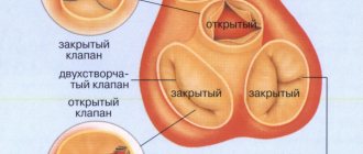
Types of valves:
bicuspid tricuspid lunate
Semilunar valves have wedge-shaped leaflets that prevent blood from returning as it leaves the heart.
There are two semilunar valves in the heart. One of these valves prevents backflow in the pulmonary artery, the other valve is located in the aorta and serves a similar purpose.
Other valves prevent blood from flowing from the lower chambers of the heart to the upper chambers. The bicuspid valve is located in the left side of the heart, the tricuspid valve is in the right. These valves have a similar structure, but one of them has two leaflets, and the other, respectively, three.
To pump blood through the heart, alternating relaxations (diastole) and contractions (systole) occur in its chambers, during which the chambers fill with blood and push it out accordingly.
Natural pacemaker
.
called the sinus node
or Kis-Flyak node, located in the upper part of the right atrium. This is an anatomical formation that controls and regulates heart rate in accordance with the activity of the body, time of day and many other factors affecting a person.
The heart's natural pacemaker produces electrical impulses that pass through the atria, causing them to contract, to the atrioventricular (that is, atrioventricular) node, located at the border of the atria and ventricles. Then the excitation spreads through the conducting tissues into the ventricles, causing them to contract. After this, the heart rests until the next impulse, which begins a new cycle.
SUBMIT AN APPLICATION FOR TREATMENT
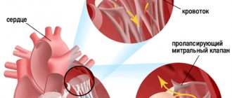
Valve prolapse
Heart valve prolapse is the most common diagnosis made by a doctor when a patient complains of malfunctions in the cardiovascular system. The mitral valve of the heart is most often affected by this pathology. Prolapse occurs due to a defect in the connective tissue that forms the valve. As a result of such defects, the valve does not close completely and blood flows in the opposite direction.
Primary and secondary valve prolapse are distinguished. Primary prolapse refers to congenital diseases when connective tissue defects are a genetic predisposition. Secondary prolapse occurs due to trauma to the chest, rheumatism or myocardial infarction.
As a rule, valve prolapse does not have serious consequences for human health and is easily treated. But in some cases, complications may occur, such as arrhythmia (disturbance in the rhythm of contractions of the heart muscle), failure and others. In such cases, treatment with medication or surgery is required.
Normal operation
As already mentioned, a normally functioning MV serves as an entrance gate for blood that flows from the left atrium into the ventricle. The heart constantly contracts, thus dispersing blood - from the atria to the ventricles, from the ventricles to the vessels, and from the vessels to all vital organs and systems. The operating principle of the snort can be summarized as follows.
When the main muscle of the heart is relaxed and the valve leaflets are open, blood from the left atrium flows into the left ventricle. The muscle then contracts and the snort valves close tightly, blocking the flow of blood from the ventricle back into the atrium. That is, blood can only flow in one direction.
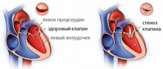
Mitral stenosis
This mechanism of operation of the entire cardiovascular system of the human body is strictly adjusted. Heart contractions occur at certain periods. If at least one part of this structure malfunctions, the working rhythm of the entire cardiovascular system as a whole is disrupted.
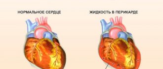
Insufficiency and stenosis of the valve apparatus
The main cause of insufficiency and stenosis is rheumatic endocarditis. Beta-hemolytic streptococcus is the cause of the inflammatory process in rheumatism, reaching the heart and changing its morphological structure. As a result of these changes, the heart valves begin to work differently. The valve walls may become shorter, causing regurgitation, or the valve opening may become narrowed (stenosis).
Due to rheumatism, mitral valve insufficiency most often occurs in adults. The aortic or mitral heart valve in children is susceptible to stenosis due to rheumatism.
There is such a thing as “relative insufficiency”. This pathology occurs if the structure of the valve remains unchanged, but its function is impaired, that is, the blood flows backward. This occurs due to impaired ability of the heart to contract, expansion of the cavity of the heart chamber, etc. Heart failure also occurs as a complication of myocardial infarction, cardiosclerosis, and tumors of the heart muscle.
The lack of qualified treatment for insufficiency and stenosis can lead to insufficiency of blood flow, degeneration of internal organs, and arterial hypertension.
Vascular valves
Veins, in addition to the structure of the walls, differ from arteries by the presence of valves, as parts of the intima, ensuring one-way blood flow. In this article we will not talk about vessels in general, but rather about specific main arteries, and about the valves between the heart and the vessels.
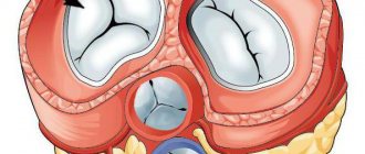
The valves of the heart and its vessels are marked in white.
Aortic valve
Its anatomy is fundamentally different from the atrioventricular valves. In its complex there is no connection with the ventricular myocardium and tension of the threads.
The exit of the left ventricle into the aorta is an arterial cone, bounded on three sides by muscular walls, while the fourth side is formed by the aortic valve. It is represented by three semilunar valves, which are attached by a fibrous ring and a triangle to the aortic mouth.
Each of the three doors (front, right and left) looks like a pocket. The initial section of the aorta, in which its valve is located, forms a bulb, which, in addition to the elastic structure of the vessel wall, is additionally strengthened by dense fibrous tissue. Thanks to the latter, it can withstand large fluctuations in blood pressure.
The integrated work of all valve elements ensures a complete result.
The average diameter of the aortic bulb is 1.5-3 cm, and the cross-sectional area corresponding to the location of the valve varies between 3.5-5.5 cm2. Changes in these indicators are similar to atrioventricular valves and lead to hemodynamic disturbances due to the formation of stenosis or regurgitation, depending on the nature of the changes.
Having understood what is between the heart and the vessels, it is important to understand how the aortic valve works, in contrast to the mitral or tricuspid valve. Its peculiarity is the presence of Arantzi nodes located in the center of each semilunar valve, on its free edge.
This allows the three valves to close tightly together during diastole, when the ventricle is filled with blood, thereby preventing blood leakage and changes in normal stroke volume.
Important! Complete closure of the aortic valve leaflets in diastole ensures complete filling of the coronary sinuses and arteries.
Pulmonary artery
This valve determines the direction of blood flow from the right ventricle into the vessels of the pulmonary circulation. It consists of three semilunar valves (anterior, right and left), which, like the aortic valve, have the form of pockets. The convex surface of the valves faces the cavity of the ventricle, and the concave surface faces the lumen of the pulmonary trunk.
The structure of the valves of the aorta and pulmonary artery.
Dense fibrous nodules located in the middle of the free edge of the valves also ensure a more dense closure between them. Between the heart and blood vessels, the leaflet valves are shaped like pockets, which plays a significant functional role. Thanks to it, blood is freely directed to the pulmonary artery during systole.
Symptoms of valve disease
The symptoms of heart disease directly depend on the severity and extent of the disease. As the pathology develops, the load on the heart muscle increases. As long as the heart copes with this load, the disease will be asymptomatic. The first signs of the disease may be:
- dyspnea;
- heart rhythm failure;
- frequent bronchitis;
- chest pain.
Heart failure is often indicated by shortness of breath and dizziness. The patient experiences weakness and increased fatigue. Congenital mitral valve prolapse is manifested in children by episodic pain in the sternum during stress or overexertion. Acquired prolapse is accompanied by rapid heartbeat, dizziness, shortness of breath, and weakness.
These symptoms may also indicate vegetative-vascular dystonia, aortic aneurysm, arterial hypertension and other heart pathologies. In this regard, it is important to make an accurate diagnosis, which will reveal that it is the heart valve that is causing the malfunction. Treatment of the disease depends entirely on the correct diagnosis.
Diagnosis of diseases
When the first signs of a heart valve defect appear, you should consult a doctor as soon as possible. The appointment is carried out by a general practitioner, and a specialist, a cardiologist, makes the final diagnosis and prescribes treatment. The therapist listens to the heart to detect murmurs and examines the medical history. Further examination is carried out by a cardiologist.
Diagnosis of heart defects is carried out using instrumental research methods. An echocardiogram is the main test that detects valve disease. It allows you to measure the size of the heart and its parts, and identify abnormalities in the functioning of the valves. An electrocardiogram records the heart rate, identifying arrhythmia, ischemia, and cardiac hypertrophy. An X-ray of the heart shows changes in the contour of the heart muscle and its size. Catheterization is important in diagnosing valve defects. A catheter is inserted into a vein and advanced into the heart, where it measures pressure.
Diagnostic methods
It is very important to correctly assess the condition of the heart valve system. For this, the following diagnostic methods are used:
- Auscultation - the heart is listened to at several specific points. This is done using a stethoscope. During normal operation, only 2 tones are heard: systolic and diastolic. Systolic occurs when the mitral and tricuspid valves close. It usually matches your heart rate. Diastolic tone does not coincide with the pulse and occurs when the semilunar valves close. With any pathologies, the tones may increase or decrease. Extraneous heart sounds may appear.
- Computed tomography - used to better visualize the affected areas. Prescribed for suspected presence of tumor formations in the atria. The tumor can also affect the mitral valve.
- Ventriculography - a special radiopaque substance is injected into the heart cavity. It is used only in cases of severe cardiac pathologies.
- Phonocardiography is the recording of heart murmurs using a phonocardiograph. Normally, only systolic and diastolic murmurs are recorded on the device.
- Echocardiography is a study of the functioning of the heart muscle using ultrasound. In this case, the state of the valves is assessed in different phases of the cardiac cycle. The number of valves, their shape and size are assessed.
Correct diagnosis is very important to identify pathological conditions that may threaten human health.
Possibility of treatment
Drug treatment includes the prescription of drugs aimed at relieving symptoms and improving heart function. Surgery is aimed at reshaping the valve or replacing it. As a rule, patients tolerate surgery to correct the shape better than replacement surgery. In addition, after replacing the heart valve, the patient is prescribed anticoagulants, which will need to be used throughout his life.
However, if the valve defect cannot be eliminated, it becomes necessary to replace it. A mechanical or biological heart valve is used as a prosthesis. The price of a prosthesis largely depends on the country of manufacture. Russian prostheses are much cheaper than foreign ones.
Several factors influence the choice of the type of artificial valve. This is the patient’s age, the presence of other diseases of the cardiovascular system, and also which valve needs to be replaced.
Mechanical implants last longer, but require lifelong coagulants. This causes difficulties in administering them to young women who plan to have children in the future, since taking such drugs is a contraindication during pregnancy. In case of tricuspid valve replacement, a biological implant is installed, which is determined by the location of the valve in the bloodstream. In other cases, if there are no other contraindications, installation of a mechanical valve is recommended.