0
Author of the article: Marina Dmitrievna
2018.10.04
727
Heart pathologies
Heart valves are damaged as a result of fusion of the leaflets. These are special holes that perform the main functions. For various pathological reasons, they can shrink and collapse, so partial or complete closure of the hole occurs. One of the main tasks is blood regurgitation.
Important! For diagnosis, doctors prescribe a Doppler echocardiogram.
It helps track the extent of valve damage. During the study, even minor deviations from the normal value are determined. This may be regurgitation through the mitral valve, which cannot be detected by laboratory tests. Heart valve disease requires urgent treatment.
If the valves are damaged, you need to undergo diagnostics
Heart Valve Disease - Symptoms, Treatment and More
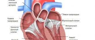
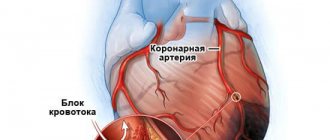
The purpose of the four heart valves (tricuspid, pulmonary, mitral and aortic valves) is to ensure that blood flows freely in the right direction when the heart beats. If one or more of the heart valves becomes diseased, either the free flow of blood through the heart is obstructed (stenosis), or blood may flow backward through the damaged valve (regurgitation)—or both. Any problem, if not monitored regularly and treated effectively, can lead to heart failure as well as other heart problems. The consequences of heart valve disease depend on which valves are involved, whether the main problem is stenosis or regurgitation, and the extent of damage to the valves. If you have cardiovascular disease, you will need to work closely with your cardiologist to ensure that the condition of your valve disorder and the need for its treatment are regularly reassessed.
Treatment methods
Damage to the heart valves leads to long-term health problems. Therapy for this pathology involves the use of an integrated approach using surgical interventions and a course of medications.
Therapeutic treatment
Therapeutic treatment involves the patient's constant stay in the hospital's cardiology department.
If there is no need for surgery, the patient receives drug treatment aimed at maintaining stable blood circulation, as well as preventing functional failure of individual segments of the heart. The duration of therapy depends on the degree of valve damage, as well as the possible presence of already developed complications.
Drug therapy
In the process of treating stenosis and functional insufficiency of heart valves, drugs of the following pharmacological groups can be used:
- antibacterial and anti-inflammatory drugs (used if the heart valves have been destroyed by rheumatic endocarditis);
- diuretics in the presence of valve dysfunction, which leads to the development of heart failure;
- antiarrhythmic medications that stabilize the heart rhythm;
- antiplatelet agents and anticoagulants that prevent embolic complications caused by increased thrombus formation.
Damaged heart valves can cause a large number of cardiac diseases, which have their own clinical manifestations. The selection of medications is carried out by the attending physician.
Surgery
The decision on the need for surgical treatment of heart valve pathologies is made by a cardiologist. The main indications for surgical intervention are severe stenosis and insufficient functioning of these areas of the heart muscle. To restore the full functions of the organ, 2 types of surgical operations are performed.
Commissurotomy
Commissurotomy is a cardiac operation that is performed on an open heart. The main goal of this type of surgical intervention is to eliminate adhesions inside the walls of the valve affected by stenosis.
After widening the narrowed hole, the previous blood flow is ensured and the patient’s normal well-being is restored. The patient returns to his previous lifestyle.
Prosthetics
Prosthetics of damaged heart valves is a more complex surgical operation that involves tissue plastic surgery. The cardiologist performing the operation restores the fibrous ring of the valve, as well as its leaflets, designed to regulate blood flow.
In this case, biological or mechanical devices can be used depending on the specific clinical case. In the absence of complications, the rehabilitation period lasts about 2-3 months. After this, the patient does not experience any discomfort.
Protection against infection due to valve defects
Patients with signs of damage to the heart valves should be protected from exposure to pathogenic microorganisms. For this purpose, patients of the melon group are placed in a separate room, in which increased sterility rules are observed.
Four heart valves and what do they do?
The heart consists of four chambers - the right and left atrium and the right and left ventricle.
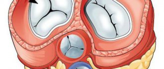
Each valve is located in an opening between two chambers and is attached to the opening by a fibrous ring called an annular. Attached to the annulus are two or three flaps (sometimes called flaps) that function as "flaps". The mitral valve has two leaflets; all other valves have three leaflets. When the heart beats, the leaflets open and close. When the leaflets are opened, blood can flow through the valve. When the leaflets are closed, blood flow through the valve stops.
The right ventricle is protected by the tricuspid and pulmonary valves. The tricuspid valve lies between the right atrium and the right ventricle. When the right atrium contracts, the tricuspid valve opens and allows blood to flow into the right ventricle. Then, as the right ventricle contracts, the tricuspid valve closes (to prevent blood from leaking back into the right atrium) and the pulmonary valve opens to allow blood pumped by the right ventricle to flow into the pulmonary artery and exit into the pulmonary artery. lungs.
The left ventricle is protected by the mitral valve and aortic valves. The mitral valve, which lies between the left atrium and the left ventricle, opens when the left atrium contracts to allow blood to flow into the left ventricle. When the left ventricle beats, the mitral valve closes and the aortic valve opens to direct pumped blood to the aorta and body tissues.
Causes of heart valve insufficiency
Congenital inferiority of the valve apparatus occurs due to connective tissue diseases. It is manifested by prolapse of the aortic or mitral valves, their parts are shortened, split or deformed.
The acquired defect develops against the background of rheumatism, endocarditis, and autoimmune diseases. At the same time, the valves thicken and then wrinkle, which causes them to close incompletely.
Relative insufficiency occurs when the mitral orifice or aortic lumen expands. Because of this, parts of the valves diverge and cannot completely block the annulus fibrosus. Aortic pathology can be caused by hypertension and aneurysm.
The causes of mitral insufficiency are:
- dilated or hypertrophic cardiomyopathy,
- severe hypertension,
- ischemic disease,
- myocarditis,
- aortic defects,
- calcification
In addition, pathology can occur acutely with rupture of the papillary muscles, chord, separation of the valves during a heart attack, trauma to the chest, severe inflammation.
We recommend reading the article about mitral heart disease. From it you will learn about the causes of pathology, diagnosis and treatment of the disease, and prognosis for patients.
And here is more information about combined heart defects.
What problems can affect the heart valves?
To reiterate, heart valves perform two fundamental jobs: They ensure that when the heart beats, blood flows freely through the heart and only moves in the right direction.
So it makes sense that when the heart valves become diseased, there are two main types of problems that arise.
First, heart valve problems can cause the valve to become partially blocked so that blood can no longer flow freely through it. This condition is called valve stenosis. When a heart valve becomes stenotic, the heart chamber that is supposed to push blood through the narrowed valve must work significantly harder to push the blood out. This causes the pressure in this chamber to increase, which ultimately causes the heart muscle to thicken (become "hypertrophic") and can eventually lead to its destruction.
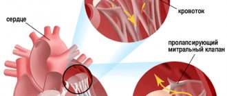
Secondly, heart valve disease can cause the valve to become incompetent; that is, the valve does not close completely and blood may flow back through the valve when it is supposed to be closed. This condition is called valvular regurgitation. Regurgitation causes the affected ventricle to pump more blood than normal, which can cause the chambers of the heart to dilate, weaken the heart muscle, and eventually heart failure. Thus, both valve stenosis and valvular regurgitation can lead to heart failure. In addition, heart valve disease is strongly associated with an increased risk of cardiac arrhythmias, especially atrial fibrillation. Any of the four heart valves can become either stenotic or regurgitating, and some diseased heart valves can display both of these problems at the same time. The severity of heart valve disease depends on the amount of stenosis or regurgitation produced. With most types of heart valve disease, the severity of the condition and its impact on heart function can progress significantly over time. Even “mild” heart valve disease should be taken seriously and periodically followed up by a competent physician.
How is a heart valve defect diagnosed?
Qualified cardiologists can detect valve defects in the heart already at the stage of listening to sounds. The most common diagnostic procedure is an examination using the so-called Doppler ultrasound. With its help, the doctor can find out the size of the heart and its chambers, as well as the thickness of the heart walls. Here the valves are checked for proper operation and closure.
In addition, the cardiologist can display the blood flow in the heart graphically in color and with acoustic noise. In this way, even complex heart valve defects can be accurately diagnosed. In addition, cardiac MRI . For some more detailed examinations, the use of a cardiac catheter is recommended in rare cases.
What conditions cause heart valve disease?
Cardiovascular disease can result from a number of different medical conditions.
The most common causes of cardiovascular disease include: Valvular calcification. Calcium deposits can form on the heart valves for no apparent reason due to aging. Valvular calcification tends to be especially likely with the mitral valve and, to a lesser extent, with the aortic valve. In many cases, valve calcification never causes any significant changes in valve function. But it can cause either stenosis or regurgitation, so anyone with valvular calcification needs to be monitored regularly to assess the condition.
Dilatation of the heart: If the chamber of the heart dilates, this can eventually cause the ring of the corresponding heart valves to dilate, which in turn can prevent the valve leaflets from closing completely. Valvular regurgitation may occur. Enlargement of the heart can occur with many types of heart disease. Typically, the dilation that may occur after a heart attack or in dilated cardiomyopathy is accompanied by dilatation of the mitral valve or tricuspid valve, resulting in mitral or tricuspid regurgitation.
Congenital heart defects: Many types of congenital heart defects involve valvular defects that can cause stenosis or regurgitation. The most common types of valve problems seen with congenital heart defects are pulmonary stenosis, bicuspid aortic valve disease (in which the aortic valve has only two leaflets instead of three, which can lead to aortic stenosis), and mitral valve prolapse.
Infective endocarditis: Infective endocarditis, an infection of the lining of the heart and especially the heart valves, can lead to heart valve damage, usually regurgitation. Valve disease caused by infective endocarditis may progress slowly, but it is one of the conditions that often leads to acute and possibly catastrophic regurgitation of the heart valves.
Rheumatic heart disease: Rheumatic heart disease is a form of heart valve damage that is sometimes seen after rheumatic fever in humans. Although rheumatic heart disease is much less common in Western countries than it was 50 years ago, it is still a major concern in many parts of the world. Preventing rheumatism by aggressively treating sore throat remains very important.
Other medical conditions: Heart valve disease can result from several other medical conditions, including syphilis, connective tissue diseases (such as lupus), various tumors, aortic aneurysm, carcinoid, and radiation therapy to the chest.
Defects and their varieties
Fibrous rings are a kind of separating tissue located between the chambers of the heart. These rings have many holes through which blood is pumped in the right direction.
This prevents disruption of natural blood circulation, which is commonly called annuloectasia. In medicine, there is a generally accepted classification of valve defects, which simplifies the identification of a specific pathology based on characteristic signs.
The designated classification is represented by the following diagram
Valve transformations, divided by location:
- Damage to the mitral valve is considered the largest type of defects and is usually found in patients of any age category. This pathology is accompanied by a change in connective tissue, which transforms into scar tissue. This disrupts the natural injection of blood masses and reduces their quantity.
- A change in the functioning of the aortic valve, accompanied by a modification of its structural basis. In the latter, the natural pumping of blood entering the small ring of blood flow is disrupted. With this disease, the activity of many organs deteriorates, and a deficiency of nutritional components necessary for normal life makes itself felt. This pathology leads to the progression of severe arrhythmia, and thromboembolism is possible.
- Pulmonary valve disease is a narrowing of the artery in the lung, in which there is a negative modification of the tissues and a deterioration in their replenishment. This pathology can only be eliminated surgically. It is accompanied by swelling of the veins in the forearm and neck. As a result, heart failure occurs, which manifests itself in a chronic form and is difficult to treat.
- Transformations in the structure of the triscupid valve. Such defects are usually represented by muscular rheumatic lesions in the myocardium. The designated valve of the cardiac system does not fully resist the blood flow, as a result of which the blood masses mix. The consequence is the onset of a heart attack.
Anatomical changes in the valve system in the form of atresia, hypoplasia, stenosis, coarthcation. This group also includes the transformation of the interventricular and interatrial structures of the heart.
Pathological transformations caused by specific organic disorders:
3.1 Obstructive changes in certain parts of the heart, in the vascular system. These pathologies occur due to narrowing of the arterial lumens.
3.2 Disadvantages detected when the structures of the cardiac septum are damaged.
- Defects that cause disturbances in the speed of blood flow. They are divided into several stages. In a complicated form, they are characterized by changes in blood pressure and are difficult to correct and treat.
- Hemodynamic values:
5.1 Blue indicators usually appear in childhood, during which mixing of arterial and venous blood occurs.
5.2 White indicators indicate a lack of mixing of blood masses from arteries and veins. With such pathologies, blood is released chaotically and in different directions.
About additional classification:
In addition to the changes indicated above, it is customary to distinguish a separate category in heart valves, it is represented by a tetrad or triad, Fallot's pentad, Ebstein's anomaly.
The most characteristic symptoms of heart valve disease are discomfort in the chest and shortness of breath, which manifests itself even with minor physical exertion. Timely detection of heart defects is the only available option to prevent their progression and subsequent aggravation.
As for therapeutic measures, they depend on the type of defect diagnosed. However, the ability to apply generally accepted health principles regarding myocardial functions in practice will help to minimize the negative consequences of any cardiac pathology.
Symptoms of heart valve disease
In most cases, heart valve disease does not cause any symptoms until the heart muscle becomes damaged enough to begin to fail.
When symptoms appear, they usually coincide with those of heart failure. These include dyspnea (shortness of breath); generalized weakness; lightheadedness; or swelling (edema) in the ankles, legs, or abdomen. Arrhythmias can also occur as a result of heart valve disease; as noted, the risk of atrial fibrillation is particularly increased. Symptoms of cardiac arrhythmia may include episodes of palpitations, lightheadedness, weakness, or poor exercise tolerance.
For most people, symptoms of valvular heart disease are usually a late manifestation of the disorder. Ideally, a person who has heart valve disease is diagnosed long before symptoms appear, so that treatment can begin before permanent damage to the heart muscle occurs.
Sometimes, however, heart valve disease can be an acute problem rather than a chronically progressive one. Acute valvular heart disease can result, for example, from damage to the heart muscle from a heart attack or from acute damage to the heart valves from infective endocarditis or rheumatic heart disease.
But more often, heart valve disease is a chronic, progressive disease that can be diagnosed long before symptoms appear. Early diagnosis is of course the key.
Symptoms
Sometimes heart valve disease does not cause any unpleasant symptoms. In such cases, the only manifestation may be a heart murmur, which can be heard through a stethoscope.
Symptoms of valvular heart defects may include:
- Shortness of breath, especially during exercise or when lying down
- Dizziness or fainting
- Chest pain or pressure
- Palpitations (feeling of the heart jumping out of the chest)
- Weakness or fatigue
- Swelling of the legs, ankles, or abdomen
Many of these symptoms can be confused with less serious conditions, the effects of aging, or physical inactivity. Similar symptoms may also occur with other diseases affecting the heart or lungs.
The severity of symptoms does not always correlate with the severity of valvular heart disease. Some patients with minor symptoms may require immediate treatment to prevent further damage to the heart.
Diagnosis of valvular heart disease
Early diagnosis of heart disease is very important for its optimal treatment.
Ideally, treatment begins before the heart muscle begins to fail and permanent damage to the heart is caused. But in order to do this, it is often important to know that valve disease is present long before any symptoms appear. Early heart valve disease is one of those silent medical problems that can usually be detected in the early stages through routine medical examination, and this is one of the reasons why we are all encouraged to undergo these routine checks.
The first sign of a heart valve problem is usually the discovery of a heart murmur during a physical examination. Valvular stenosis or valvular regurgitation causes a certain amount of turbulence in the blood flow in the heart. This turbulence creates a sound that a doctor can hear with a stethoscope (a heart murmur). However, not all heart murmurs indicate heart problems; many of them are so-called “innocent” murmurs, meaning they are caused by slight turbulence that can be present in almost any normal heart.
If your doctor detects sounds that may indicate a heart defect, an echocardiogram is very good at distinguishing between a heart valve problem and an innocent murmur. An echocardiogram can make a definitive diagnosis of almost any type of heart valve problem.
If valvular disease is present, an echocardiogram can also objectively measure the extent of the problem. Several specific measurements of blood flow patterns and the size of the heart chamber can be made, and these measurements can be compared with the results of subsequent echo tests to determine how quickly the valve problem is worsening (if it occurs at all).
Diagnostics
The following studies are usually used to determine valve defects:
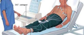
- Echocardiography: to evaluate the structure, function of the valve and blood flow through the chambers of the heart
- Electrocardiography: Helps identify heart rhythm problems
- Chest X-ray: allows you to determine the size and shape of the heart
- Coronary catheterization: identifies blocked or spasmed blood vessels
- Exercise stress test: measures the functional capacity of the heart when it is forced to work under physical stress
- Magnetic resonance imaging (MRI): takes detailed pictures of the heart's chambers and blood vessels.
Specific heart valve problems and their treatment
Mitral stenosis: In mitral stenosis, mitral valve obstruction reduces blood flow from the left atrium to the left ventricle.
Over time, pressure in the left atrium increases, eventually leading to pulmonary hypertension and heart failure, mainly affecting the right side of the heart. Treatment is surgical repair or replacement of the mitral valve, and the timing of the operation is critical. Once the need for surgery is established, the optimal type of mitral stenosis surgery must be determined for each person. Mitral regurgitation: Mitral regurgitation, which causes blood to flow back from the left ventricle into the left atrium, is the most common type of valve disease because it has so many causes. For example, the main significance of mitral valve prolapse (MVP) is that it can sometimes cause significant mitral regurgitation. Mitral regurgitation can cause dangerous dilatation of both the left atrium and the left ventricle, and if left untreated can lead to irreversible heart failure. The optimal timing of surgical treatment depends on the stage of regurgitation. Several surgical approaches are available to treat mitral regurgitation.
Aortic stenosis. In aortic stenosis, the aortic valve becomes partially blocked, making it difficult for blood to flow from the left ventricle into the body tissues. This leads to hypertrophy of the left ventricular muscles and ultimately to heart failure. Additionally, if the amount of blood the heart can pump is significantly reduced due to obstruction, fainting or even sudden death may occur. In fact, fainting due to aortic stenosis should be considered a medical emergency because it indicates a critical narrowing of the aortic valve. As with any type of cardiovascular disease, mild aortic stenosis should be closely monitored over time. If the condition becomes severe enough, aortic valve surgery will be required.
Aortic regurgitation: In aortic regurgitation, the aortic valve becomes leaky, so that blood washes back from the aorta into the left ventricle. This reverse flow of blood significantly increases the work of the left ventricle. If the regurgitation is significant, the ventricle eventually dilates greatly and heart failure occurs. Treatment of significant aortic regurgitation requires surgical replacement of the aortic valve.
Tricuspid stenosis: Tricuspid stenosis, partial obstruction of the tricuspid valve, is the least common of the major valve diseases. This is most often seen in people with rheumatic heart disease and, in most cases, is accompanied by disease of other heart valves. With significant stenosis, tricuspid syndrome causes mild fatigue and decreased exercise tolerance. However, symptoms caused by disease in one of the other valves usually appear long before tricuspid stenosis begins to produce symptoms. Consequently, surgical treatment of tricuspid stenosis (which consists of valve repair rather than replacement) almost always occurs as an additional procedure when surgery is needed to treat a more severely affected heart valve.
Tricuspid regurgitation: In tricuspid regurgitation, blood flows through the tricuspid valve from the right ventricle back into the right atrium. Tricuspid regurgitation is most often caused by dilatation of the tricuspid annulus, which occurs as a result of pulmonary hypertension, heart failure, or pulmonary embolism. Tricuspid regurgitation itself is often relatively mild and often does not require surgical treatment. A thorough evaluation to identify the underlying medical problem is important, as treatment of the underlying problem often results in significant improvement of tricuspid regurgitation.
Pulmonary stenosis: Pulmonary stenosis, an obstruction of the pulmonary valve, is usually a congenital disorder that is most often associated with tetralogy of Fallot, Noonan syndrome (a genetic disorder with cardiac abnormalities, short stature, chest deformities, and learning problems), or congenital rubella. Pulmonary stenosis is usually diagnosed at birth or soon after. If severe, it can cause the right side of the heart to fail. Mild pulmonary stenosis may be a completely benign condition that does not require therapy. If the condition is more severe, it can often be treated with balloon valvuloplasty, a minimally invasive catheterization procedure.
Pulmonary regurgitation. In pulmonary regurgitation, blood flows through the closed pulmonary valve from the pulmonary artery into the right ventricle. The most common cause of pulmonary regurgitation is pulmonary arterial hypertension, which can cause the pulmonary valve ring to dilate to the point that the pulmonary valve can no longer close completely. When severe, pulmonary regurgitation can lead to enlargement of the right heart and the development of heart failure. Typically, treatment for pulmonary regurgitation involves taking steps to reduce pulmonary artery pressure. Surgery is not often needed.
Leaking heart valves: causes, symptoms, treatment
Heart valve disease can be asymptomatic for many years. Your doctor may hear a noise during the examination and may be discovered by chance. Because valve disease is a mechanical problem, it cannot be treated with medication as it may only make the disease worse or relieve symptoms. In today's article, let's look at such a pressing topic as leaking heart valves: causes, symptoms, treatment. Review 12 causes of dark skin around the eyelids and around the eyes.
Causes
Heart valve disease can occur at birth (congenital). It can occur in adults for many reasons and conditions, such as infections and other heart conditions.
1. Regurgitation of heart defects
In this case, the heart valves do not close properly and cause blood to leak into the back of the heart.
This is usually caused by bloating, a condition called prolapse. Find out 9 causes of forehead acne you need to know.
2. Stenosis
With valve stenosis, the valves thicken or harden and may fuse together. This causes the valve opening to narrow and reduce blood flow through the valve.
3. Atresia
In this case, the valve does not form, and the hard tissue sheet prevents blood flow between the chambers of the heart.
4. Congenital heart defect
Some people have a congenital heart defect. The most common of these abnormalities is that the aortic valve must have 3 leaflets (bicuspid aortic valve). These people may develop aortic stenosis or regurgitation over time. In addition, these valves carry the risk of developing a valve infection, which we call endocarditis. Surgical treatment is possible when the valve disease is at a level that requires it. Less commonly, congenital abnormalities may be seen in other valves. Learn about hearing loss in people with diabetes
5. Rheumatic fever
Although it is rare in developed countries, it is still the leading cause of serious valve disease in our country. Rheumatic fever usually develops in children between 5 and 15 years of age after a throat infection. The main symptoms are fever, swelling and pain in the joints. Early diagnosis and treatment with penicillin are important. The valve disease this causes is felt after 20-30 years. Valve damage is the result of the immune system fighting bacteria by attacking the valve tissue. Some patients do not remember childhood rheumatic fever. Rheumatic fever is the most common cause of mitral valve stenosis in women and aortic valve regurgitation in men. It often contains multiple covers.
6. 3-mitral valve prolapse
This is a mitral valve disease that affects 2-3% of the population, mostly women, but rarely causes serious problems. Their mitral leaflets are thicker, longer and more mobile than usual; this may cause the lid to fail to close. Drug treatment is not required in most patients. However, an antibiotic is prescribed before tooth extraction and surgical procedures to prevent valve infection.
7. Calcification of the heart valve
The most common cause of heart valve disease in older adults is stenosis, caused by thickening and calcification of the valve leaflets. Some patients may require surgery.
8. Valve infection
This is a serious condition, usually seen in the presence of valve disease or a malfunctioning valve on the ground, and is called infective endocarditis. Main symptoms; fever, weakness, joint pain. To prevent this disease, patients with the valve need to take antibiotic protection before tooth extraction and surgical procedures. Read 7 consequences of swallowing food immediately without chewing
Symptoms
Complaints about valve disease vary depending on which valve has the problem. In the early stages of the disease, a person may not have any complaints, and sometimes they can live like this for years. As the valve's condition increases, symptoms begin to appear.
- rapid fatigue
- weakness
- cardiopalmus
- dyspnea
This is manifested by swelling of the legs, which is an indicator of fluid accumulation in the body. Some valve diseases, dizziness, chest pain, even fainting may be noticed.
Treatment methods
1. Operation
Techniques used for a leaking valve can be summarized as placing tissues to support the valve structure, cutting and repairing valve tissue for better closure, or releasing adhered leaflets. Read more Varicose veins of the pelvis in women
2. Heart valve replacement
Valve disease is most often treated with valve replacement surgery. During this operation, the patient's valve is removed and replaced with a mechanical or biological valve:
Bioprosthetic valves are made from bovine, porcine, or human tissue. The use of biological diluents is not necessary for the use of biological valves, but the valves may wear out after 10 to 15 years and surgery may be required. In this regard, they are not preferred for young patients.
3. Transcatheter valve treatment
Interventional cardiologists can repair or replace valves using clips or catheters that pass through large blood vessels.
A recent current application of the valve is transcatheter aortic valve replacement for aortic valves.
In this method, a catheter is inserted through a groin vein and the balloon is advanced to the level of the folded aortic valve, and a catheter containing an artificial valve is folded around it.
Take care of yourself and be healthy!
Is there a non-surgical treatment for valve disease?
Heart valve disease is mainly a mechanical problem.
To address the underlying cause, surgery is usually the only option. However, in many cases there may be medical therapy available that can help. Sometimes medications can help stabilize the heart and slow the progression of valve disease. This is especially true of many types of valvular regurgitation, where the valve problem is caused by an enlarged chamber of the heart. Aggressive medical treatment aimed at treating underlying hypertension or dilated cardiomyopathy, or preventing ventricular remodeling after a heart attack, or even controlling the heart rate in atrial fibrillation, for example, can significantly reduce the likelihood of developing significant mitral or tricuspid regurgitation.
For this reason, it is usually recommended for people with valvular heart disease to regularly visit a cardiologist, who will readily address any problems with the cardiovascular system.
Symptoms of pathologies
Damage to the heart valves leads to the following symptoms, which significantly reduce the patient’s quality of life:
- feeling of lack of air;
- heart rhythm disturbances (tachycardia, bradycardia);
- squeezing pain inside the chest;
- dizziness;
- loss of consciousness;
- attacks of severe headache;
- fainting state;
- darkening of the eyes;
- dyspnea;
- fast fatiguability;
- dry cough with a healthy respiratory system;
- pronounced cyanosis of epithelial tissues.
Patients with similar symptoms are subject to a comprehensive examination of the cardiovascular system using laboratory and instrumental research methods. If necessary, the patient is hospitalized in a hospital setting.
Living with Heart Valve Disease
If you have heart valve disease, there are several things you need to do to optimize your chances of living a long, healthy life.
You should learn all you can about the type of valve disorder you have and the extent of your valve problem. If you are taking medications that will help your heart work more efficiently, you should take them regularly and report any problems with them to your doctor. For this reason, you need to visit your doctor regularly. While this is important for everyone, it is especially important for a person who has problems with their heart valves, as heart disease often progresses over time. You should talk to your doctor about the need for antibiotic prophylaxis for endocarditis.
Finally, since you already have heart problems, you should do everything you can to reduce your chance of developing other types of heart disease: don't smoke; maintain a healthy diet and healthy weight; get plenty of exercise; and, if you have hypertension or diabetes, make sure these conditions are optimally controlled.
Introductory part
Before heart surgery, a person has many questions. Some of them we ask the doctor, and some we cannot even formulate. When we understand what is happening to our body and what we can do to restore health, it is easier for us to endure all procedures.
Acquired heart valve defects
- these are diseases that are based on morphological and/or functional disorders of the valve apparatus (valve leaflets, annulus fibrosus, chordae, papillary muscles), developed as a result of acute or chronic diseases and injuries, disrupting the function of the valves and causing changes in intracardiac hemodynamics.
Valve defects can be congenital or acquired.
Congenital defects occur when the structures of the heart are formed incorrectly during intrauterine development; sometimes they do not make themselves felt until adulthood. Acquired defects arise due to rheumatism, infection, metabolic disorders (when calcium is deposited in the valves), trauma and other reasons.
The main types of heart valve defects:
- mitral stenosis
- mitral valve insufficiency
- mitral valve prolapse
- aortic stenosis
- aortic valve insufficiency
- tricuspid stenosis
- tricupidal insufficiency
The normal functioning of the heart largely depends on the functioning of its valve apparatus.
Obstacles to the passage of blood cause overload, hypertrophy and expansion of the structures above the valve. Obstructed heart function disrupts the nutrition of the hypertrophied myocardium and leads to heart failure.
Etiology and pathogenesis
Etiology of stenosis
and combined rheumatic disease,
insufficiency
- usually rheumatic, rarely septic, atherosclerotic, traumatic, syphilitic.
Stenosis is formed as a result of cicatricial fusion or cicatricial rigidity of the valve leaflets and subvalvular structures; valve insufficiency - due to their destruction, damage or scar deformation.
Failure
valve damage occurs due to destruction or damage to its valve flaps. Valve insufficiency is characterized by incomplete closure of the leaflets and occurs as a result of their wrinkling, shortening, perforation or expansion of the fibrous valve ring, deformation or separation of the chordae and papillary muscles. In some cases, valve insufficiency develops as a result of dysfunction of the valve apparatus, in particular the papillary muscles.
Often stenosis and insufficiency develop on one valve (the so-called combined defect
).
In addition, there are cases when the defects affect two or more valves - this is called combined
heart disease.
Affected valves form an obstacle to the passage of blood - anatomical in case of stenosis, dynamic in case of insufficiency. The latter is that although some of the blood passes through the hole, it returns back in the next phase of the cardiac cycle.
A “parasitic” volume is added to the effective volume, performing a pendulum-like movement on both sides of the affected valve. Significant valvular insufficiency is complicated by relative stenosis (due to increased blood volume). Obstruction to the passage of blood leads to overload, hypertrophy and expansion of the overlying chambers of the heart.
The expansion is more significant with valve insufficiency, when the overlying chamber is stretched by additional blood. With stenosis of the atrioventricular orifice, the filling of the underlying chamber is reduced (left ventricle with mitral stenosis, right ventricle with tricuspid stenosis); There is no hypertrophy or expansion of the ventricle.
With valve insufficiency, the filling of the corresponding ventricle is increased, the ventricle is dilated and hypertrophied. Difficulty in the functioning of the heart due to improper functioning of the valve and degeneration of the hypertrophied myocardium leads to the development of heart failure.
to the top of the page
Anatomy of the heart
A healthy heart is a strong, continuously working organ, about the size of a fist and weighing about half a kilogram.
In addition to maintaining steady, normal blood flow, it quickly adapts and adapts to the body's ever-changing needs.
For example, the heart pumps more blood when it is active and less when it is at rest. During the day, the heart produces an average of 60 to 90 beats per minute - 42 million beats per year!
The heart is a two-way pump that circulates blood throughout the body. It consists of 4 chambers.
A muscular wall called the septum divides the heart into left and right halves. Each half has 2 chambers.
The upper chambers are called atria, the lower chambers are called ventricles. The right atrium receives all the blood returning from the upper and lower parts of the body.
Then, through the tricuspid valve, it sends it to the right ventricle, which in turn pumps blood through the pulmonary valve to the lungs.
In the lungs, the blood is enriched with oxygen and returns to the left atrium, which sends it through the mitral valve to the left ventricle.
The left ventricle pumps blood through the arteries through the aortic valve throughout the body, where it supplies the tissues with oxygen. Oxygen-depleted blood returns through the veins to the right atrium.
Four valves (tricuspid, pulmonary valve, mitral, aortic) act as a door between the chambers, opening in one direction.
These valves help blood move forward and prevent it from flowing in the opposite direction.
The petals of a healthy valve are thin, flexible tissue of perfect shape. They open and close when the heart contracts or relaxes.
Heart valves can become abnormal due to birth defects. They can become damaged or scarred due to rheumatic fever, infection, hereditary factors, age or heart attacks.
The mitral valves are most susceptible to such changes.
Regardless of the case, the heart valve can become stenotic (narrowed opening) or insufficient (does not close completely).
With valve stenosis, the heart must work harder to pump the required amount of blood through the narrowed opening.
Valve insufficiency causes blood to flow backwards through the valve after it closes. Once again, the heart has to work harder to pump enough blood for the body's needs to make up for the deficiency caused by the backflow of blood.
Both cases - stenosis and failure - force the heart to work harder to pump the required amount of blood. This extra work can weaken the heart, cause it to enlarge, and cause various diseases.
to the top of the page
If the heart valve does not close completely, what does it mean?
Is one of your heart valves not closing completely? The disorder is caused by certain reasons and is called valve insufficiency.
In order to understand the cause of heart valve insufficiency, you should familiarize yourself with the anatomical structure of the heart.
The heart is a hollow muscular organ with a four-chamber structure (two ventricles and two atria). The valves, tricuspid and mitral, separate the ventricles from the atria.
The vessels in the heart are also separated by valves: pulmonary and aortic. Valves are necessary to regulate blood flow. If their tissues are altered and their structure is deformed, they cannot close completely or open fully.
If the valve cannot close completely, then the blood entering the atrium is not enough for its normal functioning; it does not allow blood flow.
The valve does not open - the blood flow partially returns to the atrium or ventricle, which over time leads to disruption of the heart, causing severe heart failure.
Aortic valve stenosis
This is a rare, acquired heart defect that affects the left side of the organ. As a result of a valve defect, arrhythmia, electrical conduction disturbances, or infective endocarditis may develop.
Valve stenosis occurs as a result of a narrowing of the aortic opening that closes the valve.
The outflow of blood in the opposite direction leads to hemodynamic disturbances.
Mitral valve prolapse
What leads to prolapse:
- viral and bacterial infections;
- endocarditis;
- rheumatism;
- inflammation that disrupts the structure of connective tissue;
- sclerosis;
- myocarditis;
- aortic arteritis;
- injury to the chord and cardiac muscle.
Hypertension can also provoke incomplete closure of the valves.
Valve disease in rheumatism
Chronic inflammation of the nasopharynx in children can lead to rheumatism. The disorder is caused by persistent infection with streptococci. Rheumatic prolapse is difficult to diagnose, as there are no special laboratory tests.
In order to identify the problem, doctors use a list of symptoms, analyze patient complaints and take into account the clinical manifestations of the disorder.
If the mitral and aortic valves are damaged, patients are treated in the hospital with antibiotics and anti-inflammatory drugs.
How does valve insufficiency manifest?
Malfunction of the valves at the initial stage does not affect the functioning of the body, and patients do not complain of feeling unwell. The stage is called compensatory. Subsequently, during the decompensation stage, severe symptoms begin to appear that can lead to death.
At the moderate stage, a person is worried about constant fatigue, shortness of breath, and with pulmonary edema, hemoptysis begins. As the atrium on the left enlarges, the nerves of the larynx are compressed and the voice becomes hoarse.
Aortic valve insufficiency manifests itself as rapid heartbeat, chest pain, shortness of breath with moderate physical exertion.
Severe aortic insufficiency can lead to death due to a sharp decrease in pressure and pulmonary edema. Patients require immediate surgical intervention.
Diagnosis of valve dysfunction
The first step to diagnosing the problem is to listen to the tone of the heartbeat and detect any murmurs. Auscultation of the heart makes it possible to make a preliminary diagnosis, with which the patient is sent for further examination.
ECG, ECHO-CG of the heart and chest x-ray are the next stage of examination, allowing you to determine whether there is an increase in the heart chambers.
X-ray shows distortion of the heart shape and its enlargement.
ECHO shows the deformation of the valves, the inability to completely close or open, and also helps to find the cause of problems with the valve, the degree of its insufficiency and the possibility of compensation from the body.
At the next stage of diagnosis, a catheter is inserted for coronography and ventriculography.
Treatment methods
The main method of solving the problem today is prosthetics. For mild or moderate valve insufficiency, drug therapy is not prescribed; inhibitors are prescribed for severe disease that does not produce specific symptoms.
Two types of surgical intervention are used: valve replacement and plastic surgery. If the structure of the valve is preserved, there are no changes in the tissues and its mobility is completely preserved, patients undergo plastic surgery. Changed and thickened tissues require a different approach - prosthetics.
Plastic surgery has an advantage over prosthetics - fewer postoperative complications and a reduced risk of infective endocarditis.
Mild aortic insufficiency, which has an asymptomatic course, does not require therapy, however, patients are limited in physical activity and hard work is contraindicated for them.
Every year it is necessary to undergo examination by a cardiologist. When symptoms appear, patients are prescribed drug therapy.
Severe valve insufficiency requires constant conservative treatment; surgical intervention may be performed if indicated.
Heart valve replacement
The operation to replace the heart valve is performed when the atrioventricular opening is narrowed to 1.5 cm, the aortic opening to 1 cm. Various materials are used as prostheses. In some cases, patients receive heart valves from animals such as pigs or cows. It is believed that the risk of rejection of such prostheses is lower than that of synthetic materials. Another surgical option is to transplant the pulmonary valve to the site of the damaged aortic opening. This procedure is performed only on young patients, as it is a technically complex surgical procedure.