If a blood clot has only one point of attachment in the venous wall - the so-called head, and the tail and body are freely located in the vessel, then such a thrombus is considered floating. This is a dangerous form of thrombosis, since the clot can easily break off and turn into an embolus that closes the lumen of the pulmonary artery. To prevent complications, a vena cava filter is installed, vein plication or thrombectomy is performed, and anticoagulant therapy is prescribed.
About a floating thrombus
The size of floating thrombosis can reach 20 mm
Thrombosis is a disease characterized by impaired circulatory function and the formation of a blood clot in the vessels. As a result of slow blood flow, a layering of blood elements occurs. Increasing in size, the clot interferes with normal blood circulation.
Because of this, the organ tissues do not receive the required amount of nutrients. This affects its functioning. As a result of blood stagnation, necrosis develops, which is fraught with amputation of the organ and loss of capacity.
Floating thrombosis is characterized by its large size, which can reach 20 mm. The stalk with which the thrombus is attached to the vascular wall is too thin.
The tail of the thrombus is in constant motion due to the outflow of blood. At any moment, a blood clot can break off and cause a complete blockage of another vessel.
Symptoms of presence in the heart, in the inferior vena cava
Intracardiac thrombosis occurs quite often, its insidiousness lies in the fact that there are no manifestations of the disease for a long time, and when the clot moves or grows excessively, cardiac arrest occurs. The inferior vena cava is a large vessel with intense blood movement, so in case of thrombosis there is a high risk of complications.
Cardiac blood clots
Blood clots in the heart are most often located in the cavity of the left atrium. The onset of their development is often associated with blood stagnation with mitral stenosis, damage to the valves with endocarditis, excessive expansion of this part of the heart, as well as chaotic contractions of muscle fibers during atrial fibrillation or fibrillation.
Blood clots can be massive, round, filling the entire space, or polyp-like. The latter type belongs to floating formations, since it periodically enters the atrioventricular orifice and causes the following symptoms:
- sudden increase in heart rate,
- chest pain,
- hard breath,
- blue skin,
- fainting state.
A characteristic feature of transient thrombosis is intense cardiac activity, which can be determined by examining the apical impulse, weak pulse, and low blood pressure. The detachment of a blood clot is accompanied by intense pain in the heart, a state of shock, and the development of a stroke or heart attack is possible.
Blockage of the inferior vena cava by a clot
This vessel is rarely the site of blood clot formation. This is only possible against the background of structural anomalies that are congenital in nature or appear after extensive trauma or destruction by an ingrowth tumor. Heavyweight athletes are also susceptible to caval (venous cava) thrombosis, whose movements lead to a sharp increase in venous pressure with tearing of the inner layer of blood vessels.
Blockage of the inferior vena cava by a clot
Signs of the disease are:
- swelling of the thighs, lower half of the body, genitals;
- expansion of superficial veins in the abdomen;
- skin cyanosis.
Symptoms of a floating thrombus
The main feature of the disease is the blurred clinical picture in the initial stages of development. This significantly slows down diagnosis. Most often, the disease is detected at the stage of development of complications. In this case, the symptoms become more pronounced. The following symptoms appear:
- swelling in the area where the blood clot is located;
- increase in local temperature;
- redness of the skin in the affected area;
- pain that intensifies during palpation;
- cyanotic spots on the body;
- bursting pain.
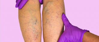
On a note! If a pathological process develops in the saphenous vein, almost immediately after the formation of a blood clot, a swelling appears, reminiscent of varicose veins. In this case, the lymph nodes also become enlarged.
Deep vein thrombosis of the lower extremities (clinical and anatomical forms)
- Deep vein thrombosis of the leg
Complaints of swelling of the foot, pain and tension in the calves, pain when pressing on the calf muscles. If thrombosis does not spread, it is almost asymptomatic. Sometimes there is thromboembolism of small branches of the pulmonary artery with cough and the development of pneumonia (pneumonia). Treatment of thrombosis of the veins of the leg can be carried out on an outpatient basis, under the supervision of a phlebologist with control ultrasound examinations.
- Thrombosis of the popliteal vein
Has a clear clinical picture. Severe swelling and tension of the lower leg, swollen saphenous veins, severe pain when walking. Thrombosis of the popliteal vein is very dangerous due to frequent pulmonary embolism, so treatment is best carried out in a vascular hospital. Most often, conservative therapy is carried out with antithrombotic drugs (heparin). If the patient had a thromboembolism, then urgent surgical treatment is necessary - ligation of the femoral vein above the thrombus.
- Clinic for deep vein thrombosis of the thigh and iliofemoral segment (ileofemoral phlebothrombosis)
It is characterized by a severe general condition, pronounced swelling of the entire lower limb, and severe pain. The saphenous veins are sharply dilated, the leg takes on a bluish color. With ascending deep venous thrombosis, thrombosis of the entire venous bed is possible with a block of venous outflow and the development of venous gangrene (blue phlegmasia), which is accompanied by high mortality. Pulmonary embolism often occurs with a fatal outcome. Treatment of ileofemoral phlebothrombosis is only in a hospital. For occlusive thrombosis, conservative treatment is possible, but it is better to remove the thrombus so that post-thrombotic disease does not develop. In case of floating thrombosis, urgent removal of the thrombus (thrombectomy) using innovative methods is necessary. A vena cava filter can be installed in cancer patients.
- Thrombosis of the inferior vena cava
The most dangerous disease. Clinically, it is manifested by a severe general condition, swelling of both legs. Kidney failure and blood in the urine often develop. With thrombosis of the liver segment, liver failure develops resulting in Budd-Chiari syndrome. Treatment of acute thrombosis of the inferior vena cava should be active. It is necessary to remove thrombotic masses, as surviving patients may develop severe inferior vena cava syndrome. For this, it is good to use our innovative methods and systemic thrombolysis. The effectiveness of this treatment
- Asymptomatic thrombosis
It should be said right away that there are silent thromboses, that is, completely asymptomatic. Therein lies a great danger. This problem is becoming more and more acute, because with the expansion of ultrasound examination of veins, signs of previous thrombosis are being found more and more often. According to some phlebologists, by old age, most people experience such asymptomatic deep vein thrombosis. In quantity they even exceed those that can be diagnosed without the use of ultrasound methods. The patient does not even feel any health problems, and serious complications occur in the midst of complete well-being, in the event of an increase in blood clot and closure of the main veins. It is not uncommon for the disease to be discovered only after the patient dies from these complications. From this position, if there are no signs of the disease and you are at risk, there is only one way out - you need to direct all your efforts to prevention.
Diagnosis of acute deep vein thrombosis of the lower extremities is very difficult. Signs of deep vein thrombosis appear only in certain localizations of the process. This is primarily due to the absence of clinical symptoms. According to some data, out of 1000 venous thromboses, only 100 have any clinical manifestations. Of these, 60 patients will develop PE, but only 10 will have clinical signs.
It should be recognized that today there is not a single clinical symptom, laboratory or instrumental sign that would indicate with absolute certainty the presence of PE and DVT. Clinical manifestations of thrombosis and ultrasound results can be the basis for the correct diagnosis of venous thrombosis. The clinical picture of deep vein thrombosis consists of a complex of symptoms that characterize a sudden disturbance of venous outflow with preserved arterial blood flow to the limb. Swelling, cyanosis of the limb, bursting pain, local increase in skin temperature, overflow of the saphenous veins, pain along the vascular bundle are characteristic to one degree or another for thrombosis of any localization. Movements in the joints of the limb and sensitivity remain virtually unchanged. General signs such as low-grade fever, weakness, adynamia, and slight leukocytosis occur in most patients. The diagnosis of thrombosis largely depends on the location of the lesion and the level of distribution of thrombotic masses.
Causes of a floating thrombus
Predisposing factors for the formation of a floating thrombus include cardiac dysfunction
The formation of a floating thrombus is associated with stagnant processes and impaired blood clotting function. Most often, the disease occurs against the background of other pathological processes. Predisposing factors include:
- lipid metabolism disorder;
- liver dysfunction;
- malignant or benign formations;
- venous diseases (thrombophlebitis, varicose veins, phlebitis);
- disruption of the heart (atrial fibrillation, cardiomyopathy, defects, etc.);
- infectious and autoimmune diseases.
Prevention
To prevent the risk of developing thrombosis and the occurrence of a floating thrombus, it is necessary to carefully monitor the health of blood vessels.
You should adhere to a healthy diet. Eliminate fatty and fried foods from your diet. Give up alcohol and cigarettes, or reduce doses to minimum.
You should add lean meat, dairy products and plant foods to your daily menu.
It is advisable to maintain the correct drinking regime, as it helps normalize blood consistency.
As sports activities, experts recommend walking and light aerobic exercise.
Timely treatment of infectious pathologies reduces the risk of developing thrombophlebitis and helps avoid vascular injury.
Following these recommendations will allow you to live a healthy, fulfilling life.
Etiology and pathogenesis
The process of complete blood circulation in the body is ensured by the coagulation system. It performs a protective function, preventing excessive blood loss due to various injuries. Thrombosis develops when blood viscosity changes.
Along with this, the development of the disease is facilitated by damage to the endothelial layer of the vascular walls. Additional factors that create favorable conditions for the development of the disease include:
- mechanical damage (including during surgery);
- pregnancy and labor;
- old age (over 60 years);
- long-term use of oral contraceptives;
- limb compression syndrome;
- excess weight and sedentary lifestyle;
- forced limitation of motor activity.
Causes of thrombosis
There are three main factors of thrombosis, which most often cause exacerbation. This is hypercoagulation, circulatory disorders and vessel rupture. Each of these conditions can be caused by different causes.
Hypercoagulation
Excessive blood clotting is caused by the following reasons:
- taking hormonal medications with estrogen;
- malignant tumors, chemotherapy;
- operations and injuries of the legs, pelvis and abdominal cavity;
- intestinal inflammation;
- infections;
- pregnancy.
Circulatory disorders
Blood flow in the vessels is disrupted due to the following factors:
- heart failure;
- phlebeurysm;
- arrhythmia;
- pressure on veins during pregnancy;
- excess weight, tumors, edema, compressive vessels;
- prolonged bed rest or staying in one position.
Damage to vascular walls
Vessel rupture can occur as a result of reasons such as:
- operations and injuries;
- repeated insertion of a needle into a vein (when administering medications or narcotic drugs);
- atherosclerosis (Atherosclerosis);
- heart valve diseases.
Diagnostic methods
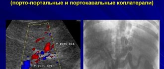
Ultrasound duplex scanning of veins will help identify the pathological process
Since the intensity of symptoms depends on the local location of the blood clot, it is impossible to identify the disease without diagnostic procedures. The main way to identify a pathological process is ultrasound duplex scanning of veins. This method helps determine the structure of the clot, its location and the condition of the vessel wall.
In some cases, MRI and X-rays are performed using a contrast agent. Of the laboratory tests, the most accurate data is provided by a blood test for the presence of D-dimer. This protein is formed as a result of the breakdown of fragments of a blood clot.
Treatment and recanalization
A mandatory condition for the treatment of patients with thrombosis of veins or cardiac cavities is immediate hospitalization. Since even with intensive drug therapy it is only possible to stop the progression of the disease, but does not prevent the danger of the blood clot breaking off and moving.
The strictness of bed rest depends on the diagnostic results. If a floating thrombus is detected, complete rest and immediate intravascular or direct surgical interventions are recommended.
Installation of a vena cava filter
Through a small puncture of the skin, a structure in the form of a metal frame is installed inside the vein. It can pass moving blood, but catches large blood clots. The method does not eliminate a blood clot, but is used to prevent thromboembolism of the pulmonary artery.
Blood clot trap (Vava filter)
Vein suturing (plication)
The inferior vena cava is divided into several tubules with a thread, stitched with wide stitches. With this method, the movement of blood clots is stopped, but blood flow is allowed to flow. An operation called plication is used in the following situations:
- the thrombus is located above the origin of the renal veins;
- when combined with tumors that need to be removed;
- repeated pulmonary embolism with inability to determine the source;
- The diameter of the vein does not allow the installation of a vena cava filter.
Thrombectomy
It is rarely used because the catheter easily passes through the clot, tearing it away from the vein wall. Therefore, there remains a high risk of vascular blockage. After removal of the blood clot, the inner lining is damaged, which leads to relapse of the disease.
Pharmacological recanalization
Indicated for all patients with floating thrombi after surgery. It includes the appointment of low molecular weight heparins (Fraxiparin, Clexane, Fragmin), and then the indirect anticoagulant Warfarin.
Taking the latter remedy is carried out under the control of a blood test for INR and lasts from 3 months to six months. If the risk of thrombosis cannot be eliminated (hereditary thrombophilia or tumor), then therapy is continued for up to 1 year.
They also use drugs to improve blood flow: Reopoliglucin, Trental, Aspirin, Curantil, as well as venotonics - Detralex, Aescin. Topical ointments with heparin (Lioton, Fastum) are recommended.
On the first day, enzymes can be injected into the area where the blood clot is located to dissolve the clots (Streptokinase, Urokinase). This method of treatment is also prescribed after thrombectomy.
Treatment of a floating thrombus
Treatment for thrombus flotation is aimed at eliminating the danger to the patient's life. Therapy helps prevent the blood clot from moving further. The required result is achieved with an integrated approach. Treatment is carried out within the walls of a medical institution.
During therapy, the patient is in a supine position. If the thrombus is concentrated in a limb, then it is fixed in an elevated position.
Important! It is strictly forbidden to take medications without consulting a doctor. The dosage is selected only after testing.
Types and symptoms of the disease
Doctors distinguish two forms of thrombosis – venous (phlebothrombosis) and arterial, which, in turn, are divided into several subtypes. In addition, there is acute and chronic thrombosis, characterized by a sluggish course with alternating periods of exacerbation and remission.
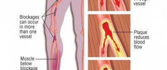
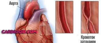
As for thrombosis of the arteries, it most often occurs against the background of blockage of the vascular lumens with atherosclerotic plaques, for this reason the disease is also called atherothrombosis, and can lead to a heart attack of any organ or a stroke of the brain.
Intestinal form of the disease
The arterial form of the disease includes intestinal thrombosis (or mesenteric thrombosis), which develops against the background of blockage of the superior mesenteric artery. It mainly affects older people and occurs with equal frequency in men and women.
The main causes of thrombosis of mesenteric vessels include heart diseases, during which myocardial infarction, endocarditis or atherosclerosis develop. The latter condition is considered one of the most dangerous, since the lack of treatment can lead to the fact that mesenteric thrombosis can develop into embolism and cause the death of the patient.
Intestinal thrombosis can be recognized by the following signs:
- sharp, sometimes paroxysmal, intense pain in the abdomen, the localization of which depends on which vessel the thrombus is located in (if the pain is in the right iliac region, intestinal thrombosis must be differentiated from acute appendicitis);
- nausea and vomiting accompanied by abdominal pain;
- cardiopalmus;
- loose stools, which may contain blood (of course, intestinal thrombosis cannot be diagnosed based on this symptom alone, but if it is accompanied by the symptoms described above, then you should immediately go to the doctor);
- elevated body temperature also indicates the development of thrombosis, the symptoms of the disease appear as it progresses, that is, at first the temperature, if raised, will not be much, and when a thrombus blocks the lumen of an artery and the onset of intestinal necrosis, it will begin to rise rapidly.
As it develops, intestinal thrombosis will increasingly resemble intestinal obstruction (bloating, severe peristaltic disturbances, vomiting), so only a doctor can make an accurate diagnosis and prescribe appropriate treatment.
Hirudotherapy
The main advantage of hirudotherapy is its natural composition.
Hirudotherapy is a method of alternative medicine that involves the use of a medicinal leech. The principle of operation of the procedure is the same as that of anticoagulants.
By sticking to the skin surface, the leech introduces a special substance into the human body - hirudin. It reduces blood clotting, preventing the formation of new blood clots.
The main advantage of hirudotherapy over synthetic drugs is its natural composition. The procedure does not have a negative impact on the functioning of internal organs.
Prevention and treatment of the disease
Effective treatment of thrombosis must be comprehensive and consist of drug therapy (medicines are prescribed by a doctor), the use of traditional medicine, minimally invasive medicinal techniques and, in some cases, surgical intervention.
Treatment of thrombosis initially takes place in a hospital setting, while for the first few days (while doctors carry out all the necessary studies), the patient is prescribed bed rest and is prohibited from performing any thermal procedures (they can lead to the detachment of a blood clot and a deterioration in the patient’s well-being). Therapy with folk remedies is also undesirable at this stage.
Inpatient treatment of thrombosis begins with intravenous administration of heparin, a natural anticoagulant that prevents the growth of an already formed blood clot and thins the blood. The course of treatment lasts seven days, after which the patient is prescribed direct-acting anticoagulants (Warfarin) and nicotinic acid (dissolves blood clots). Therapy can last from 6 to 8 months.
Surgical treatment of thrombosis is prescribed in especially severe cases, when there is a possibility of developing necrosis, gangrene or blood clot rupture.
As for treatment with folk remedies, they can be used for the sluggish nature of the disease as an addition to drug therapy. Particularly popular are the use of herbal decoctions, treatment with honey or foot baths prepared with cinquefoil root. Medicines must be taken for six months, otherwise they will not give any effect.
Prevention of thrombosis consists of periodically taking heparin (to avoid bleeding, the dose of the medicine is prescribed by the doctor), regular use of traditional medicine (nettle decoction and acacia tincture have a good effect).
Any disease is easier to prevent than to treat, so prevention of thrombosis plays a very important role in the life of every person. In any case, both treatment and prevention of the disease should be carried out by a doctor; self-medication can only harm health and aggravate the condition of the blood vessels.
Post Views: 147
Traditional methods of therapy
Onions and honey are useful for traditional treatment
Traditional methods of dissolving blood clots should be used with extreme caution. Traditional recipes cannot act as independent therapy. They complement conservative methods. It is recommended to use the following recipes:
- It is necessary to chop 2 onions and mix the resulting mass with 200 grams of honey. Then you should put the medicine in a dark place for 2 hours. Onion mixture should be taken 1 tbsp. before eating. It needs to be stored in the refrigerator.
- 2 tbsp. hop cones are poured into 500 ml of hot water. The medicine will be ready in a few hours, after infusion under the lid. It should be taken at night, half a glass.
- A mixture of garlic and lemon is valued for its ability to thin the blood and eliminate blood clots. The peeled head of garlic must be chopped. The lemon is crushed together with the peel. The resulting mixture is poured with a small amount of water and infused for 3 days. The medicine is taken up to 4 times a day, 30 minutes before meals.
Questions and answers on: deep thrombosis
Hello! June 25, 2020 I was admitted to the hospital with a diagnosis of deep vein thrombosis. In the hospital, he was given injections of heparin and droppers with Trental in the stomach. Now I take Thrombovazym, Flobofa, Wobenzym and Xarelto. I follow all the doctors’ recommendations and wear class 2 compression stockings all the time. Back in April, my husband and I bought vouchers and plane tickets, and suddenly I got sick, and now I have to decide what to do, what to do? How dangerous is the flight and why? Is it possible to go to the seaside by train for 3 days? The other day I had a control ultrasound and would like you to give advice about flying on an airplane or traveling on a train and relaxing in Anapa. I will give you the data from my last ultrasound. Left lower limb. The WBV is thrombosed non-occlusively. Reflux (+) MBVs are patent. Reflux (-) The sural and muscular veins are thrombosed non-occlusively. Reflux (+) The popliteal vein is thrombosed non-occlusively. Reflux (+) The SMV is thrombosed occlusively. Reflux (-) SV is patent, GB is patent, GSV is patent, tributaries are patent, ostial valve is intact. The SVC is patent, the inflow is patent, the ostial valve is intact. Perforating veins are passable and sufficient. The IVC is patent. The ERV is passable. OPV is passable. Conclusion: PTFS: occlusive thrombosis of the left sided vein without obvious signs of flotation, partial recanalization of the popliteal vein, left sided sided vein and muscular veins with valvular insufficiency. Moderate lymphostasis on the left leg.
The plane flight will be on August 24 and September 7 (back). The journey takes 3.5 hours. How high is the likelihood of recurrent thrombosis? Is it possible to fly with such ultrasound readings? How about a three-day train ride? How about a 2 week vacation at sea? (let's say I'll be in the shade, under a canopy). Thanks in advance for your answer!
I have acute deep vein thrombosis. I have been undergoing treatment since mid-September. They were buried in the hospital. Now I take Normoven, Sincumar, and Troxerutin gel. I live in a village and see a surgeon in the regional center. Do I need to undergo a serious examination?
Prevention of pathology
Floating can be prevented if measures are taken in time. A patient with increased coagulability in prophylactic treatments is prescribed Warfarin or its analogues. You also need to be aware of the following:
- To strengthen the structure of the vascular wall, the supply of vitamins C and K in the body should be monitored.
- If pain occurs in certain parts of the body, you should undergo a diagnostic examination. If the vascular pattern changes and the veins rise above the surface of the skin, it is necessary to visit a phlebologist.
- If you are overweight, you need to follow a diet to lose weight.
- If possible, you should stop taking oral contraceptives and drugs that affect blood clotting. If there is an urgent need for their use, regular blood donations are required to monitor clotting.
- If there is a high risk of developing thrombophlebitis, it is necessary to wear compression stockings.
- To normalize blood thickness, in some cases it is enough to establish a drinking regime. The recommended daily dose of water is 2 liters.
Treatment
Today, specialists have many techniques and hardware.
According to the method of influence, they are divided into three groups:
- conservative;
- operational;
- minimally invasive.
Drug therapy is used in the early stages of the disease. Timely initiation of treatment has a beneficial effect until complete recovery.
For this treatment, the following groups of drugs are used:
- non-steroidal anti-inflammatory drugs;
- restoring the structure of vascular walls;
- painkillers;
- enzymatic;
- disaggregants;
- gels containing heparin;
- anticoagulants.
If drug therapy does not help or has contraindications, surgical intervention is practiced. In this case, an operation is performed to remove the damaged area.
Minimally invasive methods are used in cases where the local site of the formation could not be found. A kind of filter is installed in a certain area through a puncture of the superior vena cava and the femoral vein. After collecting all blood clots in this area, the filter is removed.
If thrombophlebitis is detected, the patient is prescribed bed rest.
Forecast
You need to see a doctor in time
A floating thrombus is a rather dangerous phenomenon. With venous thrombosis of the lower extremities, there is a risk of a clot entering the pulmonary artery, which leads to death. Proper and timely treatment helps prevent complications.
The patient’s task is to see a doctor on time. The rest depends on the speed of diagnosis and compliance with medical recommendations. With the right approach, the patient becomes fully capable in 1-2 months.
Interesting! After completing the treatment course, the patient must periodically take anticoagulants to prevent relapses.
The danger posed by a blood clot
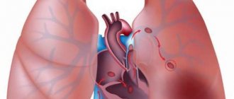
In addition to local manifestations, floating thrombi pose a serious danger if they break off. The resulting embolus from the venous network moves towards the lungs and blocks the movement of blood in the arteries. Depending on the diameter, pulmonary thromboembolism leads to the following consequences:
- sudden death (massive blockage);
- pulmonary hypertension (middle branches affected);
- respiratory failure and focal infarction-pneumonia (thrombus in small vessels).
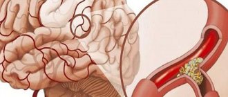
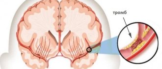
If the thrombus is located in the left atrium, then when it ruptures, parts of it enter the arterial network. They disrupt blood flow in the brain and internal organs with the development of such pathologies:
- ischemic stroke;
- heart attack;
- infarction of the kidneys, intestines;
- gangrene of the limbs.
Symptoms
It is very difficult to identify a floating thrombus by any obvious characteristic signs, since blood flow is restored on its own in most parts of the body. However, there are a number of symptoms that may indicate the presence of a floating thrombus in the bloodstream:
- Pain in the veins of the legs at the site of a possible thrombus. Moreover, the pain can be felt only upon palpation or be constant.
- Hyperemia of the skin in the area of possible location of the clot.
- Swelling of the sore leg, especially evident in the morning.
- Visualization of a diseased vein due to its distension.
- An increase in body temperature due to stagnation of a blood clot in one of the sections of the vein.
Important:
if you suspect the development of thrombophlebitis, it is advisable to seek advice from a vascular surgeon.
Diagnostics
To make an accurate diagnosis, the patient is prescribed a number of instrumental/hardware and laboratory tests:
Floating thrombosis: limbs, flotation, therapy, venous
A floating thrombus (floating) is a blood clot attached to the venous wall by a small base (head), the rest of the body moves freely in the cavity of the vessel.
When this type of blood clot forms, there is a high probability of its detachment.
A moving thrombus can enter any vital organ and cause its work to stop, which leads to death.
The appearance of a blood clot is caused by an increase in blood clotting as a result of certain pathological processes in the body. According to statistics, most often it forms in the vessels of the lower extremities.
About a floating thrombus
The size of floating thrombosis can reach 20 mm
Thrombosis is a disease characterized by impaired circulatory function and the formation of a blood clot in the vessels. As a result of slow blood flow, a layering of blood elements occurs. Increasing in size, the clot interferes with normal blood circulation.
Because of this, the organ tissues do not receive the required amount of nutrients. This affects its functioning. As a result of blood stagnation, necrosis develops, which is fraught with amputation of the organ and loss of capacity.
Floating thrombosis is characterized by its large size, which can reach 20 mm. The stalk with which the thrombus is attached to the vascular wall is too thin.
The tail of the thrombus is in constant motion due to the outflow of blood. At any moment, a blood clot can break off and cause a complete blockage of another vessel.
Symptoms of a floating thrombus
The peculiarity of the disease is the blurred clinical picture at the initial stages of development. This significantly slows down diagnosis. Most often, the disease is detected at the stage of development of complications. In this case, the symptoms become more pronounced. The following symptoms appear:
- swelling in the area where the blood clot is located;
- increase in local temperature;
- redness of the skin in the affected area;
- pain that intensifies during palpation;
- cyanotic spots on the body;
- bursting pain.
On a note! If a pathological process develops in the saphenous vein, almost immediately after the formation of a blood clot, a swelling appears, reminiscent of varicose veins. In this case, the lymph nodes also become enlarged.
Causes of a floating thrombus
Predisposing factors for the formation of a floating thrombus include cardiac dysfunction
The formation of a floating thrombus is associated with stagnant processes and impaired blood clotting function. Most often, the disease occurs against the background of other pathological processes. Predisposing factors include:
- lipid metabolism disorder;
- liver dysfunction;
- malignant or benign formations;
- venous diseases (thrombophlebitis, varicose veins, phlebitis);
- disruption of the heart (atrial fibrillation, cardiomyopathy, defects, etc.);
- infectious and autoimmune diseases.
Etiology and pathogenesis
The process of complete blood circulation in the body is ensured by the coagulation system. It performs a protective function, preventing excessive blood loss due to various injuries. Thrombosis develops when blood viscosity changes.
Along with this, the development of the disease is facilitated by damage to the endothelial layer of the vascular walls. Additional factors that create favorable conditions for the development of the disease include:
- mechanical damage (including during surgery);
- pregnancy and labor;
- old age (over 60 years);
- long-term use of oral contraceptives;
- limb compression syndrome;
- excess weight and sedentary lifestyle;
- forced limitation of motor activity.
Diagnostic methods
Ultrasound duplex scanning of veins will help identify the pathological process
Since the intensity of symptoms depends on the local location of the blood clot, it is impossible to identify the disease without diagnostic procedures. The main way to identify a pathological process is ultrasound duplex scanning of veins. This method helps determine the structure of the clot, its location and the condition of the vessel wall.
In some cases, MRI and X-rays are performed using a contrast agent. Of the laboratory tests, the most accurate data is provided by a blood test for the presence of D-dimer. This protein is formed as a result of the breakdown of fragments of a blood clot.
Treatment of a floating thrombus
Treatment for thrombus flotation is aimed at eliminating the danger to the patient's life. Therapy helps prevent the blood clot from moving further. The required result is achieved with an integrated approach. Treatment is carried out within the walls of a medical institution.
During therapy, the patient is in a supine position. If the thrombus is concentrated in a limb, then it is fixed in an elevated position.
Important! It is strictly forbidden to take medications without consulting a doctor. The dosage is selected only after testing.
Hirudotherapy
The main advantage of hirudotherapy is its natural composition.
Hirudotherapy is a method of alternative medicine that involves the use of a medicinal leech. The principle of operation of the procedure is the same as that of anticoagulants.
By sticking to the skin surface, the leech introduces a special substance into the human body - hirudin. It reduces blood clotting, preventing the formation of new blood clots.
The main advantage of hirudotherapy over synthetic drugs is its natural composition. The procedure does not have a negative impact on the functioning of internal organs.
Surgical intervention
Surgery is practiced if there is a high risk of clot rupture. The following techniques are used during the operation:
- vein suturing using endovascular catheter thromboextraction;
- vein extraction by thrombectomy;
- installation of a vena cava filter, which is a kind of trap for a blood clot;
- vascular bypass (the procedure of replacing a damaged vein with an implant).
Traditional methods of therapy
Onions and honey are useful for traditional treatment
Traditional methods of dissolving blood clots should be used with extreme caution. Traditional recipes cannot act as independent therapy. They complement conservative methods. It is recommended to use the following recipes:
- It is necessary to chop 2 onions and mix the resulting mass with 200 grams of honey. Then you should put the medicine in a dark place for 2 hours. Onion mixture should be taken 1 tbsp. before eating. It needs to be stored in the refrigerator.
- 2 tbsp. hop cones are poured into 500 ml of hot water. The medicine will be ready in a few hours, after infusion under the lid. It should be taken at night, half a glass.
- A mixture of garlic and lemon is valued for its ability to thin the blood and eliminate blood clots. The peeled head of garlic must be chopped. The lemon is crushed together with the peel. The resulting mixture is poured with a small amount of water and infused for 3 days. The medicine is taken up to 4 times a day, 30 minutes before meals.
Prevention of pathology
Floating can be prevented if measures are taken in time. A patient with increased coagulability in prophylactic treatments is prescribed Warfarin or its analogues. You also need to be aware of the following:
- To strengthen the structure of the vascular wall, the supply of vitamins C and K in the body should be monitored.
- If pain occurs in certain parts of the body, you should undergo a diagnostic examination. If the vascular pattern changes and the veins rise above the surface of the skin, it is necessary to visit a phlebologist.
- If you are overweight, you need to follow a diet to lose weight.
- If possible, you should stop taking oral contraceptives and drugs that affect blood clotting. If there is an urgent need for their use, regular blood donations are required to monitor clotting.
- If there is a high risk of developing thrombophlebitis, it is necessary to wear compression stockings.
- To normalize blood thickness, in some cases it is enough to establish a drinking regime. The recommended daily dose of water is 2 liters.
Forecast
You need to see a doctor in time
A floating thrombus is a rather dangerous phenomenon. With venous thrombosis of the lower extremities, there is a risk of a clot entering the pulmonary artery, which leads to death. Proper and timely treatment helps prevent complications.
The patient’s task is to see a doctor on time. The rest depends on the speed of diagnosis and compliance with medical recommendations. With the right approach, the patient becomes fully capable in 1-2 months.
Interesting! After completing the treatment course, the patient must periodically take anticoagulants to prevent relapses.
Danger of pathology
Complications develop if the patient does not take any measures. For example, with thromboembolism of the inferior vena cava, a clot can enter a pulmonary vessel, which will lead to respiratory arrest.
Damage to the coronary vessels provokes the development of heart attack, stroke and coronary heart disease. If a blood clot breaks off, it becomes life-threatening.
Once a floating thrombus is diagnosed, it is important to begin treatment promptly. The affected vein is removed or ligated.
In some cases, drug therapy has an adequate level of effectiveness. The speed of rehabilitation depends on the characteristics of the body and compliance with the recommendations prescribed by the doctor.
Source: https://flebolog.guru/tromboz/otlichiya-flotiruyushhego-tromba/
Floating thrombus: danger of pathology, causes and signs of thrombosis, treatment tactics
The normal functioning of the human body depends entirely on healthy blood circulation. Blood nourishes all organs and systems, moving from the heart through the arteries and returning back through the veins. However, the process can be disrupted by developing thrombosis. With this pathology, a blood clot forms in the lumen of the vein, which blocks the vessel and interferes with blood flow. A floating thrombus poses a particular danger to human health and life. That is, a thrombus that is not attached to the wall of the vessel, but moves freely along the bloodstream or is attached, but with the help of a very thin stalk that can break at any moment.
How to fix it correctly
If the presented pathology has been diagnosed, then treatment should begin immediately, otherwise serious consequences are possible. A blood clot can break off or block a vessel at any minute, causing gangrene, and treatment will be replaced by amputation of the limb. The patient should be hospitalized immediately. He needs to constantly be in a horizontal position, and the limb in which the thrombosis is located should be raised above the level of the body. This is necessary in order to maximize the outflow of blood from the affected limb.
There are three main methods of treating a floating thrombus:
- Medication.
- Instrumental.
- Surgical.
Drug treatment involves the use of thrombolytics, which dissolve blood clots. The most commonly used pharmaceutical products are Heparin and drugs based on it. They are inserted through a catheter directly into the vein in which the blood clot is located. However, these drugs have a number of side effects and can cause other undesirable symptoms, so treatment is carried out in a hospital under the supervision of specialists. Thrombolysis, as this procedure is called, is most often prescribed to treat thrombosis of the legs, arms and neck. This is because one of the side effects of the procedure is bleeding, and in the upper and lower extremities this is less dangerous.
Newer drugs for the treatment of thrombosis include Idraparinux Sodium and Fondaparinux. They have virtually no side effects, so treatment can continue at home. You need to take 1 tablet per day. Before use, you should familiarize yourself with the contraindications, otherwise you will provoke the appearance of other undesirable symptoms.
In drug treatment, drugs are used that dissolve blood clots.
In parallel with thrombolysis, the patient is prescribed decongestants aimed at normalizing blood flow. Anti-inflammatory medications are also prescribed that eliminate inflammation on the vascular walls and relieve pain. The instrumental method of treatment is considered safer. It involves the use of a vena cava filter. The procedure is reminiscent of fishing with a net: a filter similar to an umbrella is installed in the vein in which the blood clot is located, and it is removed from there after the blood clot gets into it. The filter is inserted through the superior vena cava or femoral vein.
The surgical treatment method involves bypassing the vein or artery in which the thrombus is floating. The procedure is called thrombectomy. After this, the patient must wear compression garments. After discharge, the patient must take anticoagulants throughout his life to avoid recurrence of the blood clot.
Diagnosis
Initially, the doctor listens to the patient’s complaints, based on the symptoms of the pathology, the patient is sent for diagnosis. In most cases, ultrasound is used to detect a floating thrombus. It allows you to determine both the mobility of the blood clot and its parameters. In addition, an x-ray may be prescribed. It is prescribed if the diagnosis needs confirmation. In particularly difficult cases, an MRI or CT scan is prescribed. High diagnostic accuracy is very important; the correctness of subsequent treatment depends on it.
There are four main methods for diagnosing floating thrombosis:
- Ultrasonography.
- X-ray using a contrast agent.
- Magnetic resonance imaging.
- Computed tomography – angiography.
After completing these tests, the patient is given a blood test for d-dimer. If this indicator is elevated, then a diagnosis of “floating thrombus” is made and treatment is prescribed.
Ultrasound is most often used to detect a blood clot.