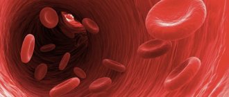
Typical symptoms of pulmonary embolism (PEA) include sudden shortness of breath or chest pain. PLE occurs when a blood clot breaks away from the vessel wall, migrates through the blood into the lungs, and causes partial or complete stenosis of the vessel.
Attention! In 90% of cases, the blood clot breaks off from a vein in the pelvis or leg. Less commonly, ELA is caused by parts of the tumor, amniotic fluid, air bubbles, or fatty plaques that enter the lungs through the bloodstream and “clog” the vessel.
What is pulmonary embolism?
In PLE, a blood vessel in the lungs is either partially or completely blocked. This occurs due to a blood clot (thrombosis) in the pelvic area or leg.
As a result, the part of the lung behind the “vascular plug” (embolus) stops working normally. To maintain blood circulation in the lungs, the chambers of the heart must pump blood harder. The pressure in the pulmonary vessels increases. If the pulmonary vessel is completely closed, it is not a pulmonary embolism, but a pulmonary infarction.
If a pulmonary embolism affects a small blood vessel in the lungs, minor symptoms may occur. But if the clot closes a large pulmonary vessel, it is life-threatening. In most cases, PEL is caused by thrombosis of the pelvis or leg.
Every year, 60 to 70 out of 100,000 people suffer from pulmonary embolism in Russia. The prognosis depends on various factors: the severity of the embolism, age, health status of the patient, and timely initiation of therapy.
Possible consequences
A broken blood clot can quickly enter the blood vessels leading to the brain, lungs, and heart. When it blocks the lumen of the vessel, an embolism is formed. Its consequences include:
- stroke;
- heart attack;
- necrosis of the intestines, lungs, etc.
This often happens so quickly that doctors do not have time to see the patient, and he dies.
Many people have probably come across the concepts of “thrombus” and “thrombosis” at least once in their lives, but not everyone has a correct idea about this phenomenon.
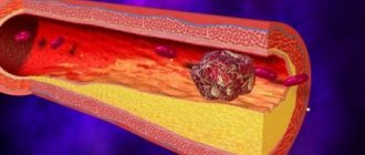
A thrombus is a pathological blood clot in a living organism, which is located in the cavity of the heart or the lumen of a blood vessel.
100% works! Varicose veins are removed by a cream made at home from three components - this is...
This is the only way VARICOSIS will disappear in 1 week! Do it every night at night...
Occurs due to impaired blood clotting function. For a blood clot to appear, the vessel wall must be damaged from the inside or have an atherosclerotic plaque.
The primary thrombus is fibrin filaments that are deposited on the altered vessel wall. Then thrombotic masses are applied to it, the clot grows. Upon reaching a critical size, the blood clot breaks off and blood flow stops.
There are three main reasons why a blood clot forms and, in many cases, breaks off:
- Damage to the vessel wall (mechanical trauma, inflammatory processes, damage to the inner wall by bacteria, toxins, viruses);
- Improper functioning of the blood clotting function (activation of coagulants and provoking platelet aggregation - joining to each other). This process is mainly associated with congenital abnormalities in the development of platelets, although sometimes changes occur at the chemical level (after exposure to bacterial, viral cells, taking certain medications);
- Slowing of blood circulation (associated with compression of arteries and veins, varicose veins, increased blood density).
Blood clots can form in any part of the circulatory system - in veins, arteries and even in the heart. The above reasons are applicable in every case.
However, there are also specific factors that affect only a certain part of the circulatory system.
A blood clot has broken off: symptoms, first signs
Complaints depend on the size of the clot and which vessels of the lung it blocks. Weak ELA does not cause symptoms.
If a blood clot breaks off, the first signs are:
- Sudden shortness of breath.
- Rapid breathing (tachypnea).
- Chest pain that increases with inspiration.
- Fainting.
- Cyanosis of the hands.
- Brain stroke.
- Cough (possibly with blood sedimentation).
- Anxiety (the patient may experience a panic attack).
- Hyperhidrosis.
- Accelerated heartbeat.
- Falling blood pressure (hypotension) and hemodynamic shock.
There are many signs that indicate ELA. Depending on the size of the occluded vessel, pulmonary embolism occurs without symptoms (in the capillaries), with obvious symptoms, or is instantly fatal. If ELA affects the larger pulmonary vessel, it affects the blood flow between the heart and lungs. Symptoms occur suddenly and can lead to cardiac arrest in severe cases.
Why does a blood clot break off?
Why does a blood clot break off and a person die? The starting point of a pulmonary embolism is a blocked vessel in the lower or upper extremities (thrombosis). Over time, the embolus breaks away from the vessel wall and circulates in the bloodstream. It passes through the inferior vena cava into the right chamber of the heart, and from there into the right or left artery.
A large embolus occludes a large vessel and causes severe symptoms. Regarding the time of day, pulmonary embolism often occurs in the morning, after a bowel movement, or with sudden physical stress. It is dangerous to create pressure in the vascular system (strong pressure during bowel movements), as it helps to dissolve the blood clot and transport the blood clot through the veins to the pulmonary arteries.
When a blood clot blocks a vessel, thrombosis develops. Often the thrombosis affects the leg or pelvic vein. Emboli are part of blood clots formed in the veins of the lower extremities. The term "embolus" is taken from the Greek word embole, which means "penetration."
Ebola does not dissolve in blood and can be solid, liquid or gaseous:
- Solid emboli: severed blood clots (the cause of 90% of all pulmonary emboli), tissue parts, parasites.
- Liquid emboli: fat droplets originating from destroyed body tissue (after a bone fracture).
- Gas emboli: air bubbles (with open damage to blood vessels).
The embolus travels with the bloodstream through the veins of the leg or pelvis through the inferior vena cava to the right chamber of the heart. From the right chamber it enters the pulmonary artery. From there it is transported to the right or left artery of the lung. A vessel may burst due to an embolus, but this condition is called a heart attack. The clot can cause expansion in the vessel. As a result, the vessel ruptures, causing internal bleeding.
Other types
In addition to the main classifications of blood clots, there are several separate types that are specific to certain groups of patients. These varieties include the following types:
- Mirantic. It occurs in weakened elderly people with long-term dehydration. The thrombus is localized mainly in the superficial veins.
- Tumorous. Formed as a result of metastasis, that is, the formation of secondary foci of a malignant tumor. Often such a clot gradually grows towards the right lobes of the heart.
- Septic. Occurs as a result of a local inflammatory process caused by infection. Localized in the veins and on the valve flaps of the heart.
Risk factors for thrombosis
There are two types of thrombosis risk factors:
- Exogenous (trauma, previous surgery, taking coagulants).
- Endogenous (congenital diseases, blood coagulation disorders).
Factors that increase risk:
- Pregnancy.
- Kidney failure with various symptoms (nephrotic syndrome).
- Stenting of the carotid arteries.
- Gender (men are more likely to suffer from ELA than young women).
- Phlebeurysm.
Factors that increase the risk moderately:
- Age over 60 years.
- Chronic heart failure.
- Heart attack in medical history.
- Obesity.
Factors that significantly increase the risk:
- Previous thrombosis or pulmonary embolism in the medical history.
- Blood poisoning (sepsis).
- Stroke with paralysis of an arm or leg.
- Thrombophlebitis.
- C-section.
- Seriously ill patients in the intensive care unit.
- Chronic obstructive pulmonary disease (COPD), requiring artificial ventilation.
The following circumstances increase the risk of thrombosis significantly:
- Therapy with female sex hormones.
- Certain drugs that block the action of sex hormones.
- Blood clotting disorders.
- Malignant diseases.
Causes
A wandering thrombus in the circulatory system
The following factors can provoke the formation of a wandering blood clot:
- passive lifestyle;
- consequences of surgery;
- chronic disorders in the gastrointestinal tract or cardiovascular system;
- being overweight;
- advanced age;
- genetic predisposition to this pathology;
- varicose veins;
- diabetes.
Also causes of a moving blood clot are:
- flow of blood circulation, provoking the separation of a blood clot;
- strong attachment of the fragment to the venous walls;
- excess of pharmacological drugs;
- improper use of compression stockings;
- active lifestyle during the rehabilitation period.
How is ELA diagnosed?
At the first stage, the doctor asks about complaints, examines the patient and examines the medical history. The doctor asks about the type and course of symptoms, about possible risk factors - previous thrombosis or pulmonary embolism.
A physical examination will provide important indications of PEL and assist in making the diagnosis:
- If the throat veins are noticeable, this means that blood is returning from the right heart to the veins.
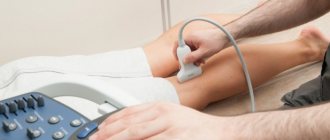
- At the same time, blood accumulates above the inferior vena cava towards the abdominal organs. This leads to swelling of the liver. Swelling is felt by the doctor during a physical examination or ultrasound examination.
- Examination of the legs is another important part of diagnosing pulmonary embolism. Blockage of deep veins is often the starting point of pulmonary embolism.
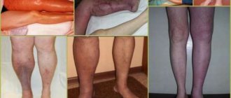
Typical signs of deep vein thrombosis (DVT):
- Edema.
- Pain.
- Muscle tension.
- Cyanosis.
- Increased visibility of superficial veins.
If complaints are detected, especially in a patient on bed rest, the diagnosis of pulmonary embolism is confirmed.
The doctor assesses the likelihood of a pulmonary embolism using the Wells-Score (named after physician Philip Wells). The score is based on seven parameters that the doctor determines through a physical examination and the patient's medical history.
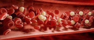
General blood analysis
Blood tests are an important part of diagnosing pulmonary embolism: laboratory tests show an increase in blood clotting. The focus is on D-dimers. These are blood clot fragments that occur when the body dissolves blood clots.
Important! Other diseases (tumor or specific coagulation disorders), trauma, surgery and certain drugs (furosemide) increase the concentration of D-dimers in the blood. The concentration of D-dimers in the blood increases during pregnancy.
Blood gas analysis (BGA) provides the doctor with information about the distribution of oxygen and carbon dioxide and the state of acid-base balance in the blood.
ECG and ultrasound examination of the heart
Recording of electrical activity (ECG) and ultrasound examination of the heart (echocardiography) visualize blood flow, rhythm, size and functional features of the heart.
Examination of the heart using ultrasound (echocardiography) helps the doctor evaluate the condition of the right side of the heart and measure blood pressure in the pulmonary vasculature. With a pulmonary embolism, the pressure in the affected vessel increases.
Echocardiography serves as a tool for differential diagnosis from other cardiac diseases that may cause symptoms similar to pulmonary embolism. Examples are a heart attack or a ruptured blood vessel (aortic rupture). The doctor uses cardiac ultrasound to make a diagnosis and to assess the patient's prognosis.
In addition to cardiac studies, the following imaging tests are especially important in diagnosing pulmonary embolism:
- Computed tomography (CT).
- X-ray examination.
- Lung scintigraphy.
Imaging procedures: Computed tomography (CT) may reveal vascular occlusion. Lung scintigraphy shows how well the lung is perfused. In addition to looking for a blood clot (embolus), it is important to find the original reason why the clot broke off.
If pulmonary embolism is suspected, action must be taken immediately. Only timely diagnosis and timely initiation of therapy improve the prospects for recovery.
Prevention measures
If a patient has a floating thrombus, complex treatment is necessary.
These may be conservative therapy measures, such as medications and other therapeutic procedures or various surgical methods.
If it is determined that the clot is floating, first of all, the actions of doctors should be aimed at preventing its further movement.
The set of therapeutic measures to eliminate the danger to the life and health of the patient is as follows:
- The patient is kept in bed around the clock, during which the limb affected by thrombosis must be fixed in an elevated position.
- The leg should be wrapped with an elastic bandage.
- Thrombosis should be treated in a medical facility, and patients are prescribed anticoagulants - blood thinning drugs. Such products, according to the instructions, have multiple side effects and contraindications. Heparin-based medications are considered the best. Heparin is injected into a vein or via a drip through a catheter. Subsequently, the patient is transferred to tablet-type anticoagulants, for example Warfarin, and often such therapy can be lifelong.
- In addition to anticoagulants, the treatment regimen includes taking anti-inflammatory drugs to prevent the risk of thrombophlebitis - inflammation of the venous wall.
- During the entire period of hospitalization, doctors monitor the rate of blood clotting by prescribing various tests: CBC, coagulogram, thrombocrit.
Sometimes a popular method such as hirudotherapy - the use of medicinal leeches - helps to successfully treat blood clots.
This method not only eliminates stagnation, but also helps reduce blood viscosity, since leech saliva contains hirudin, a natural anticoagulant with an anti-inflammatory effect.
To reduce your risk of developing blood clots, follow these guidelines:
- do not sit for a long time;
- walk more often;
- play sports;
- drink more fluids and stay hydrated;
- get checked regularly;
- do not drink alcohol or smoke tobacco.
Remember that your health is in your hands!
https://www.youtube.com/watch?v=lIFq3rUMTHg
What tests are there to detect atherosclerosis?
How to treat ELA?
First, the doctor will find out why the person’s blood clot breaks off, and then begin therapy. Since in 9 out of 10 cases the rupture of a blood clot (embolism) is the cause of pulmonary embolism, therapy is aimed at adjusting the hemostatic system. The ultimate goal of treatment is to prevent further embolism.
ELA can vary in severity. Depending on the degree of pulmonary embolism, appropriate treatment is prescribed.
First degree:
- Characteristics: The circulatory system is functioning without problems, and the right chamber of the heart is not damaged.
- First-line drugs: anticoagulants (coagulation inhibitors).
Second degree:
- Characteristics: The circulatory system functions correctly, but the function of the heart is impaired.
- The optimal treatment option is still unclear. Anticoagulant drugs are used (heparin and coumarin therapy).
Third degree:
- Characteristics: The patient has low blood pressure (hypotension) and the heart rate increases up to 100 times per minute (tachycardia).
- Treatment method: dissolving the blood clot with drugs (lysis therapy). Therapy is carried out with heparin and coumarins. Lysis therapy is not carried out only in case of absolute contraindications.
Fourth degree:
- Characteristics: cardiac arrest. The patient is in danger and must be resuscitated immediately.
- Therapy: Focuses on cardiac stimulation (cardiopulmonary resuscitation) for at least 60 minutes until the patient is stable.
- It is necessary to quickly release a clogged pulmonary vessel to restore blood circulation. Only in this way will a person survive.
Two drugs are especially important for the treatment of pulmonary embolism:
- Phenprocoumon is a vitamin K antagonist.
- Heparin.
Effective diagnostics
The best and most favorable option for diagnosis and treatment is to detect a blockage in the venous system of the lower extremities in the early stages of the disease, when a person feels pain and seeks help. It’s worse if the blood clot comes off while the patient is being treated in the hospital: the chances for timely detection of the pathology are much higher, but the risk to life is extremely high. A person has a minimum chance of survival if a blood clot bursts far from a medical facility.
In addition to assessing typical symptoms, it is necessary to quickly perform the following studies:
- duplex ultrasound scanning;
- angiographic examination;
- X-ray or computed tomography.
Laboratory tests in the context of primary care are ineffective: there is no need to wait for the result of a coagulogram analysis, so as not to waste time. The optimal type of diagnosis is endovascular methods, with which you can perform 2 main tasks - make an accurate diagnosis and eliminate the obstacle to blood flow.
Update: Why Wear Compression Stockings
Is it possible to save a person if a blood clot breaks off?
Pulmonary embolism occurs both during a hospital stay and at home. If a person suspects a pulmonary embolism, an ambulance should be called immediately.
If a pulmonary embolism is suspected, the affected person is given medication and mechanical ventilation. He is subsequently placed in a semi-sitting position and carefully transported to the clinic. Vibrations should be avoided as they may cause further embolism.
In cases of very severe pulmonary embolism, cardiac arrest may occur. Resuscitation includes cardiac massage and ventilation.