What is a blood clot in the leg
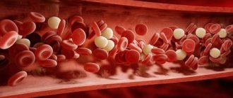
If blood clots form in the superficial veins of the lower extremities, it means thrombosis has occurred in the leg. As a rule, a blood clot completely or partially blocks the blood vessels, which can lead to extremely negative consequences. With venous thrombosis, blood does not move freely through the veins. Due to impaired outflow, stagnation occurs, accompanied by swelling and blue discoloration of the skin. A thrombus is a consequence of poor functioning of the anticoagulant system when it thickens.
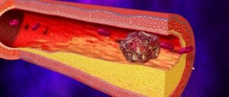
More often, clots appear in the area of damage to the vascular walls or in the area of localization of atherosclerotic plaques. Over time, fibrin threads are deposited there and microinflammation develops, which is the cause of the appearance of the first blood clot. The clot grows due to the layering of thrombotic masses. When there is an excess of them, blood flow stops in the lower limb.
Diagnostics
In order to identify a blood clot and check the vessels, several effective methods for diagnosing veins and arteries are used.
Medical examination
Before the external examination, the specialist interviews the patient. Clarifies the location of pain, determines the type of pain, collects anamnesis and indicates the individual characteristics of the patient.
The external examination also includes an indication of concomitant diseases that are provoking factors.
The initial examination is limited to superficial palpation and a presumptive diagnosis. However, a final diagnosis can be made by conducting a series of hardware studies.
Hardware diagnostics
The most accurate and effective diagnostic method used by phlebologists and vascular surgeons.
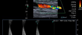
- Ultrasound of the intended area. Ultrasound diagnostics gives a complete picture of the pathological process, accurately describes the disorders and stages of the disease. But, despite its effectiveness, ultrasound is not a fundamental method of thrombosis.
- Angiography is a specific research method, the results of which provide a “verdict”. The method is radiography using a contrast agent. Using the resulting images, the doctor accurately determines the location of the clot and the degree of blockage of the veins (arteries).
- Duplex scanning is an equally effective method that allows you to identify the disease at the primary stage
- Dopplerography is a method for assessing the blood supply to organs and determining the degree of blood flow disturbance. Often used in conjunction with ultrasound examination.
To check blood vessels for patency, a modern method called electroencephalography is used.
This method is applicable to identify the pathological process in the vessels of the brain.
Laboratory research
- Biochemical blood test. The concentration of substances that aggravate the course of the pathology will be determined.
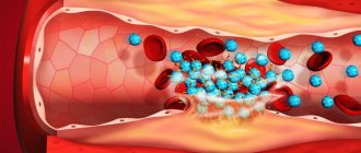
- General blood analysis. Necessary to detect possible inflammation, as well as to determine the level of hemoglobin and platelets. With thrombosis, hemoglobin and hematocrit are often increased and thrombocytosis is observed.
- Coagulogram is the main laboratory method. The purpose of the analysis is to determine blood clotting time, partial thromboplastin time activity, prothrombin index and fibrinogen concentration. Based on the data obtained, the clinical picture of the disease is supplemented and treatment tactics are selected. The analysis time is about 2 hours.
What causes a blood clot to break off in a person?
At first the clot is soft, but over time the structure becomes denser. Under the pressure of blood, the clot breaks away from the vascular wall, breaking into fragments. Some of them undergo destruction, others move to the organs, posing a serious danger to the further functioning of the body. Blood clots that form in large veins are especially dangerous. After tearing off, they migrate through the bloodstream, causing stroke, heart attack, pulmonary embolism, and death.
The risk group includes the following group of people:
- men over 40 years old;
- women during menopause;
- with obesity;
- with malnutrition;
- drinking large amounts of alcohol;
- with reduced activity (physical);
- pregnant women;
- after surgery on large joints or the abdominal cavity;
- coffee abusers;
- smokers;
- cancer patients;
- taking hormonal drugs.
Treatment and prevention of postpartum thrombosis
Correctly selected therapy will help to completely cope with this problem. It is important to remember that self-treatment and the use of alternative medicine in the postpartum period, as well as in pregnant women, are contraindicated. If you notice the symptoms described above, you should immediately visit a vascular surgeon or phlebologist. What does thrombosis look like on the legs? Photos are presented in the article. In the treatment of the disease, an integrated approach is used, including:
- Taking medications. The main anticoagulants are the heparin group, which are approved for use by nursing women. The use of non-steroidal anti-inflammatory drugs is indicated for severe thrombosis and only after stopping breastfeeding. Venotonics are also contraindicated when breastfeeding a baby.
- Wearing special underwear. Compression tights, stockings or knee socks help reduce the diameter of blood vessels, normalize and increase blood flow through the veins. As a result, venous congestion and the risk of pulmonary embolism are reduced. Your doctor will help you choose the right knitwear.
- Physiotherapy. Electrophoresis, laser therapy, cryo-wraps, and phonophoresis are effective in treating symptoms of leg thrombosis in the postpartum period. Massage, hot wraps, baths, compresses, and wave forms of physical therapy are contraindicated.
In order to exclude postpartum thrombosis, preventive measures should begin during the planning period of conception. If a woman in this position has varicose veins, then wearing compression garments and a special bandage will be mandatory. If this disease is detected in the second trimester, the use of venotonics - Detralex, Antistax, Phlebodia - is indicated. If a woman has high blood clotting, she is recommended to administer injectable anticoagulants, which significantly reduce the risk of a blood clot.
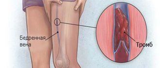
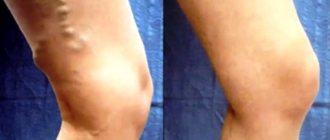
What does a blood clot look like?
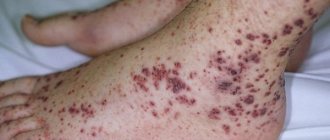
To see a blood clot in a vein in the leg, you need to carefully examine and feel the lower limbs. If redness or hardening is found in the area of the arteries, pain on palpation, then we can talk about thrombophlebitis. Sometimes the temperature in a compacted area increases significantly. Often, symptoms of a blood clot in the leg are not present at all, but the neoplasm is visually visible through small swelling and bluish areas, as in the photo.
Signs
After a blood clot forms in a vein, completely different symptoms may appear. Often the patient experiences pain in the calf muscles when flexing the feet, distension in the area of the affected vessels, inflammation of the knee joints, and severe heaviness in the legs, especially at the end of the day. Secondary symptoms are general malaise, weakness, high fever, swelling.
Bedridden patients often suffer from phlebothrombosis of the deep veins of the legs. Seeing a doctor after the first symptoms of a blood clot in the leg is a must. The doctor will evaluate the signs of pathology in the body and prescribe diagnostic procedures: ultrasound scanning and blood clotting tests. The treatment method is selected individually, depending on the type and location of the blood clot, and the presence of concomitant diseases.
Is it possible to cure deep vein thrombophlebitis?
As soon as the first symptoms of the disease appear, experts suggest the use of therapeutic therapy to eliminate them. Depending on the patient's medical history, treatment can be conservative or surgical. To make the right choice, you will need to get acquainted with the characteristics of the body (below photo and treatment ).
Measures that can significantly improve the patient’s condition:
- The affected limb should be at rest. It is better for the leg to be at some elevation in relation to the whole body.
- Anticoagulants are considered first aid drugs; they actively fight blood clots and prevent new blood clots from forming. To use such drugs, you need to have hemostasis indicators on hand. Their subsequent control is mandatory. Of the medications, doctors prefer Warfarin tablets, Fraxiparin injections and Heparin in various release forms.
- To obtain a high therapeutic effect, it is assumed to use a complex of various drugs. It is necessary to prescribe the patient pain relief capsules that relieve inflammation, antioxidants, antimicrobial agents, and vitamin formulations. Solutions for improving the rheological properties of blood are helpful.
- Prescribing Pentoxifylline will improve microcirculation.
- Thrombophlebitis of the lower extremities is a complex disease, therefore, to relieve pain and improve the quality of blood clotting, it is not enough to use drugs in the form of an ointment or gel. In addition, they always take pills and injections.
- After the above manipulations, a new stage of the treatment process begins. Compression garments are selected for the patient and the presence of active movements in his life is assumed. An elastic bandage helps relieve swelling in the problem area and reduce pain. The length is adjusted to suit individual needs, and compression is selected from 23 to 32 mm. rt. Art. If a patient has thrombophlebitis in the femoral vein, then buy long stockings, they reach the inguinal folds. If you plan to constantly wear such underwear, then the model will not depend on the type of disease. In this case, experts suggest compression underwear no higher than the knee joint.
Symptoms of a detached blood clot
How can you tell if a blood clot has broken off? Immediately after the separation of a dense clot, a person experiences an increase in heart rate and a decrease in blood pressure. The blood supply to the organs deteriorates, collapse occurs, accompanied by chest pain. Such symptoms are characteristic of myocardial infarction. The patient experiences urinary retention, difficulty pronouncing words, swallowing food, and sometimes the person loses consciousness. Due to the malfunction of the stomach and the fullness of the internal organs, abdominal pain is felt.
Lack of air and shortness of breath cause respiratory failure, which provokes cyanosis. Often, infarction pneumonia develops or pleurisy is detected, in which the body temperature increases. Sometimes the disease is accompanied by hemoptysis. If the blood clot is not treated, then after a while reactions of the immune system appear: a rash appears on the skin, reactive pleurisy develops, and the concentration of eosinophils in the blood increases.
Causes
The main cause of thrombosis of the arteries of the lower extremities is atherosclerosis - the lumen of the vessel is blocked by lipid deposits on the walls of the vessel. Leg injury and complications after surgery can also trigger the development of the disease.
Risk factors:
- heart disease: endocarditis, hypertension;
- vascular pathology: endarteritis, vasculitis;
- rheumatism;
- infectious diseases;
- endocrine pathology, including diabetes mellitus;
- performing invasive diagnostic or therapeutic procedures;
- blood pathology;
- excess weight;
- elderly age;
- being female;
- poor nutrition;
- inactive lifestyle.
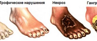
If a person suffering from diseases of the cardiovascular system experiences sharp pain in the legs, it is necessary to immediately seek medical help; this may be the first sign of artery blockage. Delay leads to the development of gangrene.
What to do if you have a blood clot in your leg
As soon as the first signs of a blood clot in the leg are noticed, the patient should immediately be provided with bed rest, complete rest and call an ambulance. It is impossible to predict the future fate of the patient, because sometimes death occurs within a few minutes. To save the patient, the doctor makes a decision based on the current situation. The location of the thrombus is important. If a person is managed to be taken to the hospital, the following measures will be taken to save his life:
- surgery to remove the stuck clot;
- installation of a venous vena cava filter, which is capable of intercepting a detached thrombus;
- injection of a large amount of anticoagulant into the vessel (Heparin is often used).
Although deep vein thrombosis is a disaster, blood clot rupture in the lower extremities is rare. For this to happen, three reasons must come together:
- Inflammation of the veins . Even the initial degree of varicose veins signals pathology. The presence of spider veins on the legs is already a mild inflammatory process. He needs timely therapy so as not to wait for a blood clot to form.
- Slowing blood flow . Occurs with a sedentary lifestyle. Without the work of the muscular system, there will be no normal tone of the venous walls. You don't have to do strength training or running. You need to walk regularly and learn to breathe from your stomach to help your blood circulation.
- Increased blood clotting . As a result of improper nutrition, blood viscosity increases and clots form. To liquefy, it is necessary to include in the diet foods such as beets, garlic, oatmeal, eggs, sunflower seeds, and sour milk products. In addition to a special diet, you can additionally take medications (Aspirin).
Treatment
Treatment of thrombosis of the arteries of the lower extremities is carried out in a hospital setting due to the need for rapid medical care and the high risk of developing tissue necrosis. Conservative therapy is possible in the initial stage of the disease (degree of ischemia IB and lower), as well as in the presence of contraindications to surgery. Arterial thrombosis is predominantly treated surgically .
Contraindications for surgery:
- age over 80 years;
- severe concomitant diseases;
- when gangrene begins (partial amputation is performed first).
Conservative treatment
Drug treatment for initial ischemia includes the use of the following groups of drugs:
- anticoagulants (Heparin, Warfarin);
- antispasmodics (No-shpa);
- thrombolytics (Streptokinase, Urokinase);
- antiplatelet agents (Aspirin, Curantil);
- means for improving tissue trophism (Reopoliglyukin).
Surgery
To perform an operation using a minimally invasive approach or close location of the blood clot to the surface of the skin, local anesthesia is used. In other cases, epidural anesthesia or general anesthesia is necessary. After restoration of vascular patency, blood pressure may drop sharply, so the presence of an anesthesiologist-resuscitator is mandatory.
The choice of surgical treatment method depends on the severity and neglect of the process. There are 4 treatment tactics:
- with zero degree of ischemia, surgical intervention (thrombectomy) can be postponed for up to 7 days, during which time the patient should be under the supervision of specialists and undergo examination;
- with ischemia of grade IA and IB, a delay of no more than 2 hours is possible;
- ischemia IIA and IIB requires a delay of no more than 1 hour; with such a degree of ischemic disorders, phlebotomy is necessary - an operation to restore blood flow and perform bloodletting, this allows the products of ischemic decay to be removed from the body;
- with degrees of ischemia IIIA and IIIB, urgent thrombectomy is indicated.
If there are irreversible changes in the tissue, amputation is indicated.
Emergency care for acute arterial thrombosis
If an acute disturbance of blood flow in the arteries of the lower limb is suspected, it is important to provide assistance to the patient as quickly as possible:
- call an ambulance;
- place the patient on a flat surface;
- fix the leg in a stationary position;
- apply cold to the affected leg;
- Give the patient Aspirin and No-shpa.
Consequences
Sometimes blood clots resolve on their own. This happens with a healthy diet, an active lifestyle, and abstinence from drinking alcohol and smoking. However, this does not happen immediately. Sometimes it takes several years to get rid of a blood clot. If the disease is not treated, poor circulation can lead to poor skin condition, dry mucous membranes, and autoimmune changes. As a result of insufficient nutrition of tissues, their death will gradually occur - gangrene will occur, which will lead to the loss of a limb.
At home
It is impossible to determine a blood clot without turning to specialists, since sometimes clinical signs are either absent or indicate another disease.
At home, one can only assume possible problems of the vascular system based on the characteristic symptoms: swelling, pain, discoloration of the limbs. The disease does not have any specific symptoms.
Thrombosis is a dangerous pathology, and determining it using unconventional methods can lead to serious complications.
In some cases, the patient risks losing a limb.
Treatment of the disease is carried out surgically, drug therapy is a preventive measure and a complement to the main treatment.