Ileofemoral thrombosis is a dangerous disease of the cardiovascular system caused by blockage of the femoral or iliac veins by blood clots. The cessation of blood circulation in the vessels of the lower extremities and pelvis leads to malnutrition and tissue swelling. Conditions are created for the development of gangrene of the leg.
Main symptoms
Thrombosis that has developed in the femoral or iliac veins is accompanied by symptoms such as:
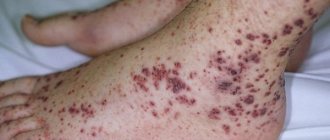
- small brown dots that protrude on the legs and do not disappear when pressed;
- increased body temperature;
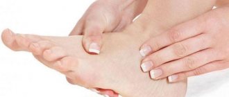
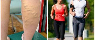
- severe swelling, which can be observed on both lower extremities or on one of them;
- purplish-red or bluish skin color;
- gradually increasing pain in one limb or both legs, as well as in the groin (sometimes).
Mechanisms of thrombus formation
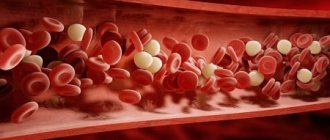
The cells of the inner lining of the veins (endothelium) participate in the formation of a blood clot. Their damage leads to increased release of interleukin, which is considered one of the platelet adhesive factors. When tissue is injured, excess thromboplastin enters the blood. He completes the formation of a blood clot.
As a rule, the wide part of the thrombus (the “head”) is fixed at the vein valve, and its “tail” can go away and fill all the large branches. It has been proven that in the first 4 days from the moment of formation, the strength of the parietal fixation is weak. It is during this period that rupture with the formation of an embolus is possible. After 6 days, inflammation of the vessel lining usually occurs and “solders” the blood clot to the wall.
The thrombus enters the femoral and common iliac vein in 89% of cases from the deep veins of the legs
In the area of the calf muscles, the venous vessels have small cavities in the wall (sural sinuses). They are filled with blood flow during muscle relaxation and open into deep veins during contraction. So, normally the muscular-venous pump is activated, pushing the blood to a higher level.
Any prolonged immobile and relaxed state of the patient (bed rest) causes stagnation of blood in the sinuses. This is where blood clots begin to form. The process is supported by the following factors that enhance coagulation.
Signs of acute thrombosis. Stages
Acute ileofemoral thrombosis has several stages of development. Depending on them, the symptoms of the disease may have their own characteristics. Let's look at them in more detail:
- Prodromal stage. At this stage of the disease, the patient experiences pain in various locations. They can be felt in the lumbosacral region, lower abdomen, and also in the affected area in the lower limb. The pain syndrome is aching and dull in nature. The patient also experiences an increase in body temperature.
- Stage of pronounced clinical symptoms. It is characterized by the manifestation of the classical triad. In other words, the patient experiences severe swelling, discoloration of the skin, and pain. By the way, the latter is felt in the calf muscle, groin area and anteromedial surface of the thigh. The nature of the pain is intense and diffuse. As for the swelling, it covers the entire area and spreads from the groin to the foot. Sometimes swelling is observed in the gluteal muscle and is combined with a feeling of fullness and a feeling of heaviness in the lower limb.
Due to the fact that edematous tissues exert strong pressure on the vessels, blood flow is disrupted, spasm occurs, and acute ischemia of the legs develops, expressed in sharp pain, lack of sensitivity and pulsation of the artery.
As mentioned above, one of the symptoms of the disease in question is a change in skin color. They may be pale or cyanotic in color. The white color occurs due to spasm of the accompanying arteries. This condition is accompanied by severe pain syndromes. A blue or cyanotic color is observed when the iliac or femoral artery does not perform its direct functions, that is, the outflow of blood through them is almost completely impaired. In this case, a very important symptom is that a pronounced pattern of saphenous veins is visible on the patient’s thigh.
In general, ileofemoral thrombosis is characterized by a satisfactory general condition of the patient. However, as an acute illness develops, it can worsen significantly. This indicates the onset of some serious complication.
Why is it dangerous?
Ileofemoral thrombosis is a dangerous pathological condition. In the absence of medical assistance, complications may develop:
- bleeding;
- pulmonary vein blockage;
- gangrene;
- death.
The prognosis for the patient is determined by the doctor, depending on the course of the disease, symptoms, effectiveness of therapy and the degree of damage to the veins and arteries.
The outcome of the disease depends on factors:
- If the patient visited the doctor in a timely manner, then complete recovery is observed in 75% of cases.
- If the patient ignored visiting the doctor and treatment. Once the process is started, chronic venous insufficiency develops.
Reasons for development
The human venous system can undergo various changes. Patients often experience deep vein thrombosis. This disease occurs due to slow blood flow and impaired blood clotting, as well as damage to the venous walls.
The development of ileofemoral thrombosis can occur in the presence of one factor or in the presence of several. Experts talk about the following triggers that contribute to the occurrence of this disease:
- getting injured;
- prolonged bed rest;
- DIC syndrome;
- infections of bacterial origin;
- period after childbirth;
- taking contraceptives;
- pregnancy;
- malignant and benign formations in the pelvis;
- aneurysms of the iliac and femoral arteries, as well as the abdominal aorta;
- popliteal cysts;
- retroperitoneal fibrosis;
- Iatrogenic deep vein injuries.
Prevention recommendations
To reduce the risk of developing ileofemoral thrombosis, doctors recommend leading an active lifestyle, eating right, and avoiding heavy physical activity.
Women should give up high-heeled shoes and switch to a comfortable model with a platform.
Simple rules for preventing relapse:
- constant wearing of compression garments;
- special exercises for legs;
- rejection of bad habits;
- taking venotonics and other medications strictly under the supervision of a doctor.
Ileofemoral thrombosis is a serious and dangerous condition.
Lack of medical assistance leads to complications that can lead to disability or even death.
At the first symptoms of the disease, you should visit a doctor for diagnosis and start timely treatment.
Diagnostics
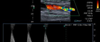
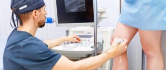
How is thrombosis of the lower extremities diagnosed? The symptoms of this disease are quite pronounced. But in addition to an external examination of the patient, doctors often resort to other diagnostic research methods, which are presented in the form of:
- fibrinogen scans;
- duplex scanning;
- radionuclide phlebography, which is carried out if the patient has intolerance to radiocontrast substances;
- descending or ascending radiopaque venography.
Why don't auxiliary vessels help?
Collateral or auxiliary veins in case of thrombosis of the femoral and mesenteric trunks are:
- superficial saphenous veins of the thigh and its tributaries;
- deep circles in the middle and lateral areas.
It is important that in the development of pathology of the ileofemoral veins, collateral circulation is insufficient. Since all additional veins, for various reasons, are not functionally prepared for the load, they are not able to provide the necessary blood flow.
The process proceeds more favorably if the thrombus spreads gradually from the peripheral veins to the center. Then some of the collaterals have time to start working. If the source of the thrombus is the great saphenous femoral vein and a transition to the common femoral vein occurs, then an acute blockage of blood flow occurs in the lower limb.
Treatment
Ileofemoral thrombosis should be treated in exactly the same way as other forms of thrombosis. Most often, treatment for this disease is carried out in a hospital setting. It is based on taking the following medications:
- anti-inflammatory drugs;
- antiplatelet agents;
- anticoagulants.
If the iliac or femoral artery is not severely affected, and the disease is in the initial stages of development, then techniques that help dissolve the blood clot can be used to treat it.
If there is a threat of thromboembolism, then its prevention is carried out. This procedure is carried out using ligation of the femoral vein, installation of a vena cava filter, or plication of the inferior vena cava.
If the patient has acute ileofemoral thrombosis, he is transported to the hospital. In this case, the patient should be in a lying position. Before the examination, he is prescribed bed rest, after which he undergoes an ultrasound scan and venography.
If there are no conditions for examining the patient, then under the supervision of specialists he is prescribed anticoagulants, which should be taken for ten days.
According to experts, acute venous thrombosis must be treated comprehensively, using three groups of drugs:
- thrombolytics or fibrinolytics;
- anticoagulants;
- disaggregants.
It should be noted that anticoagulant treatment is based on the use of the following agents:
- low molecular weight heparins;
- unfractionated heparins;
- fondaparinux pentasaccharide.
With the development of thrombolysis, which requires the use of streptokinase or urokinase, patient mortality increases significantly due to an increase in the frequency of bleeding. In this regard, this method is used only in extreme cases, for example, if widespread thrombosis developed very recently, that is, no more than a week ago.
Thrombolytic treatment for ileofemoral thrombosis is carried out after a vena cava filter is installed. This is due to the fact that such therapy promotes the transfer of a blood clot to the pulmonary artery, which can lead to the development of thromboembolism.
What did studies of surgical hospital patients show?
The fight against postoperative complications made it possible to study the process of thrombus formation in the deep veins of the lower extremities in operated patients over 40 years of age.
The method of monitoring the condition of the femoral and iliac veins was ultrasonic dynamic observation. Revealed results:
- thrombosis of the sural sinuses of the calf muscles began in 83.3% of patients already on the operating table;
- the sinuses expanded to 15 mm in diameter;
- in some cases, thrombus formation was observed in the area of the posterior tibial and small veins;
- in half of the observed patients, the period for complete formation of a blood clot was the first 7 days;
- 36.1% had it in the second week, and only 13.9% had thrombosis in the third week;
- in most cases (80% of patients), the blood clots dissolved spontaneously;
- in 1/5, thrombotic masses began to spread to the femoral, mesenteric veins and higher.
The group of patients studied included only those who underwent operations lasting more than an hour under general endotracheal anesthesia
Surgical intervention
For ileofemoral thrombosis, surgical intervention is an option. It is carried out according to vital signs and depends on the risk of developing pulmonary embolism.
It should also be noted that the operation is often performed when there is a threat of venous gangrene and when the pathological process has spread to the inferior vena cava.
It is also possible to remove a thrombus formed in the left iliac vein retrogradely. This procedure is carried out through a phlebotomy hole, which is made in the femoral vein. It should be noted that this option is not always possible, since the right iliac vein can exert significant pressure.
According to experts, an operation to remove blood clots should under no circumstances be performed when there is an adhesive process occurring in the lumen of the vein, as well as in the presence of intravascular partitions.
Complications
In the absence of timely medical care, as well as in the event of improper treatment, when symptoms are mistaken for the manifestation of another disease, a dangerous complication may develop - pulmonary embolism. In addition, acute ileofemoral phlebothrombosis in severe cases leads to gangrene of the lower limb and blockage of the inferior vena cava.
The prognosis for patients depends on the degree of vascular damage, clinical symptoms of the disease and the effectiveness of the therapy. With timely treatment, 6 months after the start of therapy, vascular patency is restored in most patients. If the disease is diagnosed in an advanced form, chronic venous insufficiency may develop.
Clinical forms of thrombosis
The onset and subsequent course of the disease most often occurs in two variants.
White painful phlegmasia or pseudoembolism occurs in cases of combination with spasm of the femoral artery or its branches. It is characterized by:
- sudden onset;
- throbbing nature of pain;
- coldness and numbness of the lower limb, reminiscent of an arterial embolism;
- rapid increase in swelling;
- limitation of sensitivity and movement in the toes;
- disappearance of pulsation on the dorsal artery of the foot.
Blue painful phlegmasia is formed due to acute complete blockade of all deep veins of the lower limb at the level of the mouth of the femoral or iliac vessels. Typical symptoms:
- very intense pain of a “tearing” nature;
- the leg is sharply increased in volume due to dense swelling;
- purple or almost black skin;
- large blisters with serous or bloody fluid appear;
- there is no pulsation in the arteries due to compression by edema.
In the severe stage, gangrene of the leg develops, symptoms of shock and increasing intoxication appear:
- inhibited consciousness;
- tachycardia;
- thready pulse;
- low blood pressure;
- body temperature rises.
Thrombosis at the level of the common mesenteric vein causes unclear peritoneal symptoms, rarely dynamic intestinal obstruction with attacks of stool and gas retention, spastic pain along the intestine.
What diseases are used for differential diagnosis?
You can also read: Thrombosis of the superficial veins of the lower extremities
Ileofemoral thrombosis has similar symptoms to a number of diseases. When examining a patient, surgeons must reject suspicion of the following processes:
- erysipelas;
- diseases of the arteries of a spastic nature;
- chronic lymphostasis (elephantiasis);
- damage to the calf muscles due to rupture of the tendons of the foot;
- pronounced cellulite;
- swelling due to heart or kidney disease;
- painful manifestations of polyneuritis, lumbosacral radiculitis.
Clinical manifestations
Symptoms of ileofemoral thrombosis include:
- The patient complains of pain on the front and inner surface of the thigh, in the groin, in the calf muscles;
- when combined with popliteal vein thrombosis, there is pain and limitation of movements in the knee joint.
Upon examination, the doctor discovers:
- increase in volume of the affected lower limb due to edema;
- swelling is widespread from the foot to the groin, and can spread to the gluteal region;
- an enhanced venous pattern on the thigh appears after 3 days, while the swelling decreases slightly (blood is “overloaded” into the superficial veins).
The skin color of the leg changes from pale to bluish
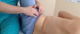
Palpation reveals maximum pain along the path of the femoral vein and in the groin area.