General information
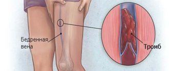
Phlebothrombosis of the lower extremities is an acute pathology that occurs against the background of blood clotting in the lumen of the vein, which creates a violation of its patency. It is important to distinguish between two concepts: phlebothrombosis and thrombophlebitis .
The latter is understood as an inflammatory process in the vascular wall as a result of local or general infectious damage. Phlebothrombosis is formed as a result of changes in blood coagulation parameters, damage to the vascular wall of the lower extremities and a decrease in blood flow speed. Phlebothrombosis is characterized by damage to the deep veins of the lower extremities. Acute phlebothrombosis affecting deep veins and thrombophlebitis of superficial veins occur in 10-20% of patients and are considered common diseases.
In 30-55% of cases, the disease is complicated by varicose veins of the lower extremities. Phlebothrombosis code according to ICD-10 is I82.9. Embolism and thrombosis of an unspecified vein.
Establishing diagnosis
Diagnosis of the disease is required to establish the affected area based on direct and indirect signs. To do this, the doctor performs an external examination and refers the patient to the appropriate examination.
- An examination by the attending physician allows you to clearly establish the fact of this disease. Upon external examination or examination of photographs of phlebothrombosis of the lower extremities, a change in the skin to a bluish or white color is noted, depending on the nature of the lesion of a particular vessel. The leg is edematous, swollen, sometimes ulcers and roughness of the epidermis can be seen;
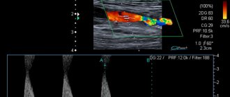
- Doppler ultrasound or duplex angioscanning can detect a specific vessel blocked by a thrombus. The monitor will show a narrowing of the lumen of the vein and stagnation of blood;
- Contrast radiography is an effective method that allows you to take pictures with the introduction of a special substance into a vein, which allows you to see the narrowing of its lumen;
- A blood test for formed elements and specific proteins is necessary to diagnose disorders of the qualitative composition of the blood, leading to the formation of blood clots.
As soon as the diagnosis of phlebothrombosis is confirmed, the patient is urgently hospitalized.
Methods for diagnosing pathology
Pathogenesis
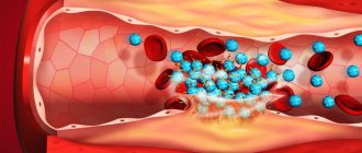
Thrombosis is normally considered a protective mechanism that is triggered in response to damage to the vascular wall in order to stop bleeding from the damaged vessel. With phlebothrombosis, blood clotting occurs in a completely healthy vein, which creates a significant obstacle to its normal functioning. Quite quickly, an inflammatory process begins to form in the wall of the blocked vessel, swelling gradually increases and nearby tissues are involved.
The deep veins of the lower extremities include the tibial veins, which are most often blocked by blood clots, the femoral and popliteal veins. The venous apparatus also includes valves located inside the venous lumen, due to which an obstacle to the reverse flow of blood is formed. The condition of the vein is also influenced by the sural sinuses, located in the muscles of the back of the leg. The sinuses are a kind of “lakes of blood” in the thickness of the muscle mass. During muscle contractions, they are drained (during running, walking), and it is from them that blood flows into the opening mouths of the deep veins.
Treatment
The most popular methods of therapy are following a diet and taking special medications. To enhance the effect, you can use folk recipes. In extreme cases, surgery is indicated.
Diet
No specific dietary regimen has been developed for this disease. Clinicians recommend adhering to certain rules that prevent the development of complications:
- Eliminate fatty, fried and flour foods from the diet;
- Eat fresh fruits - they contain antioxidants that prevent the formation of blood clots;
- Drink at least 2 liters of clean water per day;
- Stop drinking alcohol.
A very important point is to follow a diet when treating phlebothrombosis of the deep veins of the lower extremities.
Drug therapy
The use of medications is necessary to dissolve blood clots and prevent the formation of sediments. The following medications are indicated for this:
- Direct and indirect anticoagulants (Heparin, Antithrombin, Phenilin) – prevent a blood clot from forming;
- Drugs that improve blood viscosity (Curantil, Nicotinic acid, Clopidogrel);
- Preparations for dissolving blood clots (Hepatrombin, Proctosedyl);
- Anti-inflammatory ointments (Voltaren, Indovazin) - indicated for damage to superficial veins.
The use of medications is especially effective in the initial stages of phlebothrombosis - if the blood clots are small, they can be easily dissolved. As a result, the blood vessels are cleaned and the functions of the limb are restored.
Treatment methods for phlebothrombosis on the legs
Traditional methods
Traditional medicine recipes for thrombosis are used only during periods of remission and are aimed at preventing the formation of blood clots and cleaning the lumen of the veins. The most popular are rubbing the feet with apple cider vinegar, applying honey and propolis to the skin, as well as compresses with bodyaga solution.
Important!
You should not rely only on folk remedies in the treatment of thrombophlebitis. Treatment must be comprehensive. You should contact a phlebologist for professional medical help; he will determine a program of conservative therapy.
Classification
Today there are several classifications, but in the medical community only two are used: according to the stage of development of the pathological process and the location of the blocking blood clot.
Classification by stages:
- Acute phlebothrombosis . The duration of the period is 1-2 weeks from the moment of blood clotting and thrombus formation in the lumen of the vessel. It is at this stage that the greatest number of complications develop, which is explained by the high probability of the detachment of a fresh blood clot, which is weakly attached to the venous wall.
- Subacute phlebothrombosis . The duration of the period is 6-7 months. The severity of the pain syndrome is characterized by attenuation, but functional disturbances in the work of the lower extremities and their swelling remain.
- Chronic phlebothrombosis . The inflammatory process attenuates, but the formed venous insufficiency persists.
Surgical classification based on the site of thrombus formation:
- Phlebothrombosis of the veins of the leg . The deep veins in the lower leg are represented by 2-3 large vessels. If one vein is affected, others take over its functions. It is for this reason that diagnosing phlebothrombosis of the leg veins is difficult. For a long time, severe symptoms may be absent due to the compensatory capabilities of the veins. Most often, the disease is diagnosed when complications occur.
- Phlebothrombosis of the femoral vein . The severity of the pathology depends on the size of the blood clot that has closed the lumen of the vessel. With complete blockage, a rapidly increasing bluishness of the skin and severe swelling appear. On the affected limb, the subcutaneous venous network is clearly visible, severe pain appears, and the limb becomes hot to the touch. Clinical symptoms are less pronounced when the venous lumen is partially blocked by a thrombus: swelling increases gradually over several days and remains mild.
- Ileofemoral phlebothrombosis . Pathological changes are recorded not only in the lower extremities, but also in the lumbar region, gluteal region and groin. The lesion is characteristically unilateral. In all of these areas, pain, swelling and cyanosis are noted (the color of the skin may acquire a purple tint).
- Phlebothrombosis of the inferior vena cava . It is considered the most severe form, which is characterized by damage to both lower extremities at once.
Treatment options
Regardless of the cause of the manifestation, the symptoms of varicose veins of the lower extremities require complex treatment. Healing is possible only after procedures aimed at soldering (gluing) the affected veins; in severe cases, excision (removal) of the dilated vein is performed.
Phlebotonics and cooling ointments are prescribed for topical use. This course of treatment forces the vessels to contract, increasing their tone.
Phlebotics are used as a medicinal effect
Choosing a method according to the stages of the disease
The degree of development of the disease determines the method of treatment.
Latest information: Reticular varicose veins and ellipsoid - question 4488
In the initial stages, a “basic effect” is used, including phlebotropic medications that strengthen and tonify the venous walls. They can be prescribed in the form of tablets, ointments and gels. Doctors also recommend that the patient use class 1 compression support hosiery during periods of time that are dangerous for the veins:
- during increased physical activity and when putting stress on the legs;
- with forced prolonged sitting (long flights, sedentary work).
At later stages of development of varicose veins, basic treatment is used in combination with procedures that eliminate the expansion: laser coagulation, sclerotherapy. Large vessels are removed through surgery (phlebectomy).
A timely visit to a phlebologist will help avoid health problems
Drug and non-drug treatment
Treatment with medications is effective in the early stages of the disease and is also used as maintenance therapy for severe symptoms. Phlebotics are used to increase the tone of the walls of blood vessels (Deralex, Ginkor-Fort, Aescusan, etc.)
Non-drug treatment:
- Compression procedures. The therapy is effective for mild varicose veins and is used in addition to the treatment of more complex cases. Elastic bandage, workwear (knitwear). The principle of action is to weaken the manifestations of venous insufficiency.
- Sclerotherapy. The method has been known since the time of Hippocrates. Chemically active substances are injected into the vein, which, upon entering the vein, damage its internal walls with a chemical burn. As a result, the vessels “stick together” and become overgrown. Sclerosant substances (ethoxysclerol, fibrovein, thrombovar) are used for sclerotherapy. After administering the medicine, the leg is bandaged (or the patient is put on a compression stocking) to evenly distribute the fluid throughout the vascular system. The method does not protect against relapse.
- Operation Troyanov-Trendelenburg. The method involves removing the affected veins on the thighs. The leg area is bandaged, an incision is made, and the dilated veins are removed.
- Linton and Coquet method. The leg is incised in the calf area, the perforating veins are ligated under the fascia. Effective for post-thrombotic disease.
- Stripping. Removal of affected veins with a probe. The vein is dissected at both ends, a probe is inserted, and the vein is removed.
- Microphlebectomy. Removal of dilated veins is carried out using skin punctures, without incisions.
- Phlebectomy. An incision is made along the length of the vein for extraction.
- Radiofrequency and laser coagulation. The principle of operation is the treatment of varicose veins with radio frequency or laser radiation.
Surgery is a last resort in the treatment of varicose veins
The choice of treatment method depends on a number of factors:
- health status (comorbidities);
- the stage of development of the disease at which the patient presents;
- forms of the disease, symptoms, presence/absence of complications.
Each method has advantages and disadvantages: cosmetic, physiological, and associated with the risk of relapse.
Laser coagulation - a modern approach to the problem
Folk remedies
Let the grandmothers on the benches say that treatment of reticular varicose veins of the lower extremities is possible with folk remedies. It's up to you whether to believe it or not. But be prudent - if the disease manifests itself, do not expect that spells, ointments and masks from what is in the refrigerator will solve your problem. Use white magic and vegetable masks for your health, but before that, go to the doctor whose office door says “phlebologist.”
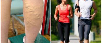
And if trouble has not yet touched you, engage in prevention. You need sports, quitting smoking and alcohol, healthy food. Swim, run, ride a bike.
Unfortunately, if you start the problem, the path through the six described stages to the surgeon's table will be completed very quickly. Take care of yourself, be healthy.
Causes
Diseases of the blood coagulation system, a sedentary lifestyle, pregnancy , as well as the recovery period after severe surgical interventions provoke stagnation of blood in the sural muscular sinuses, which creates all the conditions for thrombus formation. The formed thrombotic masses enter the venous lumen during muscle contraction. Once in the venous lumen, thrombotic masses either partially or completely close its lumen.
In response to blockage, a protective reaction is triggered in the form of an inflammatory process in the vascular wall, which negatively affects the course of the entire pathological process.
Factors provoking the development of phlebothrombosis:
- sedentary lifestyle, physical inactivity , the need to stay in one position for a long time;
- condition after extensive operations (especially abdominal operations);
- increased blood clotting, revealed by the results of a coagulogram ;
- carrying a pregnancy ;
- paraplegia ;
- condition after caesarean section;
- prolonged labor, complicated by abnormal position of the fetus;
- smoking;
- use of oral and hormonal contraceptives.
Etiology
The manifestation of symptoms of phlebopathy leads to congestion in the venous vessels. The reasons for this are:
- Prolonged stress on the venous circulation (influx or outflow of liquid mobile connective tissue). The risk of developing pathology of the venous vessels is directly related to excessive immobile sitting or prolonged stay on your feet.
- Hormonal imbalances. This applies to pregnant women or women during the period of declining ovarian function.
- Genetic predisposition.
- Excess body weight.
- New growths, adhesions, scars that appear that complicate hemodynamics.
- Injuries of large venous vessels.
According to studies, functional phlebopathy is observed in half of patients who are at risk associated with heredity.
Deterioration of the veins in the legs can occur during pregnancy.
There are 2 types of pathologies:
- Orthostatic. Manifested by deterioration of hemodynamics and overstretching of veins.
- Hormonal. The disease develops as a result of hormonal imbalance. Such changes appear while a woman is carrying a child, due to the use of certain medications.
Pathological changes occur under the influence of the active ingredients of oral contraceptives. Estrogen-containing medications provoke disruption of the nutrition of the inner lining of the venous vessels. The use of medications containing progesterone leads to the destruction of collagen and elastin fibers and, as a result, the thickness of the vascular walls decreases and the proliferation of connective fibers increases.
Symptoms
In some cases, the disease is practically asymptomatic. Conditions under which phlebothrombosis can be suspected:
- a sudden and rapidly growing feeling of fullness in one or both lower extremities;
- difficulty trying to relax the muscles of the thigh, leg, or foot;
- increasing feeling of heaviness in the lower limb;
- convulsions, involuntary twitching of the legs;
- severe swelling of the thigh, leg or foot (swelling can spread to the groin, buttocks);
- rapidly spreading appearance of spider veins;
- a pronounced venous pattern appearing under the skin (if this was not previously characteristic of the patient);
- pain in the popliteal fossa, aggravated by pressure;
- limitation of mobility in the joints of the legs.
Phlebopathy. What is this? Classification
Phlebopathy is a set of disorders of venous blood flow associated with impaired vascular tone. The disease is characterized by the presence of symptoms in the absence of damage to the walls of the veins. Phlebopathy of the lower extremities most often develops, but in some cases the retina of the eyes is affected.
There are several types of phlebopathy:
- hormone-dependent – associated with treatment with hormonal drugs and oral contraceptives;
- orthostatic – occurs in people who are in a forced position for a long time, this leads to disruption of the outflow of venous blood from the lower extremities;
- phlebopathy in pregnant women develops when the enlarged uterus puts pressure on the organs and blood vessels of the pelvis; after childbirth, signs of the disease disappear even without therapy;
- hereditary – caused by a genetic factor.
Tests and diagnostics
Phlebologists are involved in the diagnosis and treatment of the disease, but general practitioners - therapists, surgeons (including vascular surgeons) - can also suspect the pathology.
Main directions in diagnostics:
- General blood analysis . Deviation of the main indicators will indicate the presence of an inflammatory process in the body. platelet count indicates excessive clotting .
- Coagulogram . A change in indicators will indicate a pathology of the blood coagulation system; INR and APTT indicate the “intrinsic” and “extrinsic” pathways of thromboformation.
- D-dimer concentration . It is the most valuable diagnostic criterion, which indicates intravascular coagulation and the parallel process of resorption - thrombolysis.
- R-graphy of the chest organs . The examination is necessary for timely diagnosis of complications.
- Ultrasound , duplex scanning of veins . The study is considered preferable and makes it possible to determine the exact location of the thrombus, its size, the safety of the valvular apparatus of the vein, and the degree of occlusion of the vessel. In the acute form of the pathology, ultrasound cannot always show reliable information due to the low contrast of the newly formed thrombus compared to nearby adjacent tissues.
- Phlebography . An X-ray examination of the veins is performed with the addition of a contrast agent. The study allows you to accurately determine the degree of blockage of a vessel by a thrombus and identify venous obstruction.
- CT scan . Thanks to the study, it is possible to evaluate all the features of the venous network in real time.
- MRI . It is carried out when it is impossible to conduct a computed tomography (age, elderly patients, as well as pregnant women).
- Phlebotonometry . Shows the severity of valve changes in the venous system.
Diagnostics
Timely treatment of a dangerous condition is possible only after correct diagnosis. It is important not only to determine the degree of pathology, but also to determine the location of the blood clot.
If you suspect the development of deep vein pathology, consultation with a vascular surgeon is important. Based on a complex of studies, anamnesis data and analysis of the patient’s complaints, an accurate diagnosis is made.
A general blood test is taken from the finger. The laboratory technician counts the number of platelets, their clotting time, and the duration of bleeding.
Graphic determination of coagulability and thrombin generation test allow identifying thrombus-forming factors. An APTT (blood clotting indicator) and a biochemical blood test are also additionally prescribed. They can also assign:
- Doppler examination: lower leg, iliofemoral region.
- Ultrasound of the abdominal and pelvic organs.
- Venography.
- MRI, with a positive response to a biochemical blood test.
Treatment of phlebothrombosis with folk remedies
Folk remedies can only be used as an auxiliary component of the main therapy for mild, uncomplicated forms of thrombosis and thrombophlebitis . Before using unconventional methods, it is recommended to consult with your doctor.
Simple recipes for phlebothrombosis:
- Draw a bath and add an infusion of chamomile, string and St. John's wort at the rate of 500 ml of water - 2 tablespoons of herbal collection. This bath increases vascular tone and has an anti-inflammatory effect.
- Beat off a leaf of white cabbage, grease it with honey and secure it in the area of the changed veins with bandages. The procedure should be done before going to bed. It is advisable that the compress lasts all night.
- Mix 0.5 teaspoon of olive oil, 2 tablespoons of liquid honey. Apply the resulting mixture to the affected areas and cover with a gauze bandage. The compress should be kept for 6-8 hours. Can be left overnight.
The best remedies in the fight against blood clots are considered to be: olive oil, ginger, garlic, onions and flax seeds.
Treatment method
Phlebologists assure that treatment of phlebopathy of the lower extremities must be comprehensive, otherwise the improvements will be temporary. The most important task is to eliminate the root cause of the disease, therefore, before selecting a treatment regimen, it is necessary to undergo a comprehensive diagnosis.
In most cases, patients with phlebopathy are prescribed drug therapy:
- the use of external drugs with antithrombotic effects, which include heparin. The most popular medications in this pharmaceutical category are Lyoton-gel and heparin ointment;
- taking phleboprotectors. Medicines in this pharmaceutical category have an anti-inflammatory and analgesic effect, also help strengthen capillary walls and reduce excessive vascular permeability. Doctors advise patients to use Phlebolia, Detralex and Vasoket;
- taking vitamins.
If the disease is caused by an inactive lifestyle, it is recommended to introduce moderate physical activity. The minimum you need to do is exercise in the morning and take long walks in the fresh air several times a week.
Swimming will also help strengthen veins and normalize blood flow. The following are often prescribed as auxiliary methods:
- wearing compression garments. Medical knitwear will help to evenly distribute the load across the legs and reduce pressure (it can also be worn for preventive purposes);
- visiting exercise therapy;
- correction of diet. The patient must, if possible, eliminate “harmful” foods; the basis of the diet should now be vegetables and fresh fruits.
Reference! Surgical intervention for phlebopathy is prescribed extremely rarely; most often it is required if the patient sought professional help too late and the disease caused multiple complications.
First aid
If you suspect phlebothrombosis, you must call an ambulance, and before the team arrives, try to provide first aid yourself. The patient must be calmed and immobilized. Fibrinolysin and Heparin are administered parenterally . If the cardiovascular system is depressed, subcutaneous administration of Caffeine in a dose of 1 ml (10%), Cordiamin 2 ml is indicated.
Causes and risk factors for the development of phlebopathy
The main cause of this disease is a violation of the outflow of venous blood. An increase in the elasticity and distensibility of blood vessels leads to a loss of their anatomical shape. Reasons leading to this condition:
- long-term stress on blood flow;
- hormonal disorders;
- diabetes mellitus causes angioretinopathy, which provokes the development of retinal phlebopathy;
- heredity.
Long-term use of hormonal drugs causes the development of changes in the body, including in the blood vessels. The effect of estrogen is to increase the layers of the venous wall. Progesterone disrupts the functioning of elastin and collagen fibers. Also, under the influence of oral contraceptives, phleboslerosis is observed - the proliferation of connective tissue. This phenomenon complicates venous outflow.
Particular attention to the health of the veins of the lower extremities should be paid to people whose relatives suffer from phlebopathy. Half of patients with an established diagnosis have a hereditary predisposition.
Risk factors are identified, the presence of which significantly increases the likelihood of developing phlebopathy:
- nature of work activity: work associated with being in a forced position;
- sedentary lifestyle;
- changes in hormonal balance: pregnancy and menopause;
- pelvic neoplasms that impede venous outflow;
- diseases associated with impaired venous blood flow: varicose veins, venous insufficiency;
- excess weight;
- physical overload.
Most often, there is a combination of risk factors, so it is impossible to say what exactly triggered the disease in each case.
Consequences and complications
In most cases, complications of phlebothrombosis lead to life-threatening conditions.
- Phlegmasia . The entire lower limb suffers from circulatory failure, severe ischemia and subsequent necrosis .
- TELA . A dangerous condition caused by blockage of the lumen of the pulmonary artery by a detached blood clot. It is characterized by a sharp drop in blood pressure , the development of shortness of breath and loss of consciousness, including death.
Potential Complications
Phlebothrombosis can cause complications caused by prolonged disruption of the normal nutrition of blood vessels and tissues. Among the most dangerous complications, a violation of tissue trophism should be noted, which leads to gangrenous changes. Gangrene may lead to the need for lower limb amputation.
Another possible complication (and the most likely one, since it occurs in about half of cases of deep vein thrombosis) is post-thrombotic syndrome. This pathology is manifested by trophic ulcers and a severe form of chronic venous insufficiency.
However, the most dangerous complication of phlebothrombosis is a floating thrombus. Thromboembolism can cause death (if a blood clot blocks a pulmonary artery) or severe myocardial infarction.
The complications of deep vein thrombosis listed above are far from the only ones. Phlebothrombosis can lead to hypovolemic shock, hyperkalemia, myoglobinuria, coagulopathy and other serious consequences.
Prognosis and prevention
The prognosis is considered favorable with timely diagnosis, treatment and recovery. After eliminating the blood clot, blood flow and, accordingly, leg function are completely restored.
Prevention of phlebothrombosis involves timely completion of periodic medical examinations, visits to a surgeon and phlebologist if complaints arise.
Pregnant women must monitor their coagulation levels and wear compression garments (tights, stockings). Regular exercise and maintaining a healthy lifestyle can avoid the development of phlebothrombosis.
Prevention
There is no specific program developed, but clinicians offer several tips to prevent blood clots:
- Eliminate refractory fats from your diet, as well as smoked, salty or spicy foods,
- Lead an active lifestyle
- Give up bad habits.
If the patient follows these rules, the likelihood of developing pathology decreases.
Phlebothrombosis is a serious disease that leads to blockage of a vessel and disruption of the blood supply to local tissues. At the first signs of pathology, you should consult a doctor - timely treatment begins to guarantee a complete recovery.
Symptoms of phlebopathy of the lower extremities
With phlebopathy of the lower extremities, venous dilatation of the veins does not occur, the patient’s well-being may be normal, and no significant changes in health are felt. As the disease progresses, subjective symptoms appear; there may be no external or functional changes.
Exacerbation of the disease occurs more often at night or in the evening. Symptoms may worsen after a day of work. The following symptoms of phlebopathy of the lower extremities occur:
- heaviness in the legs;
- aching pain;
- fatigue even after minor exertion;
- swelling of the legs;
- seizures occur mainly at night.
With the hormonal type of phlebopathy, the first symptoms of the disease appear 2 months after starting to take the drug.
The symptoms are similar to those of venous insufficiency, but these conditions have a significant difference: with phlebopathy, the normal structure of the wall and the functioning of the vascular valves are preserved, and trophic changes in the skin are not observed.