Disorders of the veins and arteries are accompanied by severe ischemic changes in tissue. At least at the advanced stage of the pathological process. Urgent diagnosis is needed to detect deviations at an early stage. Restoration is carried out according to need.
Ultrasound of the vessels of the lower extremities is a technique for studying the arteries and veins of the legs. With its help, you can detect many pathological processes: from phlebitis to atherosclerosis and other disorders. This is an indispensable diagnostic method. Without it, there can be no quality treatment.
Unlike other methods, this one has many advantages: bloodlessness and complete safety at a minimum.
What do you need to know about the diagnostic method, when is it prescribed, and what results can be obtained? Let's take a closer look.
Ultrasound of veins - what is it
Ultrasound examination techniques mean a non-invasive intervention that allows one to evaluate the functioning of the valve apparatus, as well as superficial and deep veins. The effect is based on the principle of changing the length of ultrasonic waves, which are reflected with different deviations from the blood.
To record the received signals, sensitive sensors are used that transmit a signal or curve to the device. The method is used to diagnose diseases that occur in a latent form or early stages of pathology. During the tests, the doctor receives comprehensive data:
- gives an assessment of the general condition of the arteries and veins;
- identifies lesions in the vascular walls;
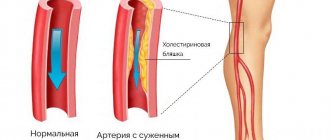
- diagnoses atherosclerotic lesions;
- determines areas of narrowing and stenosis;
- detects aneurysms;
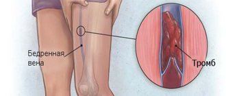
- detects thrombotic masses.
Ultrasound of the lower extremities is recommended for people with suspected varicose veins. During the manipulation, valvular insufficiency is assessed, blood clots are identified, and disturbances in the natural flow of the bloodstream are diagnosed.
The classical examination technique has disadvantages compared to triplex scanning. Ultrasound does not allow you to check the strength of blood flow and its volume. The diagnostic capabilities of the conventional technique do not allow obtaining data on the diameter of the arteries due to the lack of volumetric visualization. Triplex diagnostics have a higher cost.
Pros and cons of the study
Pros of the study:
- Ultrasound diagnosis of the condition of the vessels of the legs is a non-invasive method, the patient does not experience any discomfort during the procedure;
- Doppler is available to most patients due to its relatively low price;
- there is no harmful effect of ionizing radiation, so the study can be repeated as many times as necessary;
- Ultrasound waves can be reflected from soft tissues, due to which it is possible to obtain a complete picture of the state of the vascular system of the legs.
Disadvantages of the study:
- poor image of small arteries and veins during ultrasound examination;
- atherosclerotic plaques can interfere with the passage of sound waves, which greatly complicates diagnosis;
- Ultrasound cannot completely replace angiography, MRI or CT.
Indications
An ultrasound scan of the vessels of the lower extremities is often recommended by a phlebologist to his patients. Main indications for manipulation:
- complaints of periodic swelling in the feet and legs;
- systemic paresthesia;
- visual defects in the form of spider veins or swollen veins;
- change in skin color;
- cyanosis of the limb;
- prolonged wound healing;
- feeling of weakness;
- jumps in body temperature;
- convulsions.
Preparation stage
The ultrasound technique is simple and does not require preparation. The main rules recommended for compliance include:
- Refusal to use drugs. If possible, you should stop taking medications 3 days before starting treatment. It is important to exclude the effects of drugs that can affect blood flow. If you cannot avoid taking such medications, you should notify your doctor about their use. Such information will help adjust monitoring.
- Limited consumption of foods that affect the rhythm of the heart. Avoid drinking strong tea, coffee and other artificial stimulants.
- Quitting alcohol. It is better to avoid drinking alcoholic beverages a week before diagnosis.
- Smoking. The patient must not smoke for 3-4 hours before the intervention.
- Personal hygiene. It is better to remove hair from the legs, this will help get rid of discomfort during the process of moving the sensor.
The listed recommendations will improve the diagnostic efficiency of scanning.
How to prepare for the examination
Ultrasound examination of the veins and other vessels of the lower extremities does not provide for a separate set of preparation measures, and in emergency cases it is carried out without them at all.
For the purity of the examination, you should:
- stop taking medications, especially those that affect blood flow, three days before the procedure. If the drug belongs to the category of vital drugs, the doctor is informed about this, who makes appropriate adjustments to the examination process;
- limit daily food intake that can intensify cardiac contractility (coffee and tea, other stimulants);
- exclude alcohol consumption two days before, and smoking two to three hours before the event;
- In order to comply with hygiene standards, it is recommended to shave and wash your legs before an ultrasound scan.
Thus, preparing for an ultrasound scan of the arteries and veins of the lower extremities is an event that requires minimal time, but with significant health benefits.
Indications and contraindications
Traditional indications for ultrasound of the arteries and veins of the lower extremities are:
- swelling of the legs;
- manifestations of paresthesia;
- distinguishable structural deformations of the veins (“stars”, etc.);
- changes in the color of the epithelium, manifestations of cyanosis on the legs;
- long healing of wounds;
- undue fatigue or pain while walking (cases of intermittent claudication). Decreased temperature of the legs in comparison with similar other parts of the body;
- local cessation of hair restoration, the occurrence of itchy sensations in the legs.
Categorical contraindications to ultrasound of the legs:
- the occurrence of acute processes of an infectious nature;
- minor skin injuries (cuts, ulcerations, etc.);
- burns;
- severe conditions of patients;
- the presence of mental disorders in the subjects;
- acute emergency cases (heart attack, cerebral circulatory abnormalities, coronary circulatory deficiency, heart rhythm distortions, etc.).
At what age should you consider doing research?
In this context, the risk population includes people over 40 years of age, who are recommended to have an ultrasound scan of their legs annually.
At the same time, monitoring ultrasound of the arteries and veins of the lower extremities demonstrate a significant rejuvenation of the corresponding diseases and, in particular, atherosclerotic anomalies. Consequently, age parameters are not as relevant and sufficient as indicators of the need for ultrasound. It is important to carefully monitor the signs of possible vascular diseases of the legs and, if they are detected, you should immediately carry out the necessary diagnostics.
Quite significant arguments for doing an ultrasound of the venous vessels in the legs can be:
- diabetes mellitus (elderly diabetics are especially prone to thrombosis of not only superficial, but also deep vessels);
- high blood pressure;
- pain in the legs while walking and especially at rest;
- overweight;
- tobacco addiction;
- elevated levels of cholesterol in the blood;
- surgical intervention;
- edematous phenomena;
- leg cramps;
- vein pathologies;
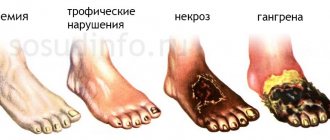
- trophic ulcers;
- obvious color changes in the skin of the legs;
- suffered heart attacks and strokes.
How is ultrasound of leg veins performed?
The examination position is determined by the doctor depending on the method and the nature of the location of the vessel being examined. Initially, the skin is lubricated with a conductive gel to eliminate background noise. Depending on the type of scan, the principle of intervention may differ.
Angiography
The procedure is performed using X-ray equipment. A contrast agent is injected into the veins being examined using a catheter. The patient is placed on the machine table and pictures are taken.
Attention! Angiography is performed using CT. This condition makes it possible to reduce the negative effects of X-ray radiation and increase the efficiency of the examination.
The duration of the procedure is 20 minutes. Angiography is performed in a hospital setting. After the procedure, the patient is given bed rest. The patient should remain under medical supervision due to the risk of a reaction to the injected substance. The images are transferred to a vascular surgeon, who forms a conclusion about the health status of the subject.
Dopplerography
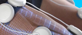
The patient removes clothing from the area being examined and lies down horizontally. A gel is used to ensure strong contact between the sensor and the skin. The doctor places sensors at the required points. The small popliteal and saphenous veins are examined in a standing position. A curve showing the indicators of the studied zones is displayed. Sometimes there is a need to clarify data. In this case, the right and left legs are examined simultaneously. The procedure lasts about 50 minutes, the patient receives results immediately.
Duplex scanning
No special preparation is required before performing ultrasound examination. The examination is carried out on an outpatient basis. Before the diagnosis, the patient removes clothing below the waist. When conducting a diagnosis, the sonologist may ask the patient to hold his breath or inhale. Such conditions are necessary to obtain the picture required for studying individual zones. At the final stage, the manipulation is performed while standing. The process is painless and takes no more than 40 minutes.
Triplex scanning
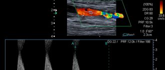
Triplex scanning is an improved variation of duplex examination. The technique is based on the Doppler mode. The ultrasound system uses frequencies to penetrate tissue and visualize an image of the vessel. To obtain accurate information, it is recommended to conduct a study with changing frequencies. To obtain accurate information about the nature of blood flow, color mapping is used.
Examination of the legs is carried out in a standing or lying position. The patient must remove clothing from the area being examined. The skin is lubricated with a gel that ensures contact between the sensor and the skin. The sensor is moved to different areas of the vessels and the device records all vibrations. After the procedure, the patient removes the remaining gel from the skin. The duration of the manipulation is no more than 60 minutes.
Preparation for the procedure and its implementation
No special preparation is required before Doppler ultrasound. The only thing the patient needs to do is perform hygiene procedures. Feet must be clean.
In the ultrasound room, you will have to take off your clothes so that the examiner can gain access to the extremities. The patient remains wearing underwear. It is advisable to bring a towel or diaper with you to lie on. They are not issued in all offices.
Having undressed, the patient lies face up on the couch, and special cuffs are put on the examined limbs to measure blood pressure. The doctor applies a transparent gel to the skin of the leg, which will improve the conductivity of ultrasound beams through soft tissue. Without this tool you will not get a clear image.
Then the process itself begins. The diagnostician moves a special sensor over the examined area, barely touching the skin or pressing harder on it. The doctor may ask the patient to hold their breath and strain very hard.
Duplex scanning is performed in different body positions. In horizontal (lying on your back, stomach, side) or vertical. This is necessary to obtain more accurate information. The doctor guides the process and tells you what position to take. When examining deep veins, as a rule, black and white Doppler ultrasonography or a color version is used.
The duration of the procedure depends on the severity of the condition and the goal. Usually it ranges from 30 minutes to an hour. Ultrasound is a completely painless examination. The only unpleasant sensation may be caused by applying cool gel to the skin. After completing the procedure, the product must be wiped off with a towel brought with you or provided in the office.
Decoding the results
After the ultrasound scan, a protocol is filled out, which includes important information. The main parameter is the ankle-brachial complex, which reflects the difference between ankle and brachial systolic pressure. The protocol data is presented in the table.
LPC indicators in the ultrasound protocol of the lower extremities
- norm – From 0.9;
- stenosis – 0.7 – 0.9;
- ischemia – 0.4 – 0.6;
- the risk of trophic ulcer formation is 0.3.
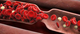
Blood flow velocity is presented as the difference in the ratio of the minimum and maximum speed for relaxation and tension.
| Blood flow speed (norms) | |
| Popliteal area | 50-75 |
| Arteries of the body of the foot | 25-50 |
| Tibia and posterior | 30-55 |
| General femoral | 70-100 |
In addition to the listed parameters, when using triplex scanning, a specialist may consider:
- main blood flow;
- turbulent blood flow;
- collateral blood flow.
To obtain accurate information, it is recommended to supplement the technique with color mapping.
Based on the data obtained, the doctor can make the following diagnoses:
- varicose veins;
- thrombophlebitis;
- phlebothrombosis;
- chronic heart failure;
- insufficiency of valves and blood vessels;
- various aortic diseases;
- Raynaud's syndrome;
- peripheral aneurysm.
If ultrasound data is insufficient, the patient is recommended to undergo a CT or MRI. Laboratory parameters can be assessed.
What does ultrasound scan help identify?
Doppler ultrasound is performed for both veins and arteries:
| Procedure | Which vessels are being viewed? | What characteristics and pathologies are visible |
| Doppler ultrasound of the veins of the lower extremities | Iliac, greater and lesser subcutaneous, popliteal, femoral, anterior tibial, posterior arch, perforator, sural, superficial circumflex iliac, dorsal plantar arch, inferior hollow. |
|
| Doppler ultrasound of the arteries of the lower extremities | All branches of large arteries, tibial, foot, femoral, iliac. |
This diagnostic method shows almost all possible changes in the condition of blood vessels, including deep ones. And, importantly, it allows you to track the speed of blood flow in the lower extremities.
Diseases that can be detected using Doppler ultrasound include:
- Varicose veins.
- Atherosclerosis.
- Deep vein thrombosis.
- Thrombophlebitis
- Other vascular changes in this location.
Contraindications
Ultrasound examination has no permanent contraindications. The patient’s poor health can affect the result of the examination, therefore it is worth rescheduling the diagnosis if there are signs of acute respiratory viral infection, high blood pressure and a significant deterioration in health.
Attention! Ultrasound diagnostics does not affect the patient’s well-being and does not aggravate the disease. The method is used to obtain information about violations.
The device does not provoke adverse reactions and does not emit radiation, therefore the use of the product is permitted by patients of different health groups.
Where is ultrasound of veins performed and how much does it cost?
An ultrasound examination allows you to determine the cause of the disorder and establish an accurate diagnosis. The cost of diagnostics may vary depending on the technique and accuracy of the ultrasound machine itself. The price of manipulation varies by region. In Moscow and St. Petersburg it ranges from 1,500 to 7,500 rubles. The exact price can be found directly at the diagnostic center.
Ultrasound examination of blood vessels is a simple and common examination technique that allows one to obtain accurate information about the condition of the arteries. This technique is recommended for patients with complaints of pain and heaviness in the legs. Phlebologists consider ultrasound scanning as a means of early prevention of varicose veins in people at risk, therefore they recommend undergoing the study once a year.