What are the main indications for the procedure? How do you need to prepare to do an ultrasound of the blood vessels of the legs? What can a diagnostic specialist see?
Author:
Dehissi-Golbrekht Muad Alfredovich
ultrasound diagnostic doctor, phlebologist
The lower limbs are an important element of the human musculoskeletal system, which can be susceptible to a number of diseases, pathologies, syndromes and altered conditions. Ultrasound of the vessels and veins of the legs allows us to identify individual or complex disorders of blood flow in the specified location and help in establishing the correct diagnosis.
What are the main indications for the procedure? How do you need to prepare to do an ultrasound of the blood vessels of the legs? What can a diagnostic specialist see? You will read about this and much more in our article.
In what cases is examination of the blood vessels of the legs carried out?
Vascular disorders are a fairly common occurrence. Swelling of the limbs, tingling, aching, numbness, pain are signs of incipient blood flow pathologies. Of course, such symptoms require careful verification using both x-rays (angiography and CT angiography), magnetic waves (venous MRI), and Doppler ultrasound examination.
The examinations are designed to check the extent of damage to the veins and arteries in the legs in the following diseases.
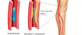
- Atherosclerosis of the arteries. Various research methods reveal how narrowed the vessels are and whether they are blocked by plaques.
- Diabetic foot syndrome, also called obliterating endarteritis. Inflammation of the spasmodic walls of small capillaries and arteries of the legs. They examine how deformed the walls of the vessels in the foot are and whether blood clots have formed in them.
- Such vascular pathologies in which protrusion of the arterial wall occurs: diverticulitis, dissecting aneurysm. Using hardware diagnostics, they look for the location and extent of the vessel wall dissection.
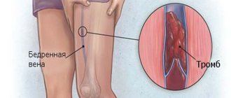
- Venous damage: thrombophlebitis, thrombosis. The study helps to identify the location of the blood clot and its size.
- Injuries of the lower extremities. The examination is carried out to determine whether compression of the vessels has occurred.
- Trophic ulcers and necrotic changes in the feet or legs.
- Instrumental diagnostics are also carried out to monitor the effectiveness of treatment before surgery or prescribing medications and after surgery or therapy.
Despite the fact that the indications for examining the veins and arteries of the legs are the same, the techniques used are different. It is important that any of the proposed types of examination will help detect damage in the veins or arteries of the legs.
CT or MRI of veins, angiography, Doppler ultrasound do not replace each other, but complement and clarify the research results.
How to treat veins and blood vessels?
For atherosclerosis
In the process of diagnosing the venous system, atherosclerosis can be diagnosed. With this pathology, atherosclerotic plaques form in the lumen of the vessels, preventing normal blood flow.
Atherosclerosis is dangerous because plaques can calcify over time and completely block the vascular lumen. In addition, lipid metabolism disorders increase the likelihood of developing thrombosis and thrombophlebitis.
In the early stages, it is customary to treat atherosclerosis conservatively. Patients are prescribed:
- Antiplatelet drugs - Aspirin, Reopoliglyukin, Aspirin Cardio.
- A nicotinic acid.
- Fibrates – Clofibrate, Bezofibrate.
- Bile acid sequestrants – Cholesteramine, Colestid.
- Statins – Atorvastatin, Lovastatin, Pravastatin.
Patients also need to follow a diet. You will have to significantly reduce the amount of fat you consume. The menu includes foods rich in fiber, Omega-3 and Omega-6 fatty acids, protein, zinc, B vitamins, and ascorbic acid.
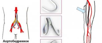
If conservative therapy is ineffective, then the disease is treated surgically. The most effective are vascular stenting, bypass surgery, balloon angioplasty, and prosthetics. After the operation, the patient needs to undergo a rehabilitation course.
With atherosclerosis, the prognosis is favorable. If treatment is started in a timely manner, the success rate of treatment measures will be 90%.
For thrombosis and thrombophlebitis
Vascular thrombosis often develops against the background of atherosclerosis or varicose veins, but sometimes the disease appears for other reasons. Thrombotic masses can be eliminated conservatively.
If the thrombus is small in size, then it is enough for the patient to take phlebotonics and blood-thinning medications. The drugs of choice are Troxevasin, Troxerutin, Aspirin, Escin, Ginkor Fort. To eliminate pain, non-steroidal anti-inflammatory drugs are prescribed - Ibuprofen, Nurofen, Diclofenac and others.
Latest information: Is walking beneficial for varicose veins in the legs?
If the blood clot is mobile or large in size, then surgery cannot be avoided. In such cases, thrombectomy is performed. During the operation, an incision is made in the area of the affected area, and the doctor removes the thrombotic masses. The procedure does not require long-term rehabilitation and is well tolerated by patients.
As for thrombophlebitis, it is also treated conservatively in the early stages. Patients are prescribed:
- Anticoagulants. These drugs prevent blood thickening, enhance the effect of fibrinolytic enzymes on blood clots, and prevent the development of various types of complications. The medications of choice are Antithrombin, Angiox, Warfarin, Viatromb, Heparin Sodium, Trombless, Fragmin.
- Fibrinolytics. Promote the resorption of thrombotic masses, prevent the development of acute thrombophlebitis. The drugs mainly used are Thrombofazim, Gemaza, Metalise, and Ukidan.
- Antiplatelet agents. They inhibit platelet activity, relieve the unpleasant symptoms of thrombophlebitis, and normalize blood circulation. The medications of choice are Zylt, Plagril, Trental, Clopidex.
If conservative measures are ineffective, thrombophlebitis is treated surgically. Crossectomy, catheter thrombectomy, or suturing of the pudendal veins of the leg are used.
Principle of angiography
Methods for assessing vascular disorders are divided into two types: invasive (with penetration into the patient’s body) and non-invasive. The first group includes the most accurate and informative research method - angiography. What is the principle of the study?
This method uses x-ray photography of the studied vessels into which a radiopaque substance has been injected. Angiography can examine arterial disorders (in this case the method is called arteriography) and the condition of the veins (phlebography). The examination is carried out in a hospital or medical centers equipped with an X-ray angiography room.
The research proceeds as follows. First, the leg area is anesthetized, an incision is made, and a thin tube, an introducer, is inserted into the vessel being examined. The catheter through which the contrast agent is supplied is connected to the introducer. The doctor injects an iodine-based substance into the vein or artery of the leg being studied. A series of x-rays are then taken as the substance spreads through the veins or arteries.
The manipulation is quite long, about an hour. After the examination, a tight bandage is applied to the incision site. Bed rest for 6–10 hours is recommended to prevent blood clots.
To quickly remove iodine from the body, it is recommended to drink at least one and a half liters of water.
Principles for deciphering results and normal indicators
The assessment of the venous bed does not have numerical values
. The sonologist analyzes the patency of the veins, the condition of the venous valves, the topography of the segment where the pathology was detected and the degree of blood flow disturbance.
Normally, ultrasound examination shows thin, smooth vascular walls, a lumen without foreign inclusions, and the valves oscillate rhythmically in rhythm with breathing. Blood flow is normally synchronized with the respiratory cycle.
Arterial blood flow has several parameters:
- The ankle-brachial ABI is the ratio of blood pressure at the ankle to blood pressure measured at the upper arm. The ABI normally should be 0.9 or higher. After loading the parameter increases. The lower the indicator, the worse the patency of the arteries in the leg. If the initial degree of stenosis is 0.9-0.7, then the critical degree is already 0.3.
- Peak blood flow velocity in the femoral artery is normally 100 cm/s, in the lower leg it is normal – 50 cm/s.
- The resistance index in the femoral artery exceeds 1 m/s.
- The pulsation index in the tibial artery exceeds 1.8 m/s. The smaller the last 2 indicators, the narrower the diameter of the vessel.
- Turbulent type of blood flow means that there is incomplete narrowing of the vessel.
- Trunk type is the norm.
- The main altered type means that there is stenosis above the area.
- Collateral blood flow is recorded below the area with complete absence of blood flow.
Thus, based on the study, the doctor can see how the veins and arteries are located, the degree of vascular patency, and the length of narrow segments.
The result of this study is a medical report on the uniformity of blood flow
, the nature of its change, which occurs due to narrowing and sometimes even blockage of the lumen, which can occur due to an atherosclerotic plaque or blood clot.
The compensatory capabilities of blood flow and pathologies of vascular structure are analyzed:
- the existence of tortuosity, aneurysms;
- degree of spasm severity;
- the possibility of compression of the artery by nearby scar tissue or, for example, spasmed muscles.
This video lecture tells more about the necessary equipment and deciphering the indicators (intended for specialists):
Indications and contraindications
Preparation for the procedure begins two weeks before it takes place. Alcohol is excluded, blood-thinning medications are suspended, general and biochemical blood tests, a coagulogram, and tests for infections (HIV, syphilis, hepatitis B and C) are taken. The day before the examination, an allergy test is performed to determine the tolerance of the radiocontrast agent. The intestines are cleansed the day before, since you will not be able to get up after the angiography. Half an hour before the test, the doctor gives intravenous injections of antihistamines and sedatives. The examination is carried out on an empty stomach, since when contrast is applied to the veins, sensations of heat and nausea are possible.
Contraindications to the procedure are:
- allergies to iodine or anesthetic drugs;
- pregnancy - due to the negative effects of x-ray radiation on the fetus;
- lactation period, because the contrast agent passes into breast milk in small doses;
- inflammation of the veins - due to the risk of blood clot rupture;
- low blood clotting – due to heavy bleeding after the procedure;
- increased blood clotting - due to the risk of blood clots;
- mental illness - angiography causes stress in most patients, and a person with a mental disorder will not be able to adequately respond to the doctor’s instructions and report his well-being.
Angiography is a relatively safe method. But in practice, there are cases of a serious allergic reaction to the contrast agent and injury to blood vessels. Less aggressive are angiography using computed tomography (CT) and magnetic resonance imaging (MRI).
Angiography is used more often as a simultaneous surgical treatment of blood vessels.
Preparation and progress of the procedure
Take with you, if available, a referral from your doctor and, if available, the results of other tests. There is no need to drink stimulants on the day of the study.
: alcohol, coffee, energy drinks, tea, do not smoke for 2 hours before the test, do not take medications.
You should not use painkillers and warm rubbing, or undergo physical exercise on this day.
It is necessary to remove clothing from the area being examined and lie on your back on a couch near the ultrasound scanner. The doctor will apply a contact gel to the skin, which improves the transmission of ultrasound waves. The doctor uses a sensor to take measurements at control points
, which correspond to the projection of the vessels under study.
Examination of the small saphenous and popliteal veins is carried out by asking the patient to stand or roll over onto his stomach.
Images of the areas under study are recorded on the monitor. It is possible that after the examination while lying down, the doctor will conduct the examination while standing. In some cases, measurements are taken on the right and left leg
to compare blood flow speed to obtain more in-depth information.
The study is equally informative for both large and small vessels, and for arterial and venous circulation.
The examination takes up to one hour, is absolutely painless and does not cause any discomfort. After the procedure is completed, the patient stands up and wipes off the gel. After 15 minutes
The results of the study will be formalized and handed over.
CT angiography and MRI angiography
Angiography can be performed using different devices. A more modern and gentle method is angiography with examination of blood vessels using a computed tomography (CT) scanner. Externally, the procedure resembles conventional angiography, only the contrast agent is administered not through a catheter, but with a syringe into a vein. The patient also lies on the tomograph table, he is fixed, connected to monitors and placed together with the table inside the scanning machine. A computer tomograph processes x-ray sections and displays a clear anatomy of the vessels of the legs in a three-dimensional image.
Another principle for studying the vessels of the lower extremities is used in MRI angiography. A person is placed in a magnetic resonance chamber and irradiated with radio waves in a magnetic field. The procedure is safe and painless, but has a number of contraindications, which include pregnancy, metal implants in the body, claustrophobia, and excessive body weight (over 135 kg).
If you compare the two methods, you get the following picture.
| Method | results | Advantages |
| CT angiography. | Allows you to see the size and location of not only blood vessels, but also atherosclerotic formations and blood clots. | Another advantage is the outpatient method of performing CT angiography and the absence of risks of surgical complications that are possible with conventional angiography. |
| MRI angiography. | The results of the study (images of blood vessels) are as accurate as possible. | The study avoids the radiation and injection of contrast agents used in other types of angiography. |
CT examination and MRI diagnostics do not require hospitalization. On the day of the procedure, the patient returns to active life. No special patient preparation is required before the MRI procedure.
MRI angiography is an alternative to vascular examination with a computed tomograph. However, diagnosticians note less clear tracking of aneurysms in the vessels during MRI examination.
Effective methods for diagnosing circulatory disorders in NK
How the vessels in the legs are checked depends on the location of the lesion.
Checking the arteries
Diagnosis of atherosclerosis and other diseases of the arteries of the legs requires an integrated approach. It is based on clinical, laboratory and instrumental data.
It should be noted that a significant part of vascular diseases of the legs are associated with the formation of cholesterol plaques in the endothelium, which, when growing, cause artery occlusion and ischemic changes in peripheral tissues.
Note! About half of patients with atherosclerosis of the lower extremities have no complaints. Often beginning pathological changes can be noticed only by the results of an instrumental examination.
During a physical examination, the specialist evaluates the existing signs of damage to the peripheral arteries:
- weakening/absence of pulse;
- systolic murmur when listening to the artery with a stethoscope;
- decreased blood pressure in the extremities;
- pale skin, bleeding of the finger under the nail plate.
The distal parts of the extremities are the first to suffer from atherosclerosis.
So, how to check the vessels of the lower extremities for patency?
Among the popular instrumental tests:
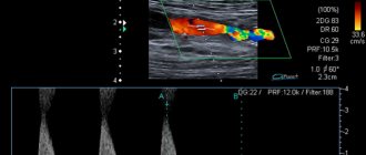
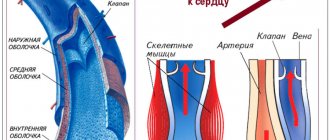
- Duplex ultrasound is a non-invasive examination method using color overlay. Typically, arterial flow is displayed in red, venous flow in blue. This allows you to assess the degree of vessel obliteration and the severity of stenosis.
- X-ray contrast angiography is one of the most accurate methods for diagnosing problems with the arteries, based on the introduction of a contrast agent into the vascular bed and the creation of a series of X-ray images. Allows you to reliably determine the localization of narrowing and the extent of the pathological process.
- Computed tomography is a more modern way of examining the arteries, allowing for a layer-by-layer image of the internal structures of the body. It is used primarily to assess the condition of the walls of the abdominal aorta, large iliac, and femoral arteries.
- MR angiography is a modern and highly accurate method for diagnosing vascular diseases, the distinctive feature of which is the absence of R-radiation. It is based on the effects of magnetic waves and computer processing of the data obtained.
Ultrasound (duplex, triplex) is a simple, safe and patient-friendly diagnostic method
Checking the veins
The study of veins is also based on characteristic symptoms, data from laboratory and instrumental tests. Moreover, in some cases the clinical picture turns out to be more informative. Thus, with varicose veins, the stage of the disease is determined based on typical symptoms.
Table: Stages of varicose veins of the legs:
| Stage (with photo) | Clinical manifestations |
| A person presents with complaints typical of varicose veins (heaviness, rumbling in the legs, night cramps), but it is not possible to identify the disease using instrumental and laboratory tests | |
| Spider veins and spider veins become noticeable on the legs. From time to time, patients complain of:
|
| Dilated vessels and venous nodules become noticeable under the skin of the leg or thigh. The patient's subjective complaints intensify. |
| The manifestations described above become more pronounced and are accompanied by swelling, which is especially noticeable in the afternoon. |
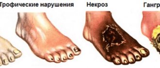
| Trophic (associated with a lack of O2 and yenhbtynjd supply) disorders are formed:
Ultimately, all the changes described above lead to the formation of a trophic ulcer. |
| The same + healed trophic ulcer. |
| Active, progressive, non-healing trophic ulcer. |
Important! Trophic ulcer is a dangerous condition that has a chronic progressive course. Without timely treatment, it can lead to limb loss.
How to check the vessels of the lower extremities using instrumental tests? Popular:
- Doppler ultrasound (USD) is a non-invasive diagnostic method based on the Doppler effect. After converting the sent and received sound waves, the doctor receives an image that allows them to evaluate the location, diameter, structure and tortuosity of the venous network.
- Angiography is used as an additional examination method.
Preparing for the study
One of the advantages of ultrasound examination is that it does not require any special preparation, except for observing banal rules of personal hygiene. However, it is advisable to observe the following rules:
- 60 minutes before the examination, avoid any significant physical activity, as it can affect vascular tone and change the condition.
- The day before the ultrasound, avoid drinking alcohol, and 30 minutes before the ultrasound, avoid smoking. These substances also affect vascular tone.
- If there is no risk to health, stop using antispasmodic (aminophylline, No-shpa, drotaverine, platifillin), anticoagulant (heparin, warfarin, syncumar), antiplatelet (aspirin) drugs, as they may affect the accuracy of the final results.
Ultrasound of the vessels of the legs should be postponed if there is cardiovascular pathology in the acute phase, acute infectious diseases, skin lesions of the lower extremities (extensive trophic ulcers, burns, injuries, purulent-inflammatory processes).
May be of interest:
|
Website about feet
Traditional Chinese Medicine: Recognizing Diseases by the FootFew of us have ever thought that if we put both feet together and look at them from above, their general features will look like a human figure. But the appearance of the legs can tell a lot about the state of health of their owner.
Thus, a large part of Chinese traditional medicine is based on recognizing diseases by external signs that can be seen in the soles of the feet. And this is not surprising - the human foot contains a huge number of nerve endings and vessels passing through the entire body.
Chinese healers offer a certain reading of the human figure based on the combined feet, or more precisely, based on the condition of the skin and blood vessels at a specific point on the foot. With a certain skill, this technique allows you to diagnose diseases of internal organs and possible health problems.
If you look at the combined legs from top to bottom, it is easy to notice that the big toes make up the head, and the transition from them to the little toes makes up the shoulders and other parts of the body.
Correspondence between parts of the foot and parts of the human body
Traditional Chinese practice suggests the following designation of internal organs and body parts on the foot.
- The head and front of the body are represented by the big toes and the front of the feet, where the lungs and heart are also located.
- The heart corresponds to the front left part of the foot, and the points of the foot associated with the eyes and our vision are located in the folds of the second and third toes.
- The outer part of the foot is part of the human silhouette - the shoulder, knee and elbow.
- The spine corresponds to the instep on the inside of the foot.
- The internal organs are the middle part of the foot where the organs of the digestive system are located, the bladder is located on the right leg, and the spleen is on the left.
- The pelvis and its internal organs are the heels.
- The frontal lobes and maxillary sinuses also have their correspondence on the foot - these are the pads of the nail phalanges of our fingers, with the left ones being on the left and the right ones on the right.
Thus, it is not difficult to build a cause-and-effect chain and draw conclusions about why we catch a cold or have heart problems immediately after acute pain in the leg or a limp.
To catch a cold or runny nose, it is enough to get your feet wet, and acute pain in the left foot serves as an immediate signal for heart problems. That is why, if you suddenly limp on your left leg for no reason, you should immediately consult a doctor, as this is the body’s warning reaction about an imminent heart attack.
Modern diagnostics of general health based on the feet
Modern traditional medicine also offers diagnosis of diseases based on the condition of the legs and, in particular, the manifestations of blood vessels and irritations on the skin.
Thus, poor condition of nails and various diseases of the nail plate - thickening and pores - indicate calcium deficiency and its poor absorption by the intestines. These same manifestations may also indicate high bone fragility, hidden osteoporosis and age-related changes in nails during menopause in women.
An excessive amount of fat on the feet and its formation, the appearance of all kinds of seals, bubbles and flagella are signals about a decrease in the performance of internal organs and a malfunction of the metabolism. The pits say the same thing - they appear in the corresponding areas of the foot as a result of the removal of internal organs or due to dysfunction of one of them.
Skin color and the condition of the skin of the leg are very important for modern diagnostics, so it is worth talking about them in more detail.
Diagnosis of diseases based on the condition of the skin on the feet
- Thyroid disorders and calcium deficiency mean cold, damp, often sweaty feet, regardless of the weather.
- Hypertension, impaired thermoregulatory function, atherosclerosis are dry, hot feet.
- Endocrine diseases, metabolic failure, dysbacteriosis - this is a uniform roughening of the skin over the entire area of the foot.
- Lack of vitamins A and B, exhaustion of strength, and nervous strain are reflected in the form of corns, dry and flaky skin with irritation.
- Greenish color of the skin of the feet indicates the presence of tumors in the body, if it is not caused by staining the skin as a result of wearing specific shoes.
- Yellowish tints are signs of poor liver function, and excessive pallor of the feet is characteristic of anemia.
- Purple hues at the base of the legs are a result of food poisoning and diabetes, while redness indicates fatigue and poor cardiovascular function.
- A purple tint to the thumb, if its cause is not a bruise or other mechanical impact, is a problem with blood circulation in the brain.
- Cobwebs of vessels appearing under the skin, their unhealthy coloring are problems of the internal organs corresponding to these parts of the foot.
Normal, healthy foot skin is a healthy white color with a red tint inside, which may make it appear pink. It should be smooth and matte, without calluses or inflammation.
Calluses and inflammatory processes on the foot are also evidence of poor health and have a certain interpretation in modern folk medicine.
Foot calluses are signs of diseases of internal organs
Depending on the location of the calluses and the degree of development of cuticulation of the epidermis (keratinization of the skin), it is determined which internal organ requires attention.
- Calluses above the heel at a biologically important point called kun-lun mean problems with the pituitary gland, adrenal glands, blood vessels and heart. They are defining indicators of blood pressure disorders.
- Calluses around the perimeter of the heels, so-called horseshoes, are a warning about changes in the joints.
- Calluses on the heel are associated with disorders of the rectum, possible hemorrhoids. In women, they give a signal that there are problems with the uterus.
- Calluses in the area of the thumb are osteochondrosis, a little lower are problems with the nasopharynx, and even lower are problems with the vocal cords and throat.
- Calluses on the pad in the area of the little finger tell smokers that it is time for them to stop this bad habit; for those who do not smoke, this is the need for gymnastics and attention to the bronchi and lungs.
- Calluses in the area under the little finger are both a sign of urinary incontinence and reverse retention, swelling.
- Calluses under the second finger are problems with blood flow, under the middle finger are possible inflammations of the lymphatic system, and under the fourth finger are prerequisites for respiratory diseases.
With proper understanding of the signals from our feet, our feet can serve as a visual medical guide to the health of our body. If we learn to promptly recognize changes in their appearance, we can prevent serious diseases and improve our overall health.