Vasoconstriction of the lower extremities, which is treated with surgical and conservative methods, occurs under the influence of many factors. The pathological condition contributes to disruption of blood flow, causing tissue ischemia. The disease is accompanied by pain and discoloration of the skin of the legs.
Diagnosis of vascular stenosis of the lower extremities abroad
In order to prevent stenosis of the blood vessels of the legs, doctors recommend that people over 45 years of age be examined annually in medical institutions. Diagnostic measures include blood tests for cholesterol, coagulation, glucose, medical history, and external examination of the patient. But to establish an accurate diagnosis, it is necessary to carry out the following procedures:
- Doppler study - helps measure blood pressure levels in the upper and lower extremities to compare the outflow of blood through them. This diagnostic method effectively identifies circulatory disorders, if present.
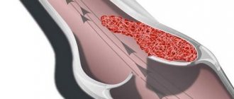
- Duplex scanning is a study using ultrasound equipment that allows us to judge stenosis or complete blockage (occlusion) of the lumens of blood vessels. The method is used before surgery.
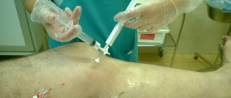
- Angiography is a diagnostic method that allows you to accurately determine the location of narrowing of the arteries in the leg. It is performed by injecting a radiopaque contrast agent into the blood vessels of the lower extremities.
- A coagulogram is a comprehensive laboratory test of a patient’s blood, done to assess the functioning of the blood coagulation system.
- An immunogram is necessary to check the state of the patient’s immune system. The weakening of the body's defenses leads to disruption of blood flow and subsequently becomes the cause of stenosing atherosclerosis of the lower extremities.
- Rheovasography is carried out to study the condition of the veins and arteries of the lower extremities. The method allows you to judge the existing changes in the stacks of blood vessels.
After studying the diagnostic results, the doctor will prescribe the correct course of treatment if the diagnosis of “stenosis of the blood vessels of the legs” is confirmed.
It is worth noting that there is no unique examination method; all diagnostic procedures have their own disadvantages and advantages.
Only an experienced neurologist, therapist or cardiologist can indicate what types of medical examination are necessary for a particular patient.
Diagnosing this disease is a complex task. It includes:
- collecting complaints and studying the history of the disease;
- objective inspection of systems. The doctor will evaluate how the heart, respiratory organs and kidneys are working;
- consultations with narrow specialists: neurologist, ophthalmologist, vascular surgeon, endocrinologist. This list will be individual for each patient;
- laboratory tests: general and biochemical blood test, hormonal panel, tests for autoimmune diseases, urine test;
- instrumental methods: ultrasound examination of blood vessels with Dopplerography, ultrasound of the heart, angiography, computed tomography and magnetic resonance imaging with and without contrast, electrocardiography.
To identify vascular stenosis of the lower extremities, the doctor performs:
interviewing the patient and taking a medical history;
examination of the patient to determine objective symptoms;
condition of the skin, veins in the legs, checks tendon and nerve reflexes,
Prescribes examinations to determine the degree of vasoconstriction:
- urine and blood analysis;
- coagulogram;
- lipid profile;
- immunogram;
- D-dimer determination;
- gas and acid composition of blood;
There is no universal diagnostic method. Each method has advantages and disadvantages, as well as contraindications. The required type of examinations and their scope are selected by the attending physician together with a neurologist and cardiologist from the list provided.
- tonometry;
- duplex (triplex) scanning;
- Doppler ultrasound;
- contrast angiography;
- ophthalmoscopy;
- CT (computed tomography);
- MSCT (multispiral CT);
- MRI (magnetic resonance imaging);
- infrared thermography;
- ECG (electrocardiography);
- ECG with a certain physical activity;
- EchoCG (echocardiography);
- bicycle ergometry, spiroergometry;
- REG (rheoencephalography);
- radionuclide scintigraphy
- EEG (electroencephalography).
The percentage of vasoconstriction is well determined using Doppler ultrasound. During the examination, a special drug test can be performed to determine the functional reserve of the vessel. Nitroglycerin is administered to cause vasospasm.
Contrast-enhanced tomography to establish the ankle-brachial index helps determine the extent of vascular damage.
Reasons for appearance
Atherosclerosis with stenosis of the arteries of the lower extremities occurs due to an imbalance of fat (lipid) metabolism in the body.
Factors that cause the appearance and growth of atherosclerotic plaques inside the vascular walls include:
- Addiction to tobacco and alcohol use. Smoking every cigarette causes spastic constriction of blood vessels, and alcoholic beverages increase blood viscosity and cholesterol deposition. If the patient suffers from both addictions, atherosclerosis and vascular stenosis can develop at a fairly early age.
- Heart and vascular diseases, high blood pressure. These diseases lead to disruption of blood flow and deterioration of the structure of arterial walls, which, in turn, contributes to the development of atherosclerotic stenosis.
- Low mobility in daily life. With physical inactivity, blood circulation throughout the body slows down, the lower extremities especially suffer. The blood supply to the calf region is increasingly limited, the structure of the blood vessels and the activity of the blood flow gradually deteriorate.
- Obesity and overweight. These factors lead to disruption of all metabolic processes in the body, especially lipid metabolism.
- Food errors. If you give preference to foods that cause the release of “bad” cholesterol: fried meats, animal fats and eggs, sweets and starchy foods, in adulthood you will have to treat stenosing atherosclerosis.
- Long stressful period. At this time, the vascular system works in enhanced mode, and if nervous tension has become permanent, vascular diseases may occur.
- Respectable age – from 50 years and older.
- Gender - according to medical data, the majority of those who presented with symptoms of stenosing vascular atherosclerosis are older or elderly men.
Symptoms of OASNK
A decrease in the venous lumen leads to impaired blood circulation. This becomes a consequence of many pathological conditions, nutrition of tissues and organs stops, and the general well-being of the patient worsens.
Limbs
- Pain in the leg at rest, depriving the patient of sleep;
- Pain or a feeling of fatigue in the leg muscles when walking (usually in the calf muscles) - this symptom is one of the early signs of atherosclerosis of the leg vessels;
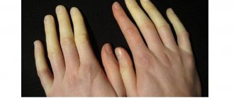
- Unusual feeling of chilliness and numbness in the foot, aggravated by physical activity (walking, climbing stairs;
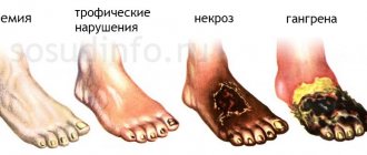
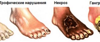
- The presence of a non-healing wound or trophic ulcer, usually located in the foot or lower third of the leg;
- Darkening of the skin, often in the form of dark brown or black necrosis of the toes (gangrene);
- Difference in skin temperature between extremities (the affected leg is cooler than the healthy leg).
Pain and other sensations
A typical localization of pain in circulatory disorders in the lower extremities caused by atherosclerosis is the calf muscles of the legs. This is due to the fact that they bear the heaviest load, and the number of arteries is minimal.
Therefore, blockage of any vessel causes pain in the legs. In the early stages, it appears only under heavy loads (running, long walking), and is accompanied by a feeling of chilliness and cold feet.
When atherosclerosis reaches a critical extent, not only the legs, but also the feet begin to hurt. Such pain is constant and is so severe that it cannot even be relieved with painkillers. Their presence is typical not only during exercise, but also at rest.
Intermittent claudication
”alt=””>
After walking a certain distance, a person is forced to stop and stand for several minutes until the muscles rest. After this, he continues to walk until the next attack of leg weakness.
This phenomenon is called intermittent claudication.
Degree of atherosclerosis
| Degree | Pain and weakness in legs when walking | Trophic disorders |
| First | Only under heavy loads, walking more than 1 km | Externally, the legs and feet do not change |
| Second | From 200–300 m to 1 km | Pale skin, impaired hair growth |
| Third | From 50 to 200–300 m | Hypotrophy, trophic ulcers |
| Fourth | Less than 50 m and at rest | Gangrene of a finger or foot |
Pay attention to the following symptoms in yourself or your loved ones:
- fatigue in the calf muscles or thigh muscles when walking 500 meters or less,
- a feeling of leaden heaviness in the legs or muscle pain that makes you stop when walking,
- changes in the skin on the legs in the form of peeling, thinning, hair loss.
These are signs of atherosclerotic damage to the arteries of the lower extremities. An examination by a vascular surgeon is required in the near future. Do not be surprised if, when diagnosing lesions of the arteries of the lower extremities, your carotid arteries are checked and a cardiological examination is prescribed. Atherosclerosis is a systemic disease, and, as a rule, different groups of blood vessels are affected.
- Sympathectomy (intersection of the nerve plexuses responsible for spasm (narrowing) of the arteries) is performed for repeated blockages of the arteries and in addition to reconstructive operations. This operation improves blood circulation in the extremities by dilating small arteries.
- Revascularization osteotomy is also an adjuvant technique that improves blood circulation by stimulating the formation of new small vessels in the lower extremities after bone injury.
- Arterialization of the venous bed is rarely used at this time, since its implementation is associated with various technical difficulties, and long-term results are worse than the methods described above.
Make an appointment by phone or on the website.
Critical arterial stenosis. Three pathologies
Obliterating atherosclerosis of the vessels of the lower extremities
This is a disease in which atherosclerotic plaques form on the inner wall of the arteries of the lower extremities, reducing its lumen. The initial signs may be pain in the calf muscles when walking (intermittent claudication), first decreasing at rest, and then constant (including night pain in the legs and feet), cramps in the legs. In advanced cases, non-healing ulcers form from lack of blood circulation, and it is even possible to develop gangrene of the fingers and toes, which requires amputation at one level or another.
The diagnosis is made on the basis of a survey, examination and ultrasound examination of blood vessels, as well as angiography - the introduction into the bloodstream of radiopaque substances that move along with the blood and show at what level there is an obstacle to its flow.
Treatment is conservative and surgical. Stenting and bypass operations are performed.
Blockage of arteries supplying the brain
In this case, an ischemic stroke occurs, that is, an acute cerebrovascular accident. Diagnosis and treatment are carried out only in an inpatient setting by a neurologist.
Stroke is an acute circulatory disorder in the brain, which manifests itself as cerebral or focal symptoms. There are two types - ischemic (when a vessel supplying any part of the brain becomes clogged) and hemorrhagic (the vessel bursts and a hematoma forms in the brain, compressing it).
Accordingly, symptoms depend on the extent of brain damage and the functions for which one or another part of it is responsible.
A. General cerebral symptoms. A person may experience dizziness, weakness, headache, impaired consciousness, accompanied by nausea and vomiting, disorientation in place, time or personality. Looks a lot like someone who has had too much to drink, doesn’t it?
b. Focal symptoms. The clinical picture is again determined by the zone in which the pathological process occurs. Impaired or complete loss of vision, hearing or smell, weakness in an arm or leg (on the same side) up to paralysis, inability to move, unsteadiness of gait. In addition, there are speech and swallowing disorders.
V. How to recognize a stroke? The diagnosis must be made by an experienced specialist - a neurologist, using the full power of modern diagnostic equipment. It is enough for you to know the simplest skills that will allow you to suspect this diagnosis. And the UZP technique will help with this: U - ask the person to smile . And puff out your cheeks. During a stroke, there will be a noticeable asymmetry in the corners of the mouth, and one of the cheeks may “sail.” Z - talk . Speech disturbances are common. However, the absence of this does not mean the absence of a stroke. P - raise both hands up or squeeze your hands at the same time. If there is an asymmetry in actions or a decrease in strength on one side, then this speaks in favor of this diagnosis.
If you have any of these symptoms, call 911 immediately This can save a person's life!
Cardiac ischemia
With a gradual narrowing of the heart arteries by more than 50% of the diameter, pain begins to appear, called angina pectoris. As a rule, they appear against the background of physical activity or stress, when the heart beats faster and needs more blood and nutrients, and clogged blood vessels can no longer pass them in the required quantity. These pains, as a rule, are relieved with nitroglycerin and other nitrates - drugs that can dilate blood vessels, primarily cardiac ones.
With further progression of the disease against the background of heavy load, ischemic necrosis of the heart muscle may begin - myocardial infarction. This condition, as a rule, is already irreversible, and the task of medical workers is only to reduce the area of myocardial damage. That is why, if heart pain does not go away after several nitroglycerin tablets, then it is necessary to call an ambulance. Doctors will take an ECG and perform a troponin test - which will show whether there is a heart attack or not.
As a rule, in all these cases the initial condition is high cholesterol in the blood - hypercholesterolemia. There is a whole group of medications - statins - that reduce the content of “bad” cholesterol in the blood. Consult your doctor about the need to take them, the choice of drug and dosage.
But let's talk a little about nutrition. I don’t like prescribing treatment on the Internet, but diet therapy is a completely different matter. Surely every person in our country already knows that there is “good” cholesterol and “bad” cholesterol.
“Good” is the one that is involved in the production of vital steroid hormones (for example, testosterone), bile acids and vitamin D. “Bad” is the one that is deposited on the walls of blood vessels, reducing their lumen and leading to the development of such unpleasant diseases such as myocardial infarction and gangrene of the lower extremities. Of course, this is a very figurative division; in reality everything is much more complicated. But still.
There are a fairly large number of medications that lower blood cholesterol. But some reduction can be achieved with therapeutic nutrition. Here are three main foods for this: 1. Avocado It contains a large amount of phytosterols (alcohols of plant origin), which reduce the amount of “bad” cholesterol and increase the level of “good” cholesterol. In addition to avocados, fresh strawberries, cranberries, chokeberries and raspberries have similar properties. 2. Cereals and legumes The addition of these crops has an extremely beneficial effect on all aspects of human health. For example, in addition to defeating hypercholesterolemia, they will help normalize the functioning of the digestive system. 3. Seafood Not all seafood is suitable for defeating “bad” cholesterol. Nutritionists recommend eating more lean fish such as sardines and red salmon. Don't forget to take a natural statin - fish oil.
Symptoms and danger of the disease
A decrease in the venous lumen leads to impaired blood circulation. This becomes a consequence of many pathological conditions, nutrition of tissues and organs stops, and the general well-being of the patient worsens.
Limbs
Vasoconstriction in the extremities does not immediately show symptoms. First of all, pain occurs in the legs when walking, when resting, especially noticeable in the area of the legs and calves. Numbness and convulsions occur.
Further, lameness, atrophy of the lower muscles, ulcers, and cracks in the feet may occur. In the case of an open wound, proper cell regeneration does not occur due to poor blood supply to the tissue. As a result, the wound begins to rot. This is dangerous due to the development of gangrene, which can lead to amputation of the leg.
Brain
The narrowing of the blood vessels in the brain causes insufficient supply of oxygen and nutrients to it. At the initial stage it manifests itself with symptoms such as:
- migraine;
- dizziness;
- nausea;
- increased irritability;
- memory impairment.
As the disease progresses, it manifests itself with symptoms such as loss of consciousness, changes in gait, and loss of coordination. If treatment is not provided, the person loses the ability to move independently and signs of dementia appear.
Heart
The causes of narrowing and spasms of the blood flow of the heart are mainly a congenital defect of the arteries and organ walls, atherosclerotic plaques, and blood pressure disorders. The disease manifests itself with the following symptoms:
- chest pain (which can radiate to the left shoulder blade and arm);
- disturbances in heart rhythm;
- rapid heartbeat.
When the condition worsens, if you bring your palm to the heart area, you may notice a slight trembling of the chest. If the attack lasts more than half an hour, is accompanied by numbness of the left half of the body, a noticeable deterioration in vision, there is a possibility of acute myocardial infarction.
Prevention and diet
To prevent the risk of developing the disease, it is important to follow the following rules:
- Perform special physical exercises every day (running, squats, race walking, etc.).
- From time to time, conduct medical examinations of the body.
- Eat healthy foods that do not contain animal fats. Fruits, vegetables, fish, lean meat are the best foods for stenosis of the leg arteries.
- Complete cessation of addictions (drugs, tobacco products, alcohol).
Vasoconstriction of the lower extremities, which is treated with surgical and conservative methods, occurs under the influence of many factors. The pathological condition contributes to disruption of blood flow, causing tissue ischemia. The disease is accompanied by pain and discoloration of the skin of the legs.
Treatment methods
The choice of therapy for stenosis of the leg arteries depends on the stage of the disease. At the initial stage, the patient is prescribed medications that will help get rid of pain in the limbs, dilate the walls of blood vessels, and improve blood circulation. But drug therapy, even in the first stages of stenosis, will be ineffective if you do not give up bad habits, unhealthy diet and change your lifestyle for the better.
When a patient, along with narrowing of the walls of blood vessels in the legs, is diagnosed with vascular heart disease, pathologies of internal organs or hypertension, doctors recommend a special diet (table No. 10). The goal of this diet is to reduce blood cholesterol levels. Also, everyone who suffers from atherosclerosis of the lower extremities should devote time to physical therapy, develop a love of sports, swim in the pool, and take daily walks.
Physiotherapy for leg vascular stenosis
There are the following types of physiotherapeutic procedures aimed at improving the health of the body with vascular stenosis of the lower extremities:
- Therapeutic gymnastics is effective in the initial stages of pathology. Special exercises strengthen the muscle tissue of blood vessels.
- Laser therapy is used in the last stages of stenosis development.
- Electrophoresis.
- Healing massage with special oils helps improve blood flow.
Drug therapy
Successful treatment of blood vessel stenosis requires the mandatory use of medications. Recommended medications include:
- Antispasmodics - improve blood circulation (Papaverine, Drotaverine, No-shpa, Eufillin).
- Disaggregants and anticoagulants - fight the formation of blood clots in the body (Heparin, Aspirin, Curantil).
- To stabilize blood pressure, antihypertensive drugs (Atenolol, Lisinopril, Bisoprolol) are prescribed.
- Diuretics - remove excess fluid from the body (Furosemide, Veroshpiron).
- Vitamin-mineral complex (ascorbic acid, preparations containing magnesium, potassium, phosphorus).
- To normalize blood cholesterol levels - lipid-lowering drugs (Zocor, Crestor).
- When stenosis is accompanied by an inflammatory process, you cannot do without taking anti-inflammatory tablets.
Surgery
Existing methods of surgical intervention include:
- Endarterectomy is the removal of atherosclerotic plaques from the affected vessels. Before the procedure, medications that improve blood clotting are prescribed.
- Bypass - placing a bypass will improve blood circulation.
- Prosthetics - the affected area of the artery is replaced with an artificial vessel (prosthesis).
- Thrombectomy - aimed at cutting out the formed blood clot. The blood clots are thoroughly crushed and then removed from the affected vessel.
Folk remedies
Stenosis of the arteries of the lower extremities requires immediate treatment. Non-traditional methods of treating pathology are effective if combined with medications and physiotherapeutic procedures. Traditional healing recipes designed to strengthen blood vessels and relieve stenosing atherosclerosis include:
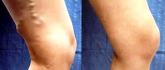
- Parsley compress - helps to improve the health of the veins in the legs. To prepare, you need to finely chop the parsley, pour boiling water (150 ml), then let the broth brew for 20 minutes, then strain and dilute with milk (proportion – 1 to 2). The resulting product is used to make compresses that should be applied to sore legs.
- Hawthorn tincture - cleanses blood vessels, strengthens the walls of veins and arteries. The product is easy to prepare: add 20 grams of hawthorn fruit to a glass of water, bring to a boil, leave for 10 minutes. The medicine is taken 3 times a day before meals.
- An excellent remedy for combating stenosis is obtained from a mixture of lemon, garlic and honey. These products improve blood circulation and prevent the accumulation of cholesterol in the blood.
general description
Obliterating (occlusive) atherosclerosis of the lower extremities (I 70.2) is a disease in which atherosclerotic plaques form and grow in the lumen of vessels, namely arteries, during life, completely or partially blocking the lumen of the vessel and disrupting blood circulation in the tissues.
This disease is more common among men over 40 years of age. The main reason for the development of this pathology is a violation of cholesterol metabolism, namely an imbalance between lipoprotein fractions, which leads to the gradual deposition of cholesterol into the vessel wall.
There are a number of factors leading to the occurrence of this pathology:
- Smoking is the main factor that accelerates the progression of the disease several times.
- Increased cholesterol levels (disorder of cholesterol metabolism).
- High blood pressure (untreated hypertension).
- Overweight. Accompanied by a violation of cholesterol metabolism.
- The presence of a hereditary predisposition.
- Diabetes. Complicates the course of the disease.
Treatment of pathology
Treatment of stenosis directly depends on the severity of the pathology and can be:
- medicinal;
- surgical.
Medicines that help cope with stenosis are quite varied and their use is individual, since vascular pathology is often accompanied by other ailments. Among the most commonly used medications by doctors are the following:
- disaggregates, that is, a group of drugs that prevent the formation of clots, blood clots and thickening in stagnation of blood flow, simple aspirin is usually used if there are no contraindications to it;
- rheological drugs, usually in the form of intravenous injections or drip solutions, the most commonly used are pentoxifylline and rheopolyglucin;
- drugs from the group of anticoagulants are prescribed for a large number of blood clots and a direct threat of vessel rupture; heparin or its analogues are usually used;
- thrombolytics are used for large blood stagnations, and a serious level of areas with thickening, as a rule, an injection of a solution of streptokinase or actilis is used.
Drug therapy must be supported by an appropriate diet and abstinence from smoking, alcohol, coffee and other substances that cause constriction and spasms in blood vessels.
Surgical treatment of stenosis can also be different; the type of intervention required is determined by the condition and type of pathology.
Open surgery and aortofemoral replacement
These operations are performed for severe and advanced stenosis. Part of the vessel lining or the entire vessel is removed; for long areas affected by stenosis, bypass surgery is used, that is, the replacement of vessels with vascular prostheses.
Leg vascular bypass surgery
Palliative surgical interventions
They are performed when there are pronounced local signs of stenosis, without a large extent and not particularly severe pathology.
These types of interventions include laser correction or laser perforation of the vessel, revascularizing osteotrepanation of the vessel and a number of other manipulations.
Among the preventive measures, folk recipes for foot baths with herbs, teas, rubdowns and compresses are very good:
- for tea you need to brew a mixture of hawthorn, rose hips, hops - these herbs prevent blood thickening, having a positive effect on blood flow;
- for foot baths there is nothing better than nettle and hops, these herbs very well stimulate all processes in the tissues of the legs, activate and strengthen blood vessels, they need to be taken at least half an hour and daily;
- rubdowns and compresses are best for a sedentary lifestyle; steamed viburnum leaves, infusions of dandelion, motherwort and mint are ideal for them.
These methods will never replace treatment if stenosis has already begun, but if there is a hereditary tendency to it, they can prevent narrowing of blood vessels and the development of pathology.
Stages of development
Stenosing vascular atherosclerosis develops gradually. In medical science, there are 4 stages (stages) of disease development, each of which has its own characteristics and characteristics.
Stage I – the patient practically does not feel dangerous symptoms. This stage is more characterized by echo signs, that is, those that can be detected by hardware diagnostics. As a rule, these are fatty streaks or spots on the inner surface of the vessel wall.
Stage II - the patient begins to experience difficulty walking for a long time, pain occurs when covering a distance of two hundred meters.
Stage III - accompanied by deterioration in the sensitivity of the legs, increased pain that bothers you even at night. The patient cannot walk 50 meters without experiencing pain.
Stage IV – characterized by a high degree of stenosis of the lumen of blood vessels. Accompanied by serious disorders of tissue trophism, the appearance of trophic ulcers, and sometimes tissue necrosis (gangrene). With this development of the disease, patients, as a rule, practically do not get out of bed and do not leave their homes.
Narrowing of blood vessels in the legs: causes
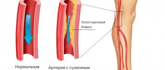
Stenosis is a pathology of the cardiovascular system, often affecting the vessels of the legs. Narrowing of the arteries and veins of the lower extremities is formed due to deposits formed inside the vascular bed. In the vast majority of clinical cases, deposits are formed due to increased concentrations of lipids in the blood plasma. An excess of these fat-like organic compounds forms fat accumulations on the vascular walls. The most famous lipid is cholesterol. By combining with calcium, which plays a structural role, and connective tissue substances, cholesterol is converted into cholesterol plaques. Therefore, stenosis, despite its local nature, is considered a manifestation of atherosclerosis.
Narrowing of the blood vessels in the legs also occurs for a number of other reasons, independent of disorders in the body’s fat metabolism. A common cause of stenosis affecting the vessels of the leg is considered to be genetics. A hereditary feature, namely a predisposition to a particular disease, manifests itself depending on the accumulation of pathological genes. As soon as their interaction with environmental factors of varying strength reaches its maximum, a person notices signs of a disease that was previously diagnosed in his close relatives.
Many other pathological processes and diseases are quite capable of causing narrowing of the arteries and veins of the legs. Among them:
- hypertension (disturbs normal blood circulation);
- thyrotoxicosis and diabetes mellitus of any type (provoke metabolic disorders);
- arteritis, vasculitis and various diseases characterized by inflammation of the arteries of the legs (reduces vascular patency);
- calculous cholecystitis in a complicated form (disturbs the functioning of blood vessels);
- diseases of the hematopoietic system;
- oncology, etc.
Narrowing of the veins of the legs can occur under the influence of harmful addictions. Smoking and alcohol abuse cause vascular spasms, which prevent the normal movement of blood through them. Alcohol negatively affects red blood cells. Under its influence, their shell is destroyed. Red blood cells stick together, forming clots and leading to partial blockage of a vein or artery. Blood cannot freely penetrate through the remaining lumen of the vessel. An incorrectly organized lifestyle (overeating, lack of exercise, unstable emotional background, etc.) leads to metabolic disorders and poor circulation.
Vessels in the legs are also narrowed due to injuries, hypothermia or overheating of the body. Age-related changes, which reduce the elasticity of the vascular walls, also affect their narrowing. The list of reasons causing stenosis of the blood vessels of the legs is very wide.
Therefore, it is extremely important to know the symptoms of the disease. More on this later in our article.
Narrowing of blood vessels in the legs - symptoms
Classification of the disease
Femoral artery occlusion is classified depending on the causes, nature, location and extent of the disease. Types of disease by nature:
- Occlusion. Impaired patency of a vessel due to damage to its walls, ensuring sealing of the pleural cavity in case of penetrating wounds.
- Stenosis. Narrowing of the lumen of a hollow structure.
- Thrombosis. The formation of blood clots inside the artery, which interfere with free blood circulation.
According to the degree of development, pathology is divided into three categories:
- Mobility and sensitivity of the limb are preserved. Coldness, numbness, and pain in the distal extremities may be felt at rest.
- Movement and sensitivity disorders occur. Some experience paresis and paralysis.
- Necrobiotic phenomena develop. These include subfascial edema, partial muscle contracture, and total muscle contracture.
The localization of occlusion can be any: both throughout the artery and in its individual sections.
Stenosis
Vascular stenosis is a disease in which there is a narrowing of the lumen of the vessels, as a result of which the blood circulation of the area to which the affected vessel leads is disrupted. This pathology often occurs with atherosclerosis, since cholesterol deposits provoke vasoconstriction.
There are several types of disease, depending on its location:
- Stenosis of the vessels of the lower extremities is usually accompanied by a complication such as occlusion of the arteries of the lower extremities, which we will discuss below. In this case, the blood circulation of the tissues is disrupted, and trophic ulcers may occur.
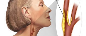
- Vascular stenosis in the neck is a serious pathology that precedes a cerebral stroke.
- Stenosis of cerebral vessels is the most dangerous pathology, as the nutrition of some parts of the brain is disrupted. As a result, a lack of coordination occurs, a person’s thoughts become confused, and a stroke may occur.
- Stenosis of the heart vessels provokes coronary artery disease, as a result a person experiences swelling, shortness of breath even with light exertion, then pain in the heart occurs, and a heart attack is possible.
- Damage to internal organs is associated with lack of blood circulation.
- Stenosis of the vessels supplying the skin is also possible; this often occurs with vibration disease in people working in hazardous industries.
Causes
The disease occurs for various reasons, most often the body is affected by several negative factors.
Vascular stenosis
Let's look at the most common ones:
- Atherosclerosis;
- Diabetes;
- Obesity;
- Sedentary lifestyle and unbalanced diet;
- Hypertension;
- Pathologies of internal organs, for example, liver, kidneys and heart;
- Presence of tumors;
- Vasculitis;
- Serious injuries;
- Hereditary predisposition;
- Alcohol abuse, smoking, drug use;
- Severe infectious diseases.
Symptoms
Vasoconstriction of the lower extremities is manifested by the following symptoms:
- Pain that can occur in the entire leg and in the lower leg area;
- Intermittent claudication;
- Severe swelling of the legs;
- The skin becomes pale and cold to the touch.
Diagnostics
Vasoconstriction is a very serious pathology, which it is advisable to diagnose as early as possible in order to prevent complications such as blockage of blood vessels, stroke and heart attack. All these conditions are serious and life-threatening for the patient, often causing disability and even death.
A phlebologist deals with the diagnosis and treatment of vascular pathologies. First of all, the doctor will examine the patient, conduct a survey, and take an anamnesis. The next step is to check the sensitivity of the skin, reflexes, and also prescribe a series of studies to identify the extent of the disease and confirm the diagnosis:
- Blood and urine tests;
- Ultrasound;
- Contrast angiography;
- ECG, ECHO;
- MRI;
- Infrared tomography, etc.
If other pathologies that often accompany vasoconstriction are suspected, the patient is referred for an appointment with specialized specialists, for example, an endocrinologist, neurologist, therapist, etc.
Treatment
Depending on the severity of the disease, the affected vessel, the age and general condition of the patient, the doctor may prescribe different treatments. In most cases, vascular obstruction is treated with conservative methods; in severe cases, surgical intervention may be prescribed.
Drug treatment consists of taking the following medications:
- lipid-lowering drugs reduce the concentration of lipids in the blood, which causes blood vessels to narrow;
- antihypertensive drugs are necessary to reduce blood pressure;
- antispasmodics relieve spasm and pain;
- anticoagulants thin the blood and prevent the formation of a blood clot;
- diuretics relieve swelling;
- vitamins are necessary to strengthen the vessel wall and immunity;
- anti-inflammatory and painkillers are also prescribed to relieve symptoms of the disease;
- Antibacterial agents are indicated in the presence of infection.
In addition, patients are prescribed physiotherapeutic treatment and diet. The patient is recommended to attend massage, physiotherapy, exercise and lead a healthy lifestyle. When vasoconstriction occurs, it is very useful to visit the pool, go to the gym, and lead a healthy and active lifestyle.
Prevention and diet
Prevention of vascular stenosis of the lower extremities involves quitting smoking and drinking alcohol. It is important to lead an active lifestyle. It is recommended to do morning exercises, dance or fitness classes. Proper nutrition helps to avoid atherosclerosis and obesity. Fatty and smoked foods, confectionery, pickled and salty foods are excluded from the diet. Fresh vegetables and fruits, dietary varieties of meat and fish, and grapes are useful.
Narrowing of the blood vessels in the legs is a dangerous pathological condition that can provoke the development of various diseases. Most often it develops due to the accumulation of atherosclerotic plaques on the inner walls of blood vessels. These plaques are formed from blood cells, cholesterol, scar tissue, etc. The larger these plaques become, the more difficult the blood flow is, and if they come off, a blockage of the vessel can occur. In a normal state, the bed of the vessel is of such a size as to provide the tissues and organs with blood in the required volume, saturating them with nutrients, blood and oxygen.
Vasoconstriction is dangerous because it does not have any clear symptoms. Most patients either do not notice differences in their condition or do not attach importance to them. Vasoconstriction can occur in the head, eyes, limbs and any other part of the body.
Diagnostics
Diagnosing this disease is a complex task. It includes:
- collecting complaints and studying the history of the disease;
- objective inspection of systems. The doctor will evaluate how the heart, respiratory organs and kidneys are working;
- consultations with narrow specialists: neurologist, ophthalmologist, vascular surgeon, endocrinologist. This list will be individual for each patient;
- laboratory tests: general and biochemical blood test, hormonal panel, tests for autoimmune diseases, urine test;
- instrumental methods: ultrasound examination of blood vessels with Dopplerography, ultrasound of the heart, angiography, computed tomography and magnetic resonance imaging with and without contrast, electrocardiography.
Consultation and examination by a cardiologist is the first step to identify narrowed vessels. Depending on the complaints, the doctor prescribes the following diagnostics:
- biochemical blood test (for cholesterol levels in the blood, which causes vascular atherosclerosis);
- ECG, ultrasound, dopplerography of the heart;
- angiography (detection of the localization of vascular obstruction, image).
Also, if necessary, arteriography, coronography, venography, lymphography are prescribed.
Through the above studies, local damage or multifocal (multi-level, multi-story) atherosclerosis of the main arteries, atherosclerosis of the aorta and arteries of the lower extremities is revealed. Areas of complete closure of the lumen (occlusion) of the artery and thrombosis may be detected.
- In the doctor’s office: special tests are performed that help assess the adequacy of peripheral circulation and its sufficiency. The color of the nail bed and various noises when listening to the femoral arteries will tell the doctor a lot.
- Instrumental methods used to diagnose atherosclerosis: Doppler ultrasound, X-ray contrast angiography, computed tomography, MRI examination and some other types.
- Laboratory research. A blood test for cholesterol and its fractions (including determination of the level of high and low density lipoproteins) will help to suspect the development of atherosclerosis in the early stages. High values of total cholesterol or low and very low density lipoproteins suggest the development of atherosclerosis.
Diagnosis of obliterating diseases of the arteries is carried out with mandatory instrumental studies. The patient is necessarily referred for ultrasound duplex scanning of blood vessels (USDS). The ultrasound scanning technique allows you to see the characteristics of blood flow in the vessel, the condition of the walls and adjacent tissues, and detect obstacles that impair blood circulation.
If surgical intervention is suspected, radiopaque arteriography is performed. A radiopaque substance is injected into the artery and a series of sequential photographs are taken along the course of the vessel. After this, the patient is left in the hospital and prescribed bed rest for 12 hours.
In controversial cases, the partial pressure of oxygen in tissues is additionally measured.
The clinical picture of atherosclerosis of the vessels of the lower extremities in the early stages resembles the manifestations of other vascular pathologies, in particular thromboangiitis and obliterating endarteritis:
- Endarteritis affects young people and usually develops against the background of frostbite, nervous strain, or severe hypothermia. Pathological changes are usually localized in the distal parts of the limb.
- Thrombangiitis is characterized by a combination of symptoms characteristic of arterial insufficiency and venous thrombophlebitis. The disease occurs in young men.
- measurement of the ankle-brachial index (the ankle-brachial index is a parameter that allows you to assess the adequacy of arterial blood flow in the lower extremities);
- duplex scanning of the arteries of the lower extremities is the “gold standard” for screening patients (detection and follow-up);
- MSCT angiography of the aorta and arteries of the lower extremities is the “gold standard” of preoperative examination and in cases where information from ultrasound scanning is insufficient;
- X-ray contrast angiography.
Occlusion of the femoropopliteal and popliteal tibial segment
- Possible causes of occlusion
- Lesions of the lower extremities
- Symptoms and diagnosis
Have you been trying to cure FUNGUS for many years?
Head of the Institute: “You will be amazed at how easy it is to cure fungus by taking the product for 147 rubles every day.
Occlusion of the popliteal artery, as well as occlusion of the femoropopliteal segment, is a cessation of blood supply to a certain area of the limb, resulting from blockage or traumatic damage to a large blood vessel. This is a common pathology that can lead to limited mobility and disability, as well as cause irreversible changes in organs and tissues, the functioning of which depends on the blood supply of this artery.
The peculiarities of damage to the blood vessels of the legs are that in the acute form of the development of the pathology, the blood supply to the lower extremities is stopped. Blockage of the vessel in this case is due to the nature of the underlying disease.
- Air (air embolism). A bubble appears in the artery, blocking the lumen and preventing blood flow. This is a common consequence of respiratory injury.
- Fat (fat embolism). Occurs when complex injuries or metabolic pathologies lead to acute disruption of natural metabolic processes.
- Arterial blood clots (arterial embolism). Appears as a result of cardiac dysfunction. Functional failures of the heart valve lead to the formation of mobile blood clots that block the artery, often at branching points.
- Atherosclerotic plaques (thrombosis in atherosclerosis). They arise from cholesterol deposits on the walls of blood vessels. This leads to complete blockage when an additional factor appears.
- Trauma and compression by nearby tissues. Occurs as a result of traumatic injury.
The cause may be aneurysms - pathological stretching or protrusion of a vessel, developing against the background of a hereditary structural abnormality or as a result of diseases present in the body. Thrombosis or embolism often occurs in the vessel itself.
A blocked artery poses an immediate threat to the life and health of the patient. Untimely assistance or improper treatment can lead to amputation of a limb or death. This is a common occurrence in older men. Impaired blood supply causes gangrene, necrosis or other irreversible changes.
The lower extremities are a common site of localization of vascular pathologies. This is due to the functional load that they constantly experience. On the legs there is occlusion of large, medium and small vessels responsible for the blood supply to the ankle.
Mixed lesions occur: obstruction occurs simultaneously in two vascular segments. The etiological factors in the leg vessels are no different from the general ones: embolism, thrombosis, trauma and aneurysms.
Popliteal occlusion occurs in chronic or acute form. Acute occurs suddenly and can be provoked by an additional obstacle in the blood flow, half-clogged with atherosclerotic plaques, when partial narrowing of the lumen is considered a chronic process.
The incidence of occlusion of the popliteal tibial segment is slightly lower than that of lesions of the femoral artery. The femoral artery is characterized by the presence of pathology in the area from the deep artery to its transition to the popliteal. Typical sites of occurrence include the area below the deep femoral artery and the place where it enters the Gunter's canal. However, occlusions of individual areas can be observed, for example, at the exit from the canal, and combined lesions, when the entire trunk of the femoral artery and part of the popliteal artery are covered.
The femoral artery is considered the main anastomosis for the occurrence of severe ischemic lesions. This is especially dangerous when the deep femoral artery is affected, while the tibial segment causes severe consequences only if all three large vessels of the leg are affected.
The arteries of the lower extremities represent an inextricable system of interaction between three segments - aortic-iliac, femoral-popliteal (from the groin to the popliteal cavity) and tibial, which includes the arteries of the ankle. They are often affected at the middle level.
Damage to even a single, isolated segment can lead to dangerous consequences. Acute occlusion rarely appears out of the blue. It is often triggered by diseases of the cardiovascular system, vascular atherosclerosis or diseases of the blood coagulation system.
Arterial occlusion is accompanied by characteristic symptoms, which vary somewhat depending on the nature of the lesion and the degree of its development. The pathology can be identified by intermittent claudication and pain in the legs, which spreads throughout the entire limb and does not subside even when taking an analgesic.
- blanching of the skin at the site of vascular blockage;
- paresthesia (numbness) and absence of pulse in the damaged area;
- cyanosis and decreased skin temperature at a later stage;
- complete paralysis and extensive ischemia with progression of the process and failure to provide timely assistance.
Diagnosis begins with external examination, palpation and medical history. After a preliminary diagnosis is made, vascular angiography is prescribed, which is performed using X-rays and a contrast agent. MSCT diagnostics are used to obtain additional information, vascular scanning and determination of the venous-brachial index, which makes it possible to assess blood flow in the upper and lower extremities.
Treatment of the pathology depends on the timeliness of seeking help and the stage of development of the disease. At the initial stage, conservative therapy and lifestyle changes are sufficient. At stages 2 and 3, surgical intervention is indicated, from thrombectomy to vascular replacement.
At stage 4 of occlusion, the only salvation for the patient may be amputation of the limb, because intervention at the vascular level in such a condition can mean death.
Removal of the obstacle that formed the vascular obstruction is dictated not only by localization. It is determined by its nature, type, stage, degree of development and background disease that provoked the negative phenomenon. This requires reliable diagnosis and complex treatment using various methods.
Obliterating atherosclerosis is a common disease that occurs most often in older men, especially in smokers. A thorough physical examination is sufficient for a correct diagnosis. If surgical restoration of blood flow is planned, angiography is necessary. When choosing treatment, both the degree of arterial damage, the patient’s profession, and the desired level of physical activity are taken into account. In approximately 50% of cases, atherosclerosis of peripheral arteries is accompanied by ischemic heart disease, including asymptomatic disease, which is the main cause of death in such patients.
A. Clinical picture. The most common complaint is intermittent claudication: pain in the affected leg that occurs when walking and disappears after stopping. As the arteries become stenotic, ischemia progresses and pain appears in the foot and toes at rest. The pain often worsens at night and decreases when lowering the leg. Even minor injuries can lead to the appearance of trophic ulcers or gangrene.
B. Physical examination. The pulse in the arteries of the affected leg is weakened or absent. Assessing the severity of ischemia: with the patient lying down, raise the leg at an angle of up to 60° and monitor the color of the skin: if it turns pale within 1 minute, then there is severe arterial occlusion. After lowering the leg, the time for restoration of normal skin color (norm - up to 10 s) and filling of superficial veins (norm - up to 15 s) is measured.
B. Non-invasive studies. Determination of the ankle-ulnar index (the ratio of systolic blood pressure on the popliteal and brachial arteries) is carried out in the office or laboratory of non-invasive diagnostics using a Doppler sensor and a conventional tonometer, while blood pressure is measured in the supine position. The ankle-ulnar index is used to confirm the diagnosis of arterial insufficiency and assess its severity. With its help, a differential diagnosis is made with pseudo-intermittent claudication in lumbar intervertebral impingement syndrome (pain in the legs caused by straightening the back when walking, disappearing in a sitting position). This syndrome can be combined with arterial insufficiency, and then the ankle-elbow index can be used to judge the extent to which lameness is caused by one or another pathology. A load test on a treadmill allows you to obtain additional data on the functional reserve of the legs (walking for 5 minutes at an incline angle of 10° and a speed of 3.2 km/h). The final choice of treatment depends on the clinic and not on the ankle-elbow index.
A. No diabetes mellitus. Without treatment, the risk of losing a leg within 5 years is 4-5%. During this period, in 50% of patients, intermittent claudication does not progress.
1) Drug treatment. General measures; 5 times a week - 30-minute walk, with stops if pain increases; pentoxifylline, 300 mg orally 3 times a day with meals. General measures include: stopping smoking, choosing the correct size and shape of shoes, foot hygiene and protecting them from any injury, treatment of hyperlipoproteinemia and diabetes mellitus, discontinuation of drugs that worsen peripheral circulation (for example, beta-blockers).
2) Surgical and endovascular methods. Carry out as planned. The main goal is to increase load tolerance.
b. Diabetes. If you have diabetes, the risk of losing a leg increases 4 times.
1) Drug treatment. The same measures and treatment of diabetes.
2. Trophic ulcers. In the presence of trophic ulcers, the risk of losing a leg within 5 years is about 12%.
1) Drug treatment. Unloading the leg (avoid putting stress on the affected leg until the ulcer heals), general measures.
1) Drug treatment. Unloading of the leg, antibiotic therapy based on the results of bacteriological examination, general measures.
3. Pain at rest. The risk of losing a leg within 5 years is about 12%.
1. Endovascular methods. For both acute and chronic arterial occlusions, endovascular interventions are increasingly being performed: balloon angioplasty, endarterectomy, laser angioplasty. With concomitant thrombosis, thrombolysis is performed in addition to them. The technique in each case is chosen depending on the location and morphology of the lesion (Curr. Opin. Cardiol. 1990; 5:666, J. Am. Coll. Cardiol. 1990; 15:1551).
1) Stenosis of the iliac artery with a length of Ј 5 cm.
2) Stenosis or occlusion of the femoral-popliteal segment, length Ј 10 cm.
1) Bilateral stenosis of the iliac arteries (one stenotic area on each side).
2) Combined stenoses of the iliac and femoral-popliteal segments (one stenotic area in each segment).
3) Aortic stenosis with a length of Ј 3 cm.
4) Stenosis of the iliac artery with a length of 5-10 cm.
5) Occlusion of the iliac artery with a length of Ј 5 cm.
6) Occlusion or stenosis of the femoral-popliteal segment with a length of 15 cm.
7) Occlusion or stenosis of the common femoral artery with a length of 5 cm.
 Occlusion or stenosis of the internal femoral artery with a length of 5 cm.
1) Stenosis of the iliac artery with a length of 5-10 cm.
2) Occlusion of the iliac artery with a length of 5-15 cm.
3) Occlusion or stenosis of the femoral-popliteal segment with a length of 15-25 cm.
A. Indications. If there is no diabetes mellitus and intermittent claudication is the only complaint, then surgical treatment is carried out as planned. The purpose of the operation is to increase load tolerance. For pain at rest, trophic ulcers, and diabetes, the purpose of the operation is to preserve the leg. With diabetes, the risk of amputation within 9 years is more than 5%. For pain at rest and trophic ulcers, the risk of amputation within 5 years is 12%.
b. Type of intervention. The choice of surgery (aortofemoral, femoropopliteal, tibiofibular bypass or endarterectomy) depends on the location and type of lesion. The final decision is made by the surgeon. If there is a high surgical risk, extracavitary operations are performed to save the leg - axillary-femoral or interfemoral bypass. Lumbar sympathectomy as a method of radical treatment is currently rarely performed: it is mainly performed either in addition to aortofemoral bypass surgery, or to accelerate the healing of trophic ulcers in cases where reconstructive operations are not feasible.
V. Preoperative examination. IHD is a leading cause of perioperative complications and death. The primary task when planning surgery is to determine the risk of intraoperative and postoperative cardiovascular complications and reduce it.
Source: M. Fried, S. Grines “Cardiology” (translated from English), Moscow, “Praktika”, 1996
Stages of development of atherosclerosis of the lower extremities
The most detailed is the modified classification of chronic arterial insufficiency of the lower extremities (CANF), which takes into account in detail the phenomena of critical limb ischemia, which is necessary when determining treatment tactics.
| Stage 1 | Muscle pain only during heavy physical activity (when walking over a distance of more than 1 km). Initial signs of stenosis appear - the skin turns pale, there is a feeling of goosebumps, it seems that the legs are always cold, fatigue quickly sets in when walking, excessive sweating is observed |
| Stage 2A | Feeling of fatigue and stiffness in the calf muscles, intermittent claudication after 200-1000 m |
| Stage 2B | Intermittent claudication in less than 200 m |
| Stage 3A | Intermittent claudication after a few steps or pain at rest when it is possible to keep the lower limb in a horizontal position for more than 2 hours |
| Stage 3B | Pain at rest, ischemic edema, inability to keep the lower limb in a horizontal position for 2 hours |
| Stage 4A | Gangrene of the fingers or part of the foot with the prospect of maintaining the supporting function of the limb |
| Stage 4B | Extensive necrotic changes in the limb without the possibility of maintaining its supporting function |
Symptoms
Each stage of stenosing atherosclerosis of the lower extremities has its own signs and characteristics.
- At stage I, the patient is practically not bothered by severe symptoms of the disease. He may feel cold in his legs, sometimes to the point of goosebumps. In addition, the color of the skin of the legs may change - from pinkish to bright red. At this stage, vasodilators and medications that normalize cholesterol balance are sufficient for cure.
- As stage II progresses, the patient becomes increasingly concerned about the feeling of chills in the legs, and pain begins to appear after physical exertion and walking distances of up to 500 meters. When one limb is affected, pain is expressed only in that limb, which gives rise to the syndrome of “intermittent claudication.”
- Stage III - vasoconstriction reaches pathological levels, which causes constant pain that bothers a person even at night. Walking is seriously difficult, the patient cannot walk even 50 m without discomfort.
- Stage IV – the patient’s temperature rises, signs of stenosing atherosclerosis become pathological: the lumen of the vessels closes completely, the blood supply to the tissues stops, and tissue necrosis (gangrene) develops. A patient in such a situation can only be treated in the most radical way - usually surgery to amputate a limb is prescribed.
Ways to combat the disease
The diagnosis requires a series of subsequent treatment procedures to eliminate disorders and causes of the disease. Today medicine practices two types of treatment.
Drug therapy helps maintain the strength and elasticity of the vessels of the lower extremities. Treatment is combined, which is characterized by a course of hypertensive and vasodilator drugs.
Thrombodissolving drugs have become widely known in recent years. Thrombolytics can effectively dissolve existing blood clots. After completion of thrombolytic therapy, relapses of thrombosis are not possible, since this pharmacological group of drugs destroys the blood clot without creating conditions for the subsequent appearance of blood clots. Therefore, the entire course of treatment is accompanied by the use of anticoagulant medications.