Which vessels are affected?
Atherosclerotic plaques are most often localized in the area of the subclavian artery. This paired vessel emerges from the aorta and supplies the head, heart and arms. According to statistics, in 2% of cases there is a violation of blood flow on both sides. Most often this phenomenon is observed on the left. Narrowing of the lumen leads to the appearance of retrograde outflow with the development of “steal syndrome” and is accompanied by numbness of the hands and vertebrobasilar abnormalities. Much less common is separate blockage of small vessels in the forearm, wrist and hand.
Important! Oxygen and nutrients partially come to the upper extremities from the brachiocephalic trunk, but if the blood flow in it is disrupted, neurological symptoms come first.
Vascular Doppler during pregnancy
Doppler ultrasound during pregnancy helps the doctor determine the quality of the blood supply to the developing fetus, find out the degree of patency and lumen of the fetal umbilical cord vessels, and also determine the fetal heartbeat. Doppler ultrasound is recommended for pregnant women suffering from hypertension, as well as eclampsia, in case of decreased motor activity of the fetus (rare movements), etc. see also Ultrasound during pregnancy.
A pregnant woman can independently determine the fetal heart rate using a special device called a fetal doppler. A fetal doppler is a small electronic device that listens to the fetal heartbeat and counts its heart rate.
Vascular ultrasound (ultrasound) is one of the methods that can be used to determine the condition of the vessels supplying the brain, arms and legs, pelvic and genital organs, kidneys, scrotum and many other organs.
Thanks to the availability of modern ultrasound diagnostic methods, it is possible to determine whether there are atherosclerotic plaques and narrowings (stenosis) of blood vessels in the lumen of the vessels, and to determine which part of the vascular lumen (as a percentage) accounts for the largest number of them. Ultrasound also allows you to identify quantitative and qualitative indicators of systemic blood flow.
Ultrasound examination of blood vessels is carried out for the purpose of:
- Identifying or excluding signs of deep vein thrombosis in the legs or arms;
- To assess the condition of primary varicose veins;
- To evaluate secondary (recurrent) varicose veins;
- To identify chronic stagnation of venous blood (chronic venous insufficiency) and postphlebetic syndrome;
- To assess the health status of patients with coronary artery disease (coronary heart disease), especially in the presence of negative symptoms from the vessels of the arms and legs;
- Diagnosis of peripheral vascular aneurysms, as well as monitoring their dynamics;
- Diagnosis of formations with pulsation.
Symptoms and signs
Atherosclerosis of the upper extremities manifests itself with nonspecific symptoms, which depend on the stage of the pathology:
- At first, a partial slowdown in blood flow appears weakly or is not noticed by the patient. Sometimes a person feels chilly in the fingers on the affected side and tolerates the cold worse.
- Gradually, the condition worsens, pain appears in the distal parts, and a transient disturbance of sensitivity in the form of itching, crawling or burning appears. Intensifies with load on the affected limb or during sleep.
- When the lumen of the artery closes by more than 50%, obliterating atherosclerosis occurs, and the hand freezes even at normal temperature. Unpleasant sensations increase and move from the fingers and hands to the forearm area and become permanent, disturbing both during exercise and at rest. Activities requiring the use of fine motor skills are performed with great difficulty (fastening buttons, writing, tying shoelaces). Nails become dull and break, hair loss begins, convulsive twitching of individual muscle fibers begins.
- The last stage is manifested by ulcers on the hand, severe swelling and the appearance of areas of necrosis, the skin becomes bluish. There is a complete loss of ability to work. An advanced form of atherosclerosis leads to the need for amputation of a limb or the death of the patient as a result of the development of gangrene.
What is atherosclerosis of the upper extremities
In fact, atherosclerotic lesions develop simultaneously in all arteries containing elastic fibers in their layers. It’s just that in different sections of the bloodstream the changes have different degrees of severity, which is how the disease is classified. And in this classification, atherosclerosis of the arteries of the upper extremities occupies a certain place, although it is not so widespread in isolated form.
If it were possible to observe changes in the vascular wall from the inside, then it would be possible to trace the alternation of stages of the process.
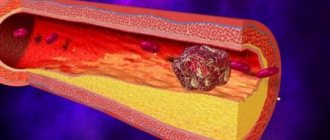
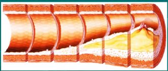
- The stage of lipid spots and stripes practically does not cause disturbances in the blood supply to the tissues of the upper extremities, since it does not change the diameter of the arterial lumen. This is the initial stage of atherosclerosis, during which fats (primarily “bad” cholesterol), proteins and their complexes accumulate in the vascular wall.
- At the stage of fibrous plaque, fatty protein deposits grow with connective tissue, and then with small vessels. The plaque is already protruding into the lumen and partially narrowing it. Fibrous formations most often appear in places where the blood flow turbulence, that is, in the initial sections of the arteries coming from larger ones.
- The next stage - atheromatosis - is caused by the disintegration of structures that have accumulated and grown in the vascular wall. The plaque deepens, right up to the outer lining of the artery, becomes saturated with immune blood cells, and becomes loose. But its top, covering layer is still intact.
- Under the influence of intense blood flow, which also increases as a result of narrowing of the lumen of the vessel, the plaque is destroyed with the formation of an ulcer . Ulceration is the next, complicated stage of atherosclerosis. At the same time, thrombotic masses actively adhere to the destroyed surface of the inner lining of the artery, seriously reducing the diameter of its lumen.
- The most dangerous stage is calcinosis . With it, lime is deposited into the affected area, giving the vessel a bone-like consistency. The arteries of the upper extremities no longer respond adequately to changes in blood pressure, they become brittle, and their lumen sharply narrows. Such atherosclerosis is called obliterating.
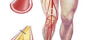
The vessels of the upper limb containing elastic fibers in the wall include the axillary and brachial. Their branches nourish not only the soft tissues of the hand, but also the bones, structures of the shoulder and elbow joints, and small joints of the fingers. Therefore, changes associated with insufficient blood flow also affect these anatomical formations.
Atherosclerosis is considered a disease of old age, but its initial stages are also observed in newborns. True, lipid spots and streaks in children subsequently resolve. In adults, the timing of the development of pathology is highly individual and depends on many factors.
Doctor's advice: how to suspect the disease
In order to avoid surgery, treatment and prevention of progression of atherosclerosis of the upper extremities should be started in the early stages. I would like to recommend contacting a doctor immediately if at least one sign of a blood supply problem in the arm occurs:
- pain and discomfort in the fingers and hands, as well as in the area where the vessel is located;
- pale skin, loss of natural color;
- when the pressure changes, the difference in the right and left arms is more than 20 mmHg. Art.;
- the appearance of convulsions, which intensify with physical stress;
- increased sensitivity to cold or temperature changes;
- deterioration of the condition of nails and hair on the hand;
- loss of sensitivity, itching, burning or crawling on the skin;
- muscle weakness in one of the limbs;
- rapid fatigue when performing ordinary activities that previously did not cause difficulties.
Weakness in the arm muscles due to atherosclerosis
One of the obvious symptoms of diseases of the cardiovascular system and atherosclerosis in particular is weakness in the hands and forearms. However, this sign is not always a signal of danger. For example, some young women may feel weak in their arms during menstruation.
Separately, it should be said about weakness in the arm muscles, which can be both pathological and physiological in nature.
Physiological weakness is natural and appears in the following cases:
- prolonged physical activity, for example, when a person holds a heavy load in his arms for a long time;
- compression of blood vessels during sleep;
- sudden change in temperature.
A symptom of atherosclerosis is pathological weakness in the limbs, which is observed for a long time. In addition to disorders of the blood vessels, weakness is caused by diabetes, arthritis and viral infections.
Diagnostics and examinations
To diagnose atherosclerosis, a patient is interviewed. I usually find out when the first sensations and changes appeared. Neurological pathologies are often accompanied by similar symptoms, but they most often develop over a shorter period of time.
Several years may pass from the moment cholesterol begins to be deposited on the vessel wall until signs of the disease appear, so you need to understand the duration of the development of the pathology.
The skin on the affected limb is paler or bluish compared to the healthy arm, and is colder to the touch. On it, the pulse is palpable weaker, blood pressure is lower. Asking the patient to stand for 3 minutes with his arm parallel to the ground is almost impossible for the patient to do. Neurological tests reveal decreased sensitivity.
A laboratory blood test for atherosclerosis will show an increase in the level of cholesterol, triglycerides and low-density lipoproteins. Additional assistance in diagnosis is provided by instrumental techniques:
- Duplex scanning. It is not highly informative in this case, but can confirm the presence of “steal syndrome”, that is, the presence of a reverse direction of blood flow.
- MRI or CT. They provide a complete picture of the lesion with high accuracy and are used to determine the need and method of surgical treatment.
Duplex scanning (ultrasound) of leg vessels
As you have already read on the main page of our website, the main activity of the Medical Center is the treatment of blood vessels using a non-surgical method. And it is first necessary to conduct an ultrasound of the blood vessels.
In Medical Lviv, st. Krushelnitskaya, 1 You will be offered an ultrasound of the vessels of the lower extremities using color duplex scanning, which allows you not only to see the vessel, its walls, surrounding tissues, but also the movement of blood through the vessel, its direction, speed, obstacles that disrupt normal blood flow (plaques, blood clots and etc.).
Ultrasound examination evaluates the severity and prevalence of varicose veins, the presence of signs of thrombosis or its consequences in the deep and superficial veins, the condition of the valve apparatus of the deep and superficial veins, the degree of valvular insufficiency and its prevalence. When visually assessed in B-mode, uneven expansion of the lumen and a change in the trajectory of the superficial veins, an increase in the echogenicity of their walls are determined; with the addition of a thrombotic lesion, the presence of blood clots in the lumen of the veins.
Using this method, we can detect diseases such as obliterating atherosclerosis with the presence of hemodynamically significant stenosis of the arteries of the upper and lower extremities with clarification of the localization of the process, obliterating endarteritis, diabetic macroangiopathies.
Studies in color and spectral Doppler modes make it possible to clarify the nature of hemodynamic disorders and assess the degree of valvular insufficiency.
Ultrasound of leg vessels (duplex scanning) is recommended:
- when you feel heaviness and pain in your legs;
- in case of rapid fatigue when walking;
- with tingling sensations on the skin of the legs;
- with increased sensitivity of the feet to cold;
- in case of ulcers appearing on the skin of the legs (skin defects that do not heal for a long time);
- in case of swelling in the legs;
- dilated veins of the legs visible under the skin (varicose veins) and in some other cases.
Ultrasound of the blood vessels of the legs can reveal the following diseases:
Atherosclerosis of the arteries of the legs (obliterating atherosclerosis of the lower extremities) is a disease of large vessels, which usually occurs after 40 years and is manifested by the following symptoms: pain and fatigue in the legs when walking, climbing uphill or stairs, cold skin of the legs, impaired growth hair (baldness) of the skin of the legs and thighs, the appearance of trophic ulcers (long-term non-healing wounds on the skin of the legs), etc.
Obliterating endarteritis is a disease of the smaller vessels of the legs, which is characterized by inflammation of areas of the arteries and their narrowing. Symptoms of obliterating endarteritis are similar to those with atherosclerosis of the arteries of the legs.
Varicose veins of the legs are a disease characterized by the appearance of areas of dilated veins and stagnation of venous blood in the legs. The main symptoms of varicose veins of the legs are: the appearance of heaviness in the legs, swelling, itching, darkening and thickening of the skin of the legs, dilated veins visible under the skin, the appearance of trophic ulcers (wounds on the skin of the legs). Using Doppler examination of the vessels of the legs, the degree of patency of the veins (lumen width), as well as stagnation of blood in the veins of the legs, will be determined.
Deep vein thrombosis of the legs is a disease characterized by the appearance of blood clots in the veins that interfere with the flow of blood from the legs. Deep vein thrombosis of the legs is manifested by swelling in the legs and pain when moving the ankle joint. Color Dopplerography (ultrasound) of the leg vessels determines the location of the blood clot, which is important for surgical treatment of the disease.
Duplex scanning is a modern, highly informative method that does not require special training, provides reliable information about the condition of blood vessels, allows you to establish or confirm a diagnosis and, most importantly, help correctly and adequately plan further treatment and, which is also very important, is affordable.
Based on the results of an ultrasound scan of the vessels of the legs, in a medical clinic, a highly qualified vascular surgeon will advise you and select an individual non-surgical treatment regimen.
ul
Medicines
I usually recommend the following groups of medications for use:
- statins - they reduce cholesterol levels by inhibiting its production in the liver (Atorvastatin, Rosuvostatin, Simvastatin);
- disaggregants and thrombolytics to prevent the formation of blood clots and complete occlusion of the site of narrowing of the artery;
- vascular agents to improve blood flow and relieve spasms;
- analgesics or local novocaine blockades for severe pain.
Methods of treatment and prevention
Therapy of atherosclerotic lesions includes the use of general agents, traditional methods, and local effects on soft tissues. Drugs prescribed:
- reducing the level of “bad” cholesterol;
- blood thinners;
- vitamins;
- analgesics (if necessary).
But treatment will be useless if a person does not change his life: does not balance his diet, protect himself from stress, does not give up bad habits and does not increase physical activity. That is, it will not do everything that is included in the concept of preventing atherosclerosis.
When is surgery needed?
Angioplasty with stenting helps restore the lumen of the artery. To do this, a catheter with a balloon is sent into the affected vessel, which contains a special durable tubular device that is installed in the area of narrowing; it strengthens the wall and does not allow it to collapse. The price in Moscow for this operation ranges from 55 to 150 thousand rubles, while the cost of the stent is negotiated separately.
A pathogenetically justified operation is endarterectomy, when the plaque is removed from the part of the inner lining of the vessel where it is located. The cost of the intervention ranges from 49 to 140 thousand rubles in different clinics in the capital.
The principle of this procedure is clearly visible in the photo:
Bypass surgery involves installing a bypass to normalize blood flow. Most often it is performed on the coronary arteries, but is often used to eliminate ischemia in the periphery. The price of such an operation can vary from 14 to 21 thousand rubles.
Any operation for atherosclerosis does not eliminate the causes of the disease. If you do not take cholesterol-lowering medications and continue to abuse smoking and fatty foods, new areas of impaired blood flow may appear.
Recommendations from specialists for treatment
Medical treatment often focuses on symptom relief. However, interventions aimed at reducing underlying atherosclerosis, as opposed to simply treating symptoms, are more effective. Non-pharmaceutical treatments are usually the first treatment, such as stopping smoking and regular exercise.
If these methods do not work, medications are usually the next step in treating cardiovascular disease and, with improvement, are increasingly becoming the most effective treatment in the long term.
The key to more effective approaches is to combine several different treatment strategies. Arteriosclerosis, or “hardening of the arteries,” usually affects the legs first. Narrowing of the arteries can lead to complete closure of the vessel.
A CT coronary angiogram films blood flow in arteries after dye is injected into a peripheral vein. Calcium deposits in artery walls contribute to narrowing and stiffness.
It is a common disorder, usually affecting men over 50 years of age. People are at higher risk if they have a personal or family history of:
- diabetes;
- heart disease;
- coronary artery disease;
- high blood pressure;
- kidney disease, hemodialysis;
- cerebrovascular diseases.
Case from practice: why you need to have surgery on time
I want to tell you an interesting case from my practice.
A 56-year-old man came with complaints of coldness in his left hand, pain and burning in the forearm, dizziness, and fainting. On examination, signs of atrophy, bluish skin, disruption of the nail structure and lack of hair were observed. The pulse is weak on both extremities, thread-like on the left, the pressure on the affected arm is 30 units lower. After examination and questioning, it turned out that the patient was diagnosed with atherosclerosis of the subclavian artery a year ago, and prescribed medications that he took for a short time. At the same time, surgery was recommended to restore blood flow (stenting), which he refused. The result was atrophy of the arm muscles. The patient is currently undergoing preparation for surgical treatment to avoid necrosis and amputation.
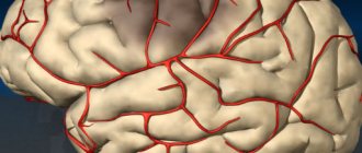
Doppler of neck and brain vessels
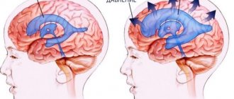
Doppler of cerebral vessels is an important method for diagnosing vascular diseases of the brain and spinal cord. Using Doppler of cerebral vessels, the quality of blood supply to the brain is assessed, and areas of narrowing or dilation of the arteries of the brain are identified. Doppler of cerebral vessels is recommended for people experiencing severe headaches, attacks of dizziness, fainting, and is also prescribed by a doctor if there is a risk of cerebrovascular accident (stroke), suspected aneurysm (expansion and thinning of a section of an artery) of a cerebral vessel, etc.
As a rule, during a session of Doppler diagnostics of cerebral vessels, a Doppler examination of the neck vessels supplying the brain (carotid arteries, vertebral arteries) is also performed.
What diseases can be detected using Doppler ultrasound of cerebral vessels?
Using Doppler examination of cerebral vessels, the doctor identifies the following diseases:
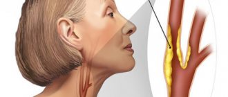
Atherosclerosis of the vessels of the neck and brain - develops, as a rule, in elderly obese people suffering from hypertension and causes the following symptoms: decreased memory and mental abilities, sleep disturbances, attacks of headaches, dizziness, weakness or numbness of the arms or legs that last up to 30 minutes and disappear without a trace. All these symptoms are harbingers of a possible stroke and therefore require careful examination, including using Doppler of the cerebral vessels. On Doppler, with atherosclerosis of cerebral vessels, a narrowing of the lumen of the cerebral arteries is noted.
Cerebral aneurysms are areas of dilation of arteries or veins that usually have a thin wall and can rupture. A ruptured aneurysm can cause massive bleeding in the brain or lining of the brain. An aneurysm of one of the cerebral vessels can be asymptomatic for a long time, or manifest itself as periodic headaches, blurred vision, etc. On Doppler ultrasound of cerebral vessels, an aneurysm appears as an area of dilatation of a cerebral artery or vein. Using Doppler ultrasound of the brain vessels, the location of the aneurysm and its size are revealed.
Cerebral artery stenosis is an area of narrowing of the arteries supplying the brain. The main symptoms of cerebral artery stenosis are headache, memory and intellectual impairment, a feeling of numbness or weakness in the arms or legs, etc. Using a Doppler study of cerebral vessels, the degree of narrowing of the vessel and the speed of blood flow in it are measured.
Doppler examination of the vessels of the neck allows us to identify diseases that lead to impaired blood supply to the brain. The study focuses on the vertebral and carotid arteries. Doppler ultrasound of the neck vessels is recommended for people experiencing attacks of dizziness, nausea (as with motion sickness), as well as headaches, fainting and other cases. To obtain a more accurate picture of the condition of the neck vessels, duplex scanning is sometimes used - this is a method that combines Doppler examination and ultrasound. Using Doppler of the neck vessels, atherosclerosis or stenosis (narrowing of the lumen) of the vertebral or carotid arteries is detected.