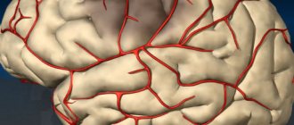
Brain vessels: blood supply system
Vascular lesions of the brain and spinal cord are the most important problem in clinical neurology
.
The blood supply to the brain is characterized by the presence of an optimal regime that ensures continuous and timely replenishment of its energy and other costs during life.
The main function of the nervous system is to regulate the physiological processes of the body, adapting to constantly changing environmental conditions.
The nervous system adapts the body to the external environment, regulates all internal processes and their constancy - constancy of body temperature, biochemical reactions, blood pressure, tissue nutrition processes and providing them with oxygen, etc.
That is why the importance of the intensity of nutrition of the nervous tissue and its sufficient enrichment with blood is very great. At rest, approximately 750 ml of blood per minute passes through the human brain. This corresponds to 15% of cardiac output.
Normal nutrition of all parts of the brain and the hemispheres themselves is ensured due to the structural organization and physiological mechanisms of regulation of brain vessels.
In the gray matter, the blood circulation of the brain is more intense than in the white matter. Compared to adults, in children under 1 year of age it is the most saturated - the nutritional intensity is 50-55% higher.
And in elderly , the intensity of blood supply is reduced by 20% or more. In this case, about one fifth of the total blood volume is pumped by the vessels of the brain.
The nervous system remains constantly active, even during sleep. At the time of dreaming (the REM sleep phase), the level of metabolism in many brain structures can be even greater than in the waking state. All this determines the extremely high need of the brain for oxygen.
With a mass of approximately 1400 g, representing about 2.% of body weight, it absorbs 20% of all oxygen and 17% of all glucose entering the human body.
How to relieve symptoms
Medicines and physical therapy are used. To improve his condition for a long period, the patient must devote time to therapeutic exercises. In some cases, if the symptoms cannot be eliminated, the attending physician prescribes surgery.
Medicines
Treatment of cervical artery syndrome involves the use of medications that improve vascular patency. If symptoms are acute and treatment needs to be accelerated, drug injections may be indicated. Non-steroidal anti-inflammatory drugs are most often prescribed. They relieve inflammation, thereby freeing the flow of blood through the artery. Platelet activity decreases and blood moves more freely.
In order not to harm the patient’s gastrointestinal tract, it is recommended to use Nimesulide or Celecoxib. Cheaper drugs such as Ibuprofen or Diclofenac have similar effects, but are dangerous for the digestive system.
Centrally acting muscle relaxants are prescribed to prevent muscle spasms and cramps, reduce pain and relieve pinched arteries. These include Tolperizone, Sirdalud, Mydocalm. Mydocalm is preferred because of its analgesic effect, plus it improves blood circulation.
To dilate blood vessels, Trental and Instenon are used. Thanks to vasodilator drugs, nutrition and metabolic processes in the brain improve. Also, taking vitamin and mineral complexes will have a positive effect on symptoms of the cervical artery.
Physiotherapy
The following procedures will relieve symptoms:
- Exposure to low frequency current;
- Shock wave therapy;
- Electrophesis with drugs that block the transmission of nerve impulses.
If the pain syndrome is severe, novocaine electrophoresis will help better. Other useful methods are acupuncture, manual therapy and massage treatments. They can only be trusted by experienced specialists, since actively working with a patient who has cervical artery syndrome can easily aggravate the situation.
Surgery is necessary if conservative treatment does not help or the cervical artery is compressed too much - up to 2 millimeters in diameter. The skin is cut into two centimeters, and the risk of damage to internal organs is minimal. The operation takes place in the following options:
- Using plastic surgery methods, the shape of the vessel is changed;
- An expanding stent is inserted into the artery.
If a tumor or hernia is found, you need to get rid of its pressure on the arteries as much as possible. If the disease proceeds without other complications, surgery becomes a very effective method of treating the syndrome, completely eliminating symptoms.
I RECOMMEND ON
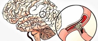
Constriction of blood vessels in the brain
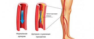
In other cases, narrowing of blood vessels in the circulatory system is called pathological stenosis of cerebral vessels, which quite often occurs due to the development of atherosclerosis in the cavity of the coronary arteries.
Due to the large amount of accumulated plaque, this leads to the closure of the arteries and obstruction of blood flow to the nerve tissue.
Atherosclerotic plaques in the head usually form in the final stages. The development of the disease can be traced over decades.
In some cases, after a long, gradual and unnoticeable development, a sudden proliferation of lipid tissues, deformation of veins and a sharp deterioration in health occur. The appearance of plaques in the brain and damage to the arteries may be a consequence of the sudden abrupt development of the disease.
The danger of their formation and growth is due to the fact that they can break away from the walls of blood vessels and move through the circulatory system, and once they get into a small vessel, completely clog it.
Signs of cerebral vasoconstriction
The amount of blood required for the organ to function properly is reduced; disturbances lead to tissue ischemia (oxygen starvation), changes in the structure of cells, and subsequently to their mass death (the appearance of foci of necrosis).
Changed or dead nerve cells of the brain are not able to perform their functions (conducting a bioelectric impulse), therefore vasoconstriction is manifested by numerous neurological symptoms:
- headaches;
- dizziness;
- insomnia.
The pathology develops slowly and is almost asymptomatic in the initial stages. If the cause of stenosis is eliminated at this moment, the disease can be cured, completely restoring brain function (92%).
The chronic form of stenosis can plague a person for years, and the initial form can lead to death at the very first stage, which is why it is so important not to miss the “signals” about the disease.
Possible signs of vasoconstriction:
- Migraine , irritability, fatigue, touchiness, tearfulness, overexcitement. functional disorders of the central nervous system;
- Memory problems Short-term forgetfulness, inability to remember what happened a few minutes ago;
- Development of dementia With dementia, cognitive processes deteriorate, and there is a depletion of emotional reactions and character traits;
- Change in gait ( person shuffles or minces heavily);
- Loss of coordination of movements, loss of balance;
- Malfunctions in the functioning of internal organs located in the pelvic area;
- Temporary loss of vision ;
- Vomiting , paresis, paralysis;
- The appearance of a false urge to urinate ;
Symptoms
Clinical symptoms may vary depending on whether the process is acute or chronic.
In case of acute pinching, the main symptom is severe pain in the neck. It may intensify when turning the head, or the person will complain of a feeling of numbness when it is physically impossible to turn the head. This happens when a person has suffered a minor injury (strained neck muscles) or slept in an uncomfortable position.
Arteries pass through the cervical vertebrae, feeding brain tissue and carrying out venous drainage. When pinched, this process is disrupted. As a rule, this process develops gradually; the body is able to compensate for a long time for the lack of oxygen and other substances entering the brain with the bloodstream. Therefore, the clinical picture increases progressively.
Initial manifestations of insufficient blood supply to the brain
The diagnosis of “Initial manifestations of insufficiency of blood supply to the brain” often presents great difficulties and cannot always be made with certainty.
However, highlighting this point is important, as it draws attention to this earliest form of vascular brain damage, when preventive and therapeutic measures are most effective.
The diagnosis of initial manifestations of vascular pathology of the brain is made based mainly on the following subjective complaints of the patient:
- headache;
- dizziness;
- noise in the head;
- memory impairment;
- decreased performance.
The basis for diagnosis is only a combination of two or more of these signs that exist for a long time and are constantly or frequently repeated.
They are characterized by their occurrence under circumstances that require increased blood supply to the brain, for example, during intense mental activity, especially if it occurs under hypoxic conditions (in a stuffy, smoky room) or against the background of great fatigue.
In most cases, the cause is atherosclerosis or hypertension.
But the same symptoms can also be caused by other reasons - chronic infections, malignant neoplasms, etc.
The assumption of the vascular origin of the described disorders is supported by rheoencephalography data, as well as the presence of signs of atherosclerotic vascular lesions in other areas (fundus vessels, coronary sclerosis, intermittent claudication, etc.) or symptoms of hypertension (rises in blood pressure, hypertensive retinopathy, left ventricular hypertrophy of the heart) and etc.).
Conditions of working capacity due to cerebral circulatory disorders
| Name | State |
| Cerebral atherosclerosis with initial manifestations of insufficiency of blood supply to the brain without organic symptoms | able to work |
| Atherosclerosis | Temporarily disabled |
| Arterial hypertension | Temporarily disabled |
| Osteochondrosis of the cervical spine | Temporarily disabled |
| Transient disturbance of cerebral circulation in the vertebral-basilar circulation | Temporarily disabled |
| Atherosclerotic stenosis of the right internal carotid artery (lumen narrowing about 75%) | Disabled |
| Infarction in the territory of the right middle cerebral artery | Disabled |
| Left-sided hemiplegia, hemianesthesia, anosognosia | Disabled |
| Hypertension SB degree | Unable to work. Needs outside care and supervision |
| Right-sided hemiparesis and motor aphasia after hemorrhage in the left hemisphere of the brain | Needs outside care and supervision |
| Dementia | Needs outside care and supervision |
| Residual effects after infarction of the anterolateral wall of the left ventricle of the heart | Needs outside care and supervision |
Causes of vasoconstriction
Cerebral vasospasm
Under normal conditions, functional constriction of cerebral vessels occurs constantly, just like their expansion, which is an important mechanism for regulating cerebral circulation.
A danger to health and life is pathological vasoconstriction - arterial spasm. A short-term spasm of the arteries of the brain occurs, for example, in response to mechanical irritation.
Prolonged, persistent spasm is a pathological reaction of the arteries to the action of breakdown products of blood and brain matter.
Among the cerebral circulatory disorders associated with changes in blood properties are an increase in blood viscosity, adhesion of platelets and other formed elements.
Platelet adhesion
Aggregation, or sticking together, of platelets can occur in various pathological conditions of the body (acute renal failure, trauma, etc.).
The small emboli formed in this case, entering the brain vessels, disrupt blood flow and lead to the development of hypoxia and foci of necrosis in the brain substance.
Embolus is any unbound intravascular substrate (solid, liquid or gaseous) circulating through the bloodstream, not found there under normal conditions, capable of causing blockage of an arterial vessel at a sufficiently large distance from the site of occurrence.
Hypertensive diseases
Vascular crises in hypertensive diseases are manifested by spasm and paralysis of intracerebral arteries and arterioles. Both of these processes can take place within the same vascular region of the brain.
Vascular hypertensive crises
play a major role in the occurrence of cerebral hemorrhage.
At the same time, permeability increases, processes of tissue necrosis develop, amylar aneurysms (protrusion of vessel walls up to 3 mm) and ruptures of the walls of intracerebral arteries are formed.
In mild cases, this manifests itself in increased vascular permeability. In more severe cases , oxygen starvation of the walls of the brain vessels occurs, aneurysms appear and hemorrhages occur around the vessels.
In the most severe situation, with a hypertensive crisis, tissue necrosis, rupture of the vessel wall and massive hemorrhage into the brain substance develop.
Drug treatment.
It is difficult to treat a severely pinched nerve in the neck, so the therapeutic regimen is selected by a doctor, and self-medication is unacceptable.
First of all, it is important to eliminate pain, swelling and discomfort. For this purpose, non-steroidal anti-inflammatory drugs are prescribed:
- "Nimesil";
- "Diclofenac";
- "Nise";
- "Ibuprofen";
In the acute period, when symptoms are severe, it is recommended to give injections. When the condition returns to normal, you can switch to tablets. To relieve muscle spasms and release nerve fibers, muscle relaxants are prescribed:
- "Structum";
- "Mydocalm";
- "Baclofen";
During treatment, it is useful to take vitamin complexes containing nicotinic acid, B vitamins, calcium, magnesium and manganese. To reduce inflammation and relieve pain symptoms, you can use an anti-inflammatory ointment. Proven:
- "Dolobene";
- "Finalgon";
- "Voltaren";
- "Fastum gel";
Methods for studying cerebral circulatory disorders
Among these methods, angiography has acquired the greatest importance, i.e., radiography of the head after the introduction of a radiopaque substance into the arteries that carry blood to the head.
Various contrast agents are used (diodon, gaipak, conrey, etc.), which can be administered in different ways.
The most commonly used is carotid angiography, in which a contrast agent is injected into the carotid artery in the neck by puncturing it. However, in this case, vessels are identified only in the basin of the punctured artery.
Meanwhile, there is often a need to get an idea of the entire vascular system of the brain, starting from the place where the arteries originate from the aortic arch to their final branches in the series - “total” angiography or “panangiography” of the head.
For this purpose, two methods are used - catheterization (according to Seldinger) and puncture. In the first method, by percutaneous puncture of the femoral or brachial artery, a thin catheter is passed through a cannula needle to the aortic arch and a contrast agent is injected through it - an aortogram.
Aortogram. Vessels extending from the aortic arch are visible.
1 - right common carotid artery, 2 - pelvic-cephalic trunk, 3 - origin of the left common carotid artery, which is not visible due to its blockage, 4 - left subclavian artery (arrow indicates concentric stenosis)
With the puncture method, a contrast agent is injected into the right and left axillary arteries using a special syringe, which makes it possible to identify the subclavian and vertebral arteries on the left, and the carotid artery on the right; To judge the left carotid artery, puncture angiography is additionally performed.
In case of cerebral hemorrhage or large infarction with cerebral edema, in addition to the avascular zone, vascular displacement is detected.
In the case of blockage of large vessels supplying the brain, angiography makes it possible to judge the paths of collateral blood flow, with the help of which the shutdown of the affected vessel is compensated. The state of collateral circulation must be taken into account when deciding on surgical intervention.
The use of special X-ray machines that produce a series of images within a few seconds, as well as X-ray filming, makes it possible not only to obtain an image of all parts of the vascular system of the brain, but also to identify the characteristics of blood flow in them.
The production of substances that have high contrast and do not give undesirable vascular reactions, as well as improvements in angiography techniques, have minimized the number of complications and made it possible to expand the scope of its application.
But serious complications, although rare, are observed. Therefore, angiography should be performed only when there is a question about surgery or uncertainty in the diagnosis does not allow the application of appropriate treatment (for example, the use of anticoagulants with a presumptive diagnosis of developing cerebral infarction).
Rheoencephalography is widely used. Its essence lies in the fact that with the help of special amplifiers, changes in the electrical resistance of the head are recorded, which depend mainly on changes in the blood supply to the brain. With a certain arrangement of electrodes, one can judge the blood supply to various parts of the brain.
A normal rheoencephalogram curve is characterized by relatively regular, regularly repeating waves, resembling pulse waves in shape. In young healthy people the upward curve is steep. With atherosclerosis, the sharp apex of the curve becomes rounded, and sometimes a plateau appears in place of the apex. Blockage of a vessel leads to a decrease in the amplitude of rheographic waves in its basin. Using various leads, one can judge the blood supply to different parts of the brain.
Surgery.
When conservative methods fail, a decision is made to perform surgical intervention, the main objectives of which are:
- remove the causes of pinching;
- maintain the physiological functionality of the neck;
- align the spine and normalize stability;
A herniated disc can only be treated surgically. During the operation, bone and soft pathological structures, such as hernias, are removed. Thus, nerves and blood vessels are released, inflammation and associated symptoms are eliminated. The following surgical treatment methods are used:
- anterior discectomy;
- replacement of discs with an artificial implant;
- increase in intervertebral space;
Source
Treatment of cerebral vessels with atherosclerosis
When choosing a treatment method, it is necessary to take into account all the factors that contributed to the development of the disease.
If atherosclerosis occurs as a result of physical inactivity, you need to increase the intensity of physical activity.
If the development of the disease was caused by hypoxia, lack of oxygen, walks in the fresh air
, oxygen baths and cocktails.
For obesity
, which provoked atherosclerosis, it is necessary to make adjustments to the diet, excluding from it foods containing large amounts of cholesterol and carbohydrates, etc.
However, these measures can only be effective in the early stages of the disease.
Atherosclerosis diagnosed at later stages requires drug therapy and, in some cases, surgical intervention.
Conservative therapy can only be prescribed by the attending physician; he is also responsible for monitoring the treatment and, if necessary, adjusting the dosages of medications taken.
Diagnostic methods.
You need to contact a neurologist and undergo instrumental diagnostics.
Doctor:
- will conduct an initial examination,
- ask the patient about signs of concern,
- will try to find out the cause of the development of the disorder.
To determine in which segment the nerve is pinched, you need to undergo an MRI, in which the doctor can examine the condition of the soft and bone structures.
If a nerve is pinched due to osteochondrosis, the patient must undergo an X-ray examination. The pictures will show the size of the bone growths and the degree of their growth.
The patient will also be referred for laboratory tests. This will help identify concomitant pathologies and prescribe an adequate treatment regimen.
Traditional methods of treating cerebral vessels
Quitting smoking and alcohol
The first thing you need to do is quit smoking. The development of the disease is also facilitated by the consumption of alcoholic beverages, fatty, smoked, salty and spicy foods, sweets and baked goods made from white wheat flour.
Deprive your body of these “joys” and the first step towards recovery will be taken.
Garlic
As you know, garlic is a good “cleaner” of blood vessels, so prepare this infusion. Grind 4 medium lemons with peel and 4 medium heads of garlic (peeled, of course) in a meat grinder.
Mix the whole mixture, transfer it to a 2-liter jar and fill it to the top with cold boiled water. Place the jar in the refrigerator to infuse for 10 days, shake the contents daily.
Then filter the contents of the jar, squeeze out the cake and throw it away, and take 1 tablespoon of the infusion in the morning and evening before meals.
Cold and hot shower
This is an ideal remedy for cerebral vascular spasms.
When you wake up in the morning and in the evening before going to bed, you need to pour your head alternately with tolerably hot water, and then immediately with very cold water.
It roughly looks like this: You stand in the shower for one minute under very hot water, and then pour cold water from a bucket over your head.
Sudden changes in temperature can quickly bring relief from constricted blood vessels in the head. It is clear that it is difficult to decide on such a feat, but the effect of the shower is almost instantaneous.
Herbal infusions
To strengthen blood vessels in the body, replace your usual tea and coffee with herbal decoctions or infusions of medicinal herbs: peppermint, fireweed, black currant leaves, and hawthorn are suitable.
Pharmacy tinctures
To strengthen the nervous system, take a mixture of the following tinctures: mix pharmaceutical tinctures of valerian, motherwort, Corvalol, peony and hawthorn in equal quantities.
Dilute one teaspoon of the resulting tincture in a glass of water and drink in the morning and before bed.
An infusion of St. John's wort helps improve blood supply to the brain.
The recipe is simple: 1 tablespoon of herb is poured into a glass of boiling water, infused for 10 - 15 minutes, filtered. Drink 100 ml 3 times a day.
Hawthorn can also be used separately as an infusion, as it has an excellent vasodilator effect, thereby improving cerebral circulation. Buy hawthorn berries and brew 2 tablespoons with a glass of boiling water. Drink this infusion one sip at a time throughout the day to strengthen blood vessels.
Treatment of atherosclerosis with clover
Treatment of cerebral atherosclerosis at the initial stage can be successfully carried out with clover. Fill a liter jar with flowering clover heads to capacity and fill it with moonshine or vodka. Leave for 2 weeks in a dark place at room temperature. Drink 10 drops in half a glass of water 2 times a day. With regular use, memory improves, noise and ringing in the ears decreases.
Garlic oil
To cleanse cholesterol and strengthen blood vessels, make garlic oil. Peel and grind a large head of garlic in a meat grinder, transfer it to a jar and fill it slightly above the level with olive oil (you can also use flaxseed, sunflower, corn, soybean). After 3 days of infusion, carefully filter. You need to take 1 teaspoon along with a teaspoon of lemon juice half an hour before each meal.
Sea buckthorn oil
An excellent assistant in the fight against cerebral atherosclerosis is sea buckthorn oil. Half an hour before each meal, drink 1 teaspoon of oil for two weeks. After a month, repeat the course.
Cleansing brain vessels with lemon
Lemon will help cleanse the blood vessels of the brain. The substances contained in it have an antioxidant effect, strengthen the walls of blood vessels and activate the outflow of lymph.
How to relieve symptoms
Medicines and physical therapy are used. To improve his condition for a long period, the patient must devote time to therapeutic exercises. In some cases, if the symptoms cannot be eliminated, the attending physician prescribes surgery.
Medicines
Treatment of cervical artery syndrome involves the use of medications that improve vascular patency. If symptoms are acute and treatment needs to be accelerated, drug injections may be indicated. Non-steroidal anti-inflammatory drugs are most often prescribed. They relieve inflammation, thereby freeing the flow of blood through the artery. Platelet activity decreases and blood moves more freely.
In order not to harm the patient’s gastrointestinal tract, it is recommended to use Nimesulide or Celecoxib. Cheaper drugs such as Ibuprofen or Diclofenac have similar effects, but are dangerous for the digestive system.
Centrally acting muscle relaxants are prescribed to prevent muscle spasms and cramps, reduce pain and relieve pinched arteries. These include Tolperizone, Sirdalud, Mydocalm. Mydocalm is preferred because of its analgesic effect, plus it improves blood circulation.
To dilate blood vessels, Trental and Instenon are used. Thanks to vasodilator drugs, nutrition and metabolic processes in the brain improve. Also, taking vitamin and mineral complexes will have a positive effect on symptoms of the cervical artery.
Physiotherapy
The following procedures will relieve symptoms:
- Exposure to low frequency current;
- Shock wave therapy;
- Electrophesis with drugs that block the transmission of nerve impulses.
If the pain syndrome is severe, novocaine electrophoresis will help better. Other useful methods are acupuncture, manual therapy and massage treatments. They can only be trusted by experienced specialists, since actively working with a patient who has cervical artery syndrome can easily aggravate the situation.
Surgery is necessary if conservative treatment does not help or the cervical artery is compressed too much - up to 2 millimeters in diameter. The skin is cut into two centimeters, and the risk of damage to internal organs is minimal. The operation takes place in the following options:
- Using plastic surgery methods, the shape of the vessel is changed;
- An expanding stent is inserted into the artery.
If a tumor or hernia is found, you need to get rid of its pressure on the arteries as much as possible. If the disease proceeds without other complications, surgery becomes a very effective method of treating the syndrome, completely eliminating symptoms.
I RECOMMEND ON