Essence and features of venous insufficiency
Many doctors believe that pathologies of the venous vessels are present in every person. The reason for this phenomenon lies in the fact that the blood in the veins constantly has to overcome gravity. Its effect is enhanced by upright walking. The vascular system of any person consists of subcutaneous lines (10%) and deep vessels (90%). It is the deep veins that form the basis of blood flow. For the same reason, people’s panic fear of removing dilated vessels due to varicose veins is unfounded, because blood supply, on the contrary, is normalized.
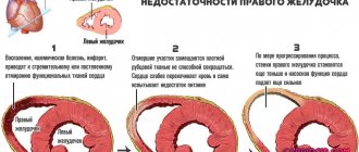
The development of venous insufficiency is accompanied by abnormal movement of blood to the lower part of the body, i.e. at the feet. This process is provoked by stretching of the vascular walls and disturbances in the functioning of the valves. In normal condition, the valve closes when blood flows upward. When a person develops insufficiency, the valve stops holding back blood. It starts to seep down.
Features of physiology
Perforators provide blood flow from superficial to deep vessels. Valve insufficiency leads to reverse blood flow and the development of varicose veins.
There are many perforating vessels located in the lower leg. They pass through muscle tissue and connect deep veins to superficial ones. Duplex examination does not allow them to be seen. Most vessels of this type are equipped with valves. With their help, blood moves from the limbs up to the heart. If the valves are malfunctioning, blood flow moves in the opposite direction. This phenomenon is called venous incompetence. Poor circulation leads to stagnation and the development of varicose veins, thrombophlebitis and thrombosis.
Causes
Incompetent valves in the veins of the lower extremities require immediate treatment, but the problem is that the etiology of its development is not entirely clear. Common reasons that influence the appearance of diseases of the vascular system include excess weight, lack of physical activity and excessive stress on the veins. Sometimes the catalyst for the development of the disease is hormonal imbalances associated with the development of the body or pregnancy. Potential causes of venous insufficiency include:
- age-related changes in the body;
- problems in the gastrointestinal tract (constipation);
- hypertension;
- taking medications that affect blood viscosity and hormone levels.
Symptoms and manifestations of pathology
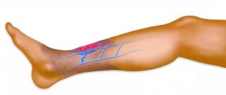
The disease manifests itself similarly to varicose veins. The patient complains of a feeling of constant heaviness in the legs and swelling. A patient develops spider veins on his legs. As the disease progresses, pain is added to these manifestations. Due to lack of nutrition, the skin becomes whitish-yellowish in color. Some patients experience seizures. Most often, patients encounter them at night, which leads to chronic fatigue. If the valves are insufficient, a person may experience the following symptoms:
- hair loss on the legs;
- severe itching of the skin;
- a sudden feeling of numbness in the limbs;
- excessively low foot temperature;
- sudden feeling of heat.
Features of the manifestation of the disease directly depend on the stage of deficiency. In some patients, the disease may take years to develop, so they do not pay attention to the symptoms present. During a medical examination, when visually assessing the patient’s condition, a surgeon can notice signs of venous insufficiency, but it is better if the patient himself goes to a phlebologist if there is frequent swelling of the legs and a persistent feeling of heaviness in the limbs.
Diagnosis of venous insufficiency
When determining the disease, the patient's complaints and examination results play a role. In addition to a visual assessment of the condition of the vessels of the lower extremities, laboratory diagnostics and hardware examination of the legs will be required. The doctor assesses the patient’s general condition and then gives directions for the necessary tests. Hardware methods for diagnosing valve insufficiency include:
- Ultrasound with Doppler sonography;
- phlebography;
- lymphography.
A general blood test is mandatory. Based on the indicators of platelets, hemoglobin, and red blood cells, the phlebologist can draw a conclusion about the indicators of blood viscosity. If they deviate significantly from the norm, the patient is referred for evaluation of hemostasis. This procedure will help determine the likelihood of blood clots in a particular patient.
Diagnosis of incompetence of the ostial valve of the great saphenous vein
To establish an accurate diagnosis, the doctor collects an anamnesis of complaints, a history of concomitant diseases and conducts an external examination of the affected limbs. Further, the following laboratory and instrumental studies are used:
- Biochemistry of blood. Establishes the quantitative ratio of important elements and plasma coagulability indicators.
- Duplex scanning of veins. Determines an increase in the lumen of blood vessels and a decrease in blood circulation rate, as well as thickening of the valve walls.
- Vascular ultrasound. There is a structural change in the veins, a violation of patency and a change in the closure of the valves.
Types of disease
When it comes to problems with valves, doctors talk about acute and chronic venous insufficiency. The disease can also be functional and lymphovenous. Acute venous insufficiency is considered the most life-threatening. It is characterized by complete blockage of the vessel. The development of the chronic form of the disease can be completely stopped with timely treatment.
Chronic
Chronic circulatory failure is characterized by damage to the saphenous veins. If left untreated, it continues to progress, which can lead to damage to the deep veins and disability of the patient. The disease is accompanied by swelling in the lower leg. Not only the muscles of the lower leg suffer, but also the ankles. At the second stage of the disease, the patient begins to lose hair on his legs, and there is a violation of tissue pigmentation. At the third stage of chronic insufficiency, the patient is faced with ulcers and trophic disorders.
Acute
When a vein becomes blocked, blood flow is severely disrupted. Doctors call this condition acute vascular insufficiency. This condition is dangerous for 2 reasons. Firstly, it always develops in the deep veins, which in itself is very bad, because They provide the body with 90% of the blood. Secondly, this disease most often develops under the influence of leg injuries. Based on this, we can say that the vessel is damaged or pinched.
The disease is accompanied by severe pain in the limbs. When you try to stand up or move your leg, the pain will intensify. If you suspect acute venous insufficiency, you should immediately call an ambulance. Cold compresses can relieve the patient's condition. They use dense natural fabric. It is dipped in cold water and then applied to the affected area. This manipulation must be repeated for at least 60 minutes. You cannot take any medications before the ambulance arrives.
Lymphovenous
This type of illness affects about 40% of all middle-aged people. The disease manifests itself in mild and severe forms. As the name implies, in patients with this disease, not only the flow of blood, but also lymph is hampered. This disease is treated with medications, but surgical intervention for this type of disease rarely has a positive effect.
Functional
A distinctive feature of this form of insufficiency is that it occurs in people with completely healthy veins. Most often, the cause of its appearance is a person’s height, excess weight and taking hormonal medications. Surgeons, teachers, hairdressers and other specialists who have to be on their feet for long periods of time face functional impairment. Treatment is mostly conservative. Patients take medications and wear compression stockings to prevent the disease from affecting the veins.
Classification
In Russia, phlebologists – vein specialists – have adopted the following classification of CVI, structuring this disease depending on its stage:
- Grade 0. No symptoms of CVI;
- Degree 1. Patients complain of pain in the legs, a feeling of heaviness, periodic cramps and swelling;
- Degree 2. Edema becomes more pronounced and persistent, increased pigmentation is noticeable, changes in the subcutaneous tissue of a degenerative-dystrophic nature (lipodermatosclerosis or “indurative cellulite”), dry or weeping eczema may appear.
- Degree 3. Expressed by the appearance of an open or healed trophic ulcer in the patient.
The “zero” degree was designated by Russian specialists in order to correctly determine the treatment of symptoms of chronic venous insufficiency of the lower extremities, as well as the disease itself. In this case, it will differ from the therapy required for CVI of the 1st or 2nd degree.
In medical practice, another structuring of venous diseases can be used, which is called CEAP and is international. It implies a “symptomatic” division of CVI according to the following criteria:
- There are no symptoms of the disease; upon palpation (feeling), CVI also does not make itself felt.
- Sustained dilation of small vessels is noticeable, but the inflammatory process is not started.
- Varicose veins are observed.
- Swelling is observed.
- Increased pigmentation of the skin is noticeable, eczema and degenerative-dystrophic changes in the subcutaneous tissue are possible.
- The symptoms listed in the previous paragraph are present in the presence of a healed trophic ulcer.
- The requirements are similar to the previous paragraph, but subject to a fresh trophic ulcer.
Each of the listed signs in this classification is considered separately, and depending on the degree of manifestation, it is given the corresponding score - “0”, “1” or “2”.
Under the auspices of this classification, the degree of disability due to venous insufficiency is also established:
- Degree 0. The patient is fully able to work, there are no symptoms of the disease, the patient does not need special therapy;
- Grade 1. The patient has some symptoms of CVI, but does not have any disability. Such patients also do not require special treatment;
- Degree 2. The patient is able to perform work only if treated with prescribed medications;
- Degree 3. Complete loss of ability to work.
Venous insufficiency of the lower extremities, as well as its symptoms and treatment, continue to be studied by doctors. New techniques appear regularly that allow you to quickly cope with the disease. Although before choosing the appropriate option, the specialist conducts a detailed examination. Through it, it is possible to identify a certain type of illness.
- Chronic failure;
- Valve insufficiency;
- Acute failure.
In all three cases, medications are used. Through them, it is possible to quickly remove the manifestation of changes in order to restore normal blood flow. Before studying it, you should familiarize yourself with a detailed description of each type.
Chronic failure
Chronic venous insufficiency of the lower extremities is considered virtually harmless, but it is a common mistake. It develops over time, so it is not immediately detected. Only a detailed examination provides an accurate picture, allowing one to understand the causes of a serious disease.
CVI is a serious problem. Its development is accompanied by the formation of trophic ulcers, and after them one usually has to deal with new ailments. They are a consequence of improper blood circulation in the legs, which leads to complications. Doctors advise attending an examination at the first suspicion, so as not to aggravate the situation.
Valve insufficiency
Doctors rarely detect valvular venous insufficiency of perforating veins. It occurs in a small number of patients, but is also a serious pathology of the musculoskeletal system. In this case, the saphenous veins become blocked, which not only disrupts proper blood flow, but in some cases leads to movement in the opposite direction.
Treatment of vein valves remains one of the most complex processes. The course of medications remains part of the long-term recovery of the legs, which occurs along with special exercises. Otherwise, it is not possible to undergo complete rehabilitation, which negatively affects the person’s movement.
Acute failure
Acute vascular insufficiency is the most striking manifestation of a serious disease. A sudden blockage of the veins leads to dire consequences, in particular, some forms of thrombosis. Development occurs in a short period of time, so only certain patients are able to undergo the examination in a timely manner. Otherwise, doctors have to rely on common symptoms.
Acute insufficiency of the veins of the lower extremities quickly develops into other diseases. Medications are used to cure, but long-term rehabilitation will be required after them. So the patient will have to stay in hospital for a long time.
Forms of valve insufficiency
This disease is difficult to detect in the acute stage. The patient may suffer from just one symptom or face the full range of manifestations of the disease. The standard classification of CVI (chronic venous insufficiency) includes the following types of disease:
- deep vein insufficiency;
- insufficiency of the saphenous veins;
- insufficiency of perforating veins.
Subcutaneous and perforating vessels are most often affected. Deep veins are very rarely affected. Doctors in such cases speak of a secondary form of the disease.
Deep vein insufficiency
This pathology occurs in patients with a genetic predisposition to varicose veins. The vascular walls of the legs experience constant high pressure. Blood begins to leak through the veins to other adjacent tissues, which causes severe swelling and the appearance of compacted areas. Smaller vessels also suffer from this disease, because The swollen tissues begin to put pressure on them. Treatment of deep vein insufficiency is often conservative. Patients are advised to move more and use heparin-based ointments.
Insufficiency of saphenous veins
Most often, patients are diagnosed with this type of disease. The valves of the subcutaneous channels are destroyed, which is accompanied by vertical reflux. Numerous studies confirm that absolutely all valves can be damaged due to the disease. Blood can flow into the hip and greater subcutaneous line. The disease is easily detected using ultrasound examination.
Insufficiency of perforating veins
Perforating vessels connect the deep and saphenous veins. Valve failure of this segment of the circulatory system is accompanied by horizontal blood reflux. This pathology can only be eliminated through surgery. Most often, the disease develops in the lower leg.
Insufficiency of perforating veins is accompanied by the destruction of muscle, collagen and elastic tissues. As a result, the blood begins to stagnate and the blood vessels become denser. Due to the fact that the valves do not work well, the outflow of blood is disrupted. Fluid enters the subcutaneous vascular network, which leads to pain, bulging veins and a feeling of heaviness. The disease is accompanied by trophic ulcers and thrombophlebitis.
Cause of perforating insufficiency of the veins of the lower extremities
The main factor provoking diseases of the blood vessels is considered to be genetic predisposition. Even if one of the parents suffered from varicose veins, this significantly increases the risk of developing the disease in children. In addition to burdened heredity, the following reasons for the development of pathology are distinguished:
Pregnancy can provoke the development of pathology.
- Pregnancy. Bearing a child and giving birth may be accompanied by an increase in intraperitoneal pressure, complicated by the formation of blood clots.
- Excess body weight. Every extra kilogram increases the load on the vessels of the legs.
- Gastrointestinal diseases. Incompetent perforating veins of the legs are diagnosed in people suffering from chronic constipation.
- Use of hormonal drugs. Self-medication with steroids or long-term use of oral contraceptives negatively affects the condition of the entire body.
- Excessive physical activity. The use of anabolic steroids by athletes, which provoke blood stagnation, is dangerous.
- Physical inactivity. Sedentary work leads to decreased venous tone and stagnation. If a person works while standing, the mechanisms that help move blood upward are depleted.
- Age-related changes. In older people, the elasticity of the venous walls is reduced.
- Hormonal imbalance. May lead to perforator insufficiency in adolescents during puberty.
Degrees of valve insufficiency
You need to start treating the disease from its first symptoms, but patients rarely turn to the doctor so quickly. More often, the patient comes when the valves are severely damaged and do not perform their functions well. Formally, there are 4 degrees of severity of the disease. Their detailed description is presented in the table below.
| Degrees | Features of manifestation |
| Zero | Patients have virtually no signs characteristic of valvular insufficiency. |
| Spider veins begin to appear on the skin. | |
| After prolonged or heavy physical activity, veins appear in the legs. | |
| At the end of the day there is a feeling of heaviness in the limbs. | |
| First | The heaviness in the legs increases. |
| A mild pain syndrome appears. | |
| The patient begins to get tired faster. | |
| Second | Swelling of the legs occurs not only at the end of the day or after physical activity, but also in the morning. |
| Pigment spots appear on the surface of the skin. | |
| The pain in the limbs becomes severe. | |
| There are problems with movement. | |
| Third | Trophic ulcers are added to the symptoms listed above. |
| Blood clots form in the veins, which leads to an increased risk of thromboembolism. |
Symptoms
The main reason for the development of incompetence of perforating type vessels is disruption of the valves and, as a result, reverse blood flow. Thus, stagnation of blood is formed, which after a certain time expands the walls of the perforating veins.
Due to this process, we can observe subcutaneous collections of blood. This is the first stage of varicose veins. If treatment is not timely and the problem is neglected, such areas become denser, and in the future the formation of trophic ulcers on the legs is possible.
The main symptoms of this disease are:
- severe swelling and pain in the lower extremities, especially at the end of the working day;
- spider veins on the legs;
- frequent cramps during sleep.
Insufficiency of the vein valves leads to the patient's skin becoming pale, dry, and peeling. The patient complains of discomfort in the form of pressure and heaviness, which intensifies after physical activity or prolonged standing. There is a feeling of numbness and crawling on the skin, which is associated with a disruption in the nutrition of nerve tissue.
The limbs become pale, cold to the touch and covered with pigment spots. The skin itches, hair falls out, and if the pathology continues for a long time, non-healing trophic ulcers may appear. The patient complains of periodic fever, fatigue and general weakness.
The disease can be complicated by pulmonary embolism due to a thrombus.
- embolism with a blood clot with the development of venous insufficiency;
- inflammation of the vessel;
- ulcers;
- pyoderma;
- prolonged bleeding from affected veins;
- heart overload;
- pulmonary embolism;
- poisoning of the body by venous blood accumulated in the vessels;
- gangrene of the limb;
- thrombosis of deep or superficial veins;
- sepsis.
CVI is distinguished by different symptoms at different stages of the disease. At the initial stage of its course, symptoms of venous insufficiency may either be completely absent or appear to a minor extent. Patients in this case express the following complaints:
- a feeling of heaviness in the legs, which intensifies with prolonged standing;
- increased swelling;
- periodically short-term convulsions, usually occurring at night;
- increased pigmentation of the skin in the area remote from the lower leg.
In the first stages of this disease, varicose veins are the exception rather than the rule, but sometimes they can also appear. At deeper stages of CVI, such a disorder, on the contrary, occurs in almost all patients.
As the pathology develops, the following symptoms may be added to the above symptoms:
- impairment of the ability of the circulatory system to deliver blood to tissues located in the lower
- limbs (in the affected area);
- the appearance of trophic ulcers;
- dizziness (sometimes accompanied by fainting) caused by excessive accumulation of blood in any of the vascular areas;
- the appearance of signs of heart failure.
Usually, with the disease “venous insufficiency,” the symptoms do not appear simultaneously, but complement each other gradually.
» Articles from an expert » Treatment
According to statistics, venous insufficiency of the lower extremities, the symptoms and treatment of which are determined by a doctor - a vascular surgeon or phlebologist, occurs in approximately 15-17% of the population.
For the convenience of differential diagnosis, CVI, in accordance with the existing symptoms, is divided into separate classes and stages.
Sometimes patients report slight tingling in the damaged area, heaviness and fatigue in the legs after physical fatigue. At the end of the day, there may be swelling in the lower extremities.
The second stage of the disease is characterized by more noticeable manifestations: the veins swell and protrude from under the skin. The disease is actively progressing.
If venous insufficiency of the lower extremities occurs, symptoms and treatment will become the main issue for patients.
Over time, blood flow is disrupted, which changes the shape of the veins and also disrupts normal muscle function.
Venous insufficiency of the lower extremities, as well as its symptoms and treatment, continue to be studied by doctors.
New techniques appear regularly that allow you to quickly cope with the disease. Although before choosing the appropriate option, the specialist conducts a detailed examination.
Chronic venous insufficiency of the lower extremities is considered virtually harmless, but it is a common mistake.
It develops over time, so it is not immediately detected.
Only a detailed examination provides an accurate picture, allowing one to understand the causes of a serious disease.
CVI is a serious problem. Its development is accompanied by the formation of trophic ulcers, and after them one usually has to deal with new ailments.
Doctors advise attending an examination at the first suspicion, so as not to aggravate the situation.
Doctors rarely detect valvular venous insufficiency of perforating veins.
It occurs in a small number of patients, but is also a serious pathology of the musculoskeletal system.
In this case, the saphenous veins become blocked, which not only disrupts proper blood flow, but in some cases leads to movement in the opposite direction.
Treatment of vein valves remains one of the most complex processes.
The course of medications remains part of the long-term recovery of the legs, which occurs along with special exercises.
Acute vascular insufficiency is the most striking manifestation of a serious disease. A sudden blockage of the veins leads to dire consequences, in particular, some forms of thrombosis.
Development occurs in a short period of time, so only certain patients are able to undergo the examination in a timely manner. Otherwise, doctors have to rely on common symptoms.
Signs of the disease are not so difficult to detect.
Pathology can have several types of course, and the chronic form is characterized by 4 successive stages.
What causes lead to the development of pathology, the main symptoms, signs and methods of treating venous insufficiency of the legs, including folk remedies, will be discussed below.
The main symptoms indicating the development of chronic venous insufficiency include: a feeling of heaviness in the lower extremities, bursting pain in the projection of the lower leg, the presence of paresthesia and convulsions. Depending on the duration of the disease, these symptoms have varying degrees of severity.
The main sign of chronic venous insufficiency is that all of the above clinical manifestations bother the patient after prolonged standing and an improvement in the condition is observed even after short-term rest of the limbs.
Clinical picture and diagnosis • Complaints •• No complaints - 4.5% •• Episodes of angina - 20.1% •• Syncope episodes - 1.0% •• Dyspnea on exertion - 31.4% •• Orthopnea - 2.8 % •• Symptoms of systemic venous hypertension - 25.6% •• Paroxysmal nocturnal dyspnea (cardiac asthma) or pulmonary edema - 32.4% •• Combination of dyspnea with syncope and episodes of angina - 0.8% •• Other symptoms - 1 .3%.
• Peripheral symptoms are caused by low diastolic and high pulse blood pressure. It should be borne in mind that all peripheral symptoms are nonspecific and are possible with neuroses, anemia, thyrotoxicosis, arteriovenous malformations, etc. •• Corrigen's sign (carotid dance) - pronounced pulsation of the carotid arteries •• High and fast pulse •• Musset's sign - shaking of the head with each pulse wave •• Müller's sign - pulsating uvula •• Pulsation of retinal arterioles •• Quincke's sign - pulsating color change lips or nail bed, synchronous with pulse;
determined by pressing on them with a glass slide •• Hill's symptom - the difference between blood pressure in the arms and legs is more than 20 mm Hg •• Double Traube tone - listening to loud (similar to a pistol shot) tones on the femoral arteries •• Durosier's symptom - systolic murmur on the femoral artery when it is clamped proximal to the site of auscultation and diastolic murmur when the femoral artery is clamped distal to the site of auscultation •• Listening to heart sounds on the palmar surface of the hand •• Listening to heart sounds on the palmar surface of the hand when raising the hand up •• Listening to the pulse above the superficial palmar arch .
• Valve symptoms •• Soft (flowing) diastolic decreasing murmur, following immediately after the aortic component of the second sound (best heard in the second intercostal space to the right of the sternum during exhalation when the patient’s torso is tilted forward), carried out to Botkin’s point •• Rough musical murmur (murmur “cooing pigeon”) occurs when the valve flaps or perforates the valve •• With decompensation and a pronounced increase in end-diastolic pressure of the left ventricle, the intensity of the murmur of aortic regurgitation weakens •• Austin Flint murmur is a mesodiastolic low-frequency murmur of relative mitral stenosis, arising in connection with the covering of the anterolateral mitral leaflet valve with a stream of regurgitation in severe aortic insufficiency.
Treatment options
There are different treatments for deep vein valve insufficiency in the legs. Unfortunately, surgery cannot cure the disease. Removing veins is effective if they are damaged by varicose veins, but valve problems will remain with you. The main goal of treatment is to normalize the functioning of vascular valves. This can be achieved in the following ways:
- taking medications that normalize tissue trophism and blood composition;
- performing specialized exercises for the legs;
- normalizing nutrition;
- resorting to physiotherapeutic techniques;
- by surgically removing severely damaged veins.
Methods of treating the disease directly depend on the health status of the individual patient. Compression therapy is not suitable for everyone, nor is taking anticoagulants. If your doctor allows it, you can use herbal infusions to normalize your condition.
Medicines
The basis of drug treatment is drugs that improve blood flow in tissues and help restore vascular elasticity. Doctors often prescribe anti-inflammatory drugs to patients, which not only help relieve pain, but also reduce general swelling of the limbs. When blood thickens, patients should take Aspirin and other antiplatelet agents. Most often, patients are prescribed the following medications:
- Lyoton;
- Chlorhexidine;
- Diclofenac;
- Troxerutin;
- Aescusan.
The type of medications prescribed depends on the patient's condition. For trophic ulcers, external preparations with antiseptics are prescribed, which will accelerate wound healing. For patients with dermatitis and eczema, doctors recommend using local glucocorticosteroids. Absolutely all patients are prescribed phlebotonics.
Magnetotherapy
As the name suggests, this physiotherapeutic treatment is based on the use of magnetic fields. It is believed that this effect reduces blood viscosity, eliminates joint pain, etc. The patient is placed in a special apparatus (magnetic turbotron), which generates magnetic fields. During the procedure, the characteristics of the magnetic field are determined individually for each patient. The course of treatment is 10-15 sessions. Doctors recommend undergoing procedures at the same time.
Compression therapy
This treatment method is based on increasing the ambient pressure around the veins. Compression therapy helps to enhance venous outflow, normalize the functioning of vascular valves, reduce blood viscosity, reduce the amount of interstitial fluid and relieve edema. This treatment helps get rid of leg cramps and promotes the healing of trophic ulcers. Compression therapy includes:
- wearing special jersey;
- use of elastic bandaging;
- hardware procedures for creating variable compression.
It is convenient for patients to use compression hosiery in everyday life. Putting them on is as easy as regular tights or stockings. All products are developed taking into account the anatomical structure of the legs, so they do not cause discomfort when worn. Compression products can be preventive and therapeutic. Their type is determined by the doctor. To prevent complications from valve insufficiency and in the absence of funds to purchase compression hosiery, you can practice elastic bandaging. The rules for applying a bandage are as follows:
- The roll is always unrolled outward.
- They begin to wrap the leg from the toes.
- The heel should be tightly wrapped with a bandage.
- Special pads are placed on the flat areas of the legs and pads on the ankles to prevent the formation of bedsores.
- Toward the hip, the pressure decreases rather than increases.
- If bandaging is done correctly, the fingertips will turn blue when lying down, but when walking they will return to their natural color.
Compression with the help of special devices is indicated for progressive valvular insufficiency. Pneumatic compression is most often used. Devices of this type can also be purchased for home use. Mercury compression is used if the patient has a severe form of deficiency, accompanied by trophic disorders in the tissues.
Folk remedies
Herbal treatment is considered complementary. By itself, it cannot help cope with valvular insufficiency, but it can eliminate some of the unpleasant symptoms of this disease. Healers advise people suffering from this disease to take a decoction of rowan bark. To prepare it, you will need 200 grams of dry product and 1 liter of boiling water. The bark is placed in a thermos, filled with liquid, and left to infuse for 10 hours. The resulting decoction is taken 30 ml/day 3 times a day. The following remedies are considered effective:
- Infusion of hazelnut bark and leaves. 1 teaspoon of each type of raw material is mixed, and then 250 ml of boiling water is poured. Take the resulting decoction 75 ml 3 times a day.
- Pine foot bath. 1 kilogram of crushed pine needles is poured with 5 liters of boiling water. The solution should sit for 1-2 hours. Use the bath mixture. The temperature of the liquid should not be too high.
Treatment of varicose veins.
The arsenal of treatments for varicose veins includes conservative therapy, sclerotherapy, and surgical treatment.
Conservative therapy.
What drugs are used to treat varicose veins and venous insufficiency?
The wide range of drugs available for sale today will amaze with its breadth. In general, the effect of the drugs is aimed at strengthening the vascular wall and reducing swelling of soft tissues.
Preparations of a number of flavonoids of plant origin - aescusan - an extract from horse chestnut, asklezan - an extract from the fruits of wild hazel, Detralex - an extract from the fruits of wild green orange, Ginkofort - an extract from tree fern and many others. It should be remembered that drugs help slow down or stop further deformation of the vein wall.
Eating a diet rich in fruits and vegetables leads to saturation with flavonoids, which helps strengthen the vascular wall, improves intestinal motility, and as a result, eliminates resistance to venous blood flow in the abdominal cavity. Eating at least 0.5 kg of vegetables and fruits per day for a person weighing 70-80 kg is considered sufficient.
Wearing elastic knitwear is strongly recommended - it is useful both as a means of treatment and as a means of prevention after surgery. The selection of elastic knitwear should be carried out by a doctor, since this underwear is both therapeutic and prophylactic, which needs to be worn for a long time. It is necessary to measure the circumference at the level of the ankle, under the kneecap, the middle of the belly, the thigh circumference below 5 cm from the perineum and three lengths, select the degree of compression - only a doctor can determine this.
Sclerotherapy.
A method of treating varicose veins, which consists in injecting a substance into the lumen of the vein that causes denaturation of the vascular endothelium and closure of the lumen of the varicose vein. Initially, iodine, alcohol, a solution of sublimate, and quinine were used for sclerotherapy. It was quite painful and often left scars on the skin. Later, a new generation of drugs for slkerotherapy appeared - salts of fatty acids: varicocid, thrombovar. This group of drugs is currently used. For sclerotherapy, we prefer Fibrovein - an English drug, highly purified, easily tolerated by patients, allergic reactions have not been noted. Despite the higher price, fiberglass has proven itself only from the best side.
Latest information: Ayurveda causes of varicose veins
Sclerotherapy is an outpatient procedure, but should be performed by an experienced specialist in a hospital. The manipulation is painless, after 2 hours you can go home. For a patient with slerotharpia, it is like an intravenous injection with the drug injected into a vein. Afterwards you will need to wear elastic knitwear or elastic bandages for 7 to 10 days.
Surgery.
Severe varicose veins will require the use of a surgical operation - phlebectomy, the operation consists of removing the affected veins from small accesses. The operation is performed under local anesthesia in case of local damage; in case of damage to the main veins, it is preferable to perform the operation under anesthesia. Usually the operation lasts about 1 – 2 hours. Already on the second day you will be able to walk. A hospital stay is required for 3 days. After surgery, it is strongly recommended to wear elastic bandages, and after 2 weeks you can switch to elastic knitwear.
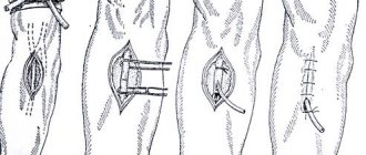
Phlebectomy. This operation is aimed at complete removal of the great saphenous vein. The surgeon creates two accesses - the first in the upper part of the thigh, the second at the level of the ankle; a probe is inserted into the lumen of the vein, with the help of which the affected vein is removed. When using cosmetic suture material, the seam is practically invisible and will completely disappear after tanning.
Outpatient phlebectomy. In the event that only tributaries to the main veins have undergone varicose transformation, you can use the technique of outpatient phlebectomy using a hook. This technique does not require an incision in the skin and does not require sutures; it is performed under local anesthesia. Using a hook, the vein is grabbed and pulled to the surface, then the surgeon performs ligation of the vein, division and removal. Only puncture points remain on the skin. After the operation, observation for 2 hours, then you can go home. After surgery, you must wear an elastic bandage for 2 weeks.
The choice of treatment method depends on many reasons - the level of the lesion, the extent of the lesion, the presence or absence of complications. therefore, only a doctor will help to correctly determine the possibilities and scope of treatment.
Consequences and complications
One of the common complications of valvular insufficiency is trophic changes in the skin. There are several negative points here. Firstly, it will not be possible to completely get rid of ulcers until blood flow is restored. Secondly, viruses, bacteria and other harmful microorganisms can enter the body through trophic damage to the skin. The most dangerous complication is considered to be thrombosis, accompanied by an embolus. A blood clot can travel through the circulatory system to the pulmonary artery, which will lead to thromboembolism. Possible complications of venous valve insufficiency include:
- dermatitis;
- erysipelas of the lower leg;
- formation of blood clots in deep and superficial veins.
What it is
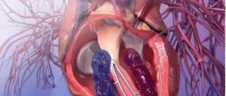
Venous valves are folds of the inner parietal membrane, consisting of muscle fibers. In the connecting vessels, they ensure the movement of blood from the subcutaneous to the deep, preventing reverse flow.
They are not evenly distributed in the circulatory system. They are found in large numbers in the lower leg - about 30, and the smallest of them are in the thigh - 2-3. There are no valves in arteries. There is only one in the body - the aortic.
The structure of the valve walls is specific. If their work is disrupted, a dangerous disease occurs. Violation entails not only complex therapy, but also possible amputation of the limb.
Prevention
People with a genetic predisposition to vascular diseases should take more walks in the fresh air. Walking promotes the natural flow of blood from the limbs. Regular warm-up will help avoid swelling and heaviness in the legs. In the office, you shouldn’t sit at the computer all the time either. Doctors advise doing light exercise or at least walking around the office every 2-3 hours. Prevention measures include:
- wearing therapeutic knitwear with a slight degree of compression;
- taking multivitamins;
- eating large amounts of plant foods;
- normalization of body weight;
- reducing the amount of salt consumed.
Doctors recommend that people with valvular insufficiency avoid heavy physical activity. Such patients should not get carried away with tanning, because... Ultraviolet radiation negatively affects not only the condition of the skin, but also the condition of blood vessels. If you have varicose veins, you should avoid baths, saunas and hot baths. They should be replaced with a contrast shower, which has a tonic effect on the whole body.
Functional tests for diagnostics
To determine valvular insufficiency in different parts of the venous network, venography and ultrasound in duplex scanning mode are used. But a phlebologist can obtain a preliminary conclusion about the state of the valve apparatus by performing functional tests.
The following tests are used to examine superficial veins:
- Troyanov-Trendelenburg - the patient lies on the bed, he is asked to raise his leg and strokes are carried out from the foot to the thigh to drain blood from the saphenous veins. In the groin area, the venous vessel is compressed with a tourniquet. After moving to a vertical position, the tourniquet is removed, and the doctor examines the rate of filling of the veins.
- Cough test - the surgeon’s hand is on the vein, the patient coughs. If there is valve insufficiency, then a push is felt at this time.
- Tapping - the doctor places his fingers along the dilated vessel, and taps with the other hand in the area of the great saphenous vein. When the valves are not closed, shocks are felt.
For perforating veins you can use:
- Test with a tourniquet and two bandages - in a lying position, an elastic bandage is applied to the leg from bottom to top. A tourniquet is fixed in the upper third of the thigh to compress the superficial veins. The patient stands up, and the doctor gradually removes the turns of the bandage, and a second bandage is wound on the free areas. Violation of the valves in a certain area can be detected between two bandages.
- Three tourniquet test - the patient raises the leg from a lying position. Install 3 tourniquets - the top of the thigh, above and below the knee. After getting out of bed, congested veins appear in the spaces between the tourniquets.