Contrasting coronary vessels is the most reliable method for choosing treatment tactics for patients with myocardial ischemia. Complications with this procedure are quite rare. Diagnostics involves the insertion of a catheter into the vessels of the heart and the supply of a contrast agent through it, so it can pose a potential danger to the patient. To prevent undesirable consequences, careful examination and preparation is necessary.
Consequences from the cardiovascular system
Medicine is constantly moving forward.
Those methods that a few years ago were available only to a limited circle of people with access to medical institutions abroad are gradually beginning to be introduced into domestic medicine. Such an unusual term as “coronary angiography” is increasingly heard in our hospitals. However, not all patients and their relatives understand the meaning behind these words, and in a stressful situation, when they need to make a decision quickly, they cannot always adequately evaluate the information that the doctor provides them. And very rarely do patients realize the possible risks and complications that may arise during or after coronary angiography.
Vascular access complications are one of the most common and severe complications of coronary angiography. The most striking symptom of these complications is bleeding from the puncture site of the artery.
It is important to remember that coronary angiography is performed through the arterial bed, in which the pressure reaches high values (above 100 mm Hg), so stopping bleeding from such a vessel is not so easy, especially if it is the femoral artery. After all, it is impossible to squeeze it above the puncture site.
In the first days after coronary angiography, the incidence of vascular complications is 0.7%-11.7%. Major bleeding and blood product transfusions are associated with longer hospital stays and decreased survival.
The use of small-diameter introducers, their early removal, control over the doses of anticoagulants, and the use of hemostasis devices allow doctors to reduce the risk of developing vascular complications of coronary angiography.
Although the incidence of complications is not very high, there are recommendations that, if followed, can reduce the risk of their development.
It should be remembered that the main way to prevent the development of complications is to choose experienced medical personnel. According to foreign colleagues, a doctor who performs more than 100 coronary angiographies per year can be considered experienced.
In some cases, coronary angiography is performed very urgently - in the first hours of myocardial infarction. In these conditions, preparation takes a minimum of time and boils down to the fact that the medical staff quickly asks the patient about complaints and medical history, conducts the minimum necessary examination, takes an ECG and draws blood for tests.
In addition, the patient receives the necessary medications for the treatment of acute coronary syndrome, and he undergoes catheterization of a peripheral vein. After this, the patient is transported to the operating room. This urgency is due to the fact that the time before surgery for acute myocardial infarction plays a huge role - the sooner it is performed, the better the result.
In most cases, coronary angiography is performed routinely. In order to prepare for it, the patient undergoes a detailed examination by a doctor, who conducts a survey and examination of the patient, evaluates laboratory and instrumental data. The patient should inform the doctor about his diseases that may affect the performance and complications of coronary angiography (for example, diabetes mellitus and kidney disease);
allergies to medications and food products; the medications he is taking. A laboratory (complete blood count, urinalysis, coagulogram, biochemical blood test) and instrumental (ECG, echocardiography) examination is carried out, which allows diagnosing concomitant pathologies.
Typically, before the procedure, the patient must:
- Follow the doctor's recommendations; You cannot independently use medications that were not prescribed to the patient.
- Do not eat or drink after midnight the day before your coronary angiography; Take prescribed tablets with a small sip of water.
- Shave the groin area and/or forearm through which the intervention will be performed. It is better to carry out this procedure using an electric razor so as not to damage the skin - this will reduce the risk of developing infectious complications.
- Take a hygienic shower the day before coronary angiography.
- Ask your doctor about the possibility of performing diagnostic surgery through the radial artery.
Carrying out coronary angiography through the radial artery can reduce the incidence of severe complications and mortality after the procedure.
Most often, the patient is prescribed sedatives before surgery, which will allow him to relax and unwind a little.
After the procedure, the patient remains in the hospital for at least one more day. At this time, his blood pressure and pulse are monitored, and medication correction is carried out.
Immediately after coronary angiography, the patient must strictly follow the doctor's recommendations about bed rest. The duration of the supine position depends on the site of surgical access (femoral or radial artery), whether the sheath was removed, and the method of hemostasis.
If hemostasis was carried out by pressing the femoral artery, you need to lie down for 6-8 hours; if a special device was used to stop bleeding, the patient can sit down after 1-2 hours.
Since the contrast agent is excreted in the urine, the patient should drink enough water, unless he has contraindications to this, and monitor diuresis (count the amount of urine).
You should immediately report any complaints or complications to medical personnel.
The intravenous catheter is removed a few hours after surgery, and the bandage over the arterial puncture site is removed the next day.
Most patients after planned coronary angiography are discharged home the next day. They may feel tired. A hematoma may remain at the site of arterial puncture for two weeks.
Upon discharge, the patient is advised to:
- Avoid bathing or showering for 1-2 days. In this case, you need to keep the wound dry.
- You should not drive a car for 3 days.
- No heavy lifting; Excessive physical activity should be avoided for 2-3 days.
A doctor should be consulted if the patient:
- bleeding appeared from the wound at the site of arterial catheterization;
- there is an increase in pain, swelling, redness and/or discharge at the site of arterial puncture;
- there is a hard, sensitive formation (larger than a pea) under the skin near the surgical access site;
- increased body temperature;
- discoloration, feeling cold, numbness in the leg or arm on the side of the body where the artery was catheterized;
- weakness or fatigue appeared;
- developed chest pain or shortness of breath.
Coronary angiography is the gold standard for detecting the presence and extent of atherosclerotic lesions of the coronary arteries. Fortunately, this is a relatively safe procedure with few complications. The use of modern equipment and medications, proper preparation of the patient before surgery, and the patient’s compliance with the postoperative recommendations of doctors - all this allows us to reduce the risks of coronary angiography to a minimum. And, of course, the greatest importance in preventing the development of complications belongs to the experience of the medical personnel who perform the operation.
Despite the fact that coronary angiography of the heart is one of the most indispensable methods in modern diagnostics and many experts consider this procedure to be the safest for the patient, in practice there are various consequences of coronary angiography. The probability of mortality after surgery is 1-1.5 out of 1000 people.
Despite the fact that the probability is very low, complications after surgery are extremely serious. Complications of coronary angiography include damage to the skin, possible allergic reactions (including to a contrast agent), and other complications that are more common in disease statistics, such as:
- Myocardial infarction. The fatal outcome is 0.5 per 1000 people. The most susceptible to it are the elderly, overweight patients, smokers and alcoholics, people with high cholesterol, as well as patients with diabetes. Myocardial infarction leads to heart failure and arrhythmia.
- Disabling complications. The fatal outcome is 0.4 per 1000 patients. These consequences of coronary angiography of the heart include: disturbances in the rhythm of the heartbeat, conduction, vascular and capillary diseases; disruption of nerve cells and damage to the human peripheral nervous system.
- Vascular complications. The fatal outcome is 0.6 per 1000 patients. This disease includes the appearance of scars on blood vessels and their narrowing. Vascular complications most often affect the older population. Since in the process of aging of the human body, blood vessels are more susceptible to various diseases.
- Transient ischemic attack. When an ischemic attack lasts longer than 24 hours, it is classified as a stroke. It is most often observed in patients with myocardial infarction and disorders of the heart valve apparatus.
- Heart rhythm disturbances (arrhythmia). Most often, this disease affects children due to overexcitement or fear. This complication can occur in a person without pathologies, so doctors cannot find out about arrhythmia in advance.
- Skin damage after coronography is not uncommon. Patients complain that their arm or leg hurts or that a hematoma has formed. During coronary angiography, the contrast agent is most often administered through the inguinal vein or brachial artery, so the pain is localized by puncture. A hematoma forms on the joints. The most serious symptom is when the arm seems to be “lost.” In this case, it is necessary to urgently contact a surgeon for a referral for an ultrasound scan and subsequent procedures. Often the consequence of coronary angiography is bleeding at the puncture site, even after removing the bandage and weight.
- Allergic reaction. The most common X-ray contrast agent is iodine. A runny nose and sore throat, watery eyes, as well as hives or a rash may occur.
In order to minimize the risk of the above complications, a competent examination and collection of the patient’s medical history is required. Unfortunately, neither specialists nor high-tech equipment are able to completely prevent the consequences of coronary angiography of the heart. However, the listed complications are extremely rare and therefore coronary angiography of the heart remains one of the advanced diagnostic techniques.
Possible complications of coronary angiography
Contrasting coronary vessels is the most reliable method for choosing treatment tactics for patients with myocardial ischemia.
Complications with this procedure are quite rare. Diagnostics involves the insertion of a catheter into the vessels of the heart and the supply of a contrast agent through it, so it can pose a potential danger to the patient.
To prevent undesirable consequences, careful examination and preparation is necessary.
Risks of coronary angiography for the patient
Since diagnosing coronary blood flow involves puncturing a peripheral artery of the thigh or shoulder, inserting a catheter through it, moving it through the aorta and coronary vessels, and supplying a contrast iodine-containing substance, this may be accompanied by a negative reaction of the body.
The risk of complications increases if the patient suffers from:
Depending on the stage of coronary angiography, it can cause the following complications:
- puncture of a peripheral artery - bleeding, hematoma, false aneurysm, fistula between artery and vein, wall dissection, thrombosis, embolism, vascular spasm, infection, allergy to painkillers;
- contrast – allergy, anaphylaxis, intoxication, kidney damage;
- administration of heparin - a decrease in blood clotting ability and, as a result, bleeding;
- insertion of a catheter - arrhythmia, embolism with parts of a cholesterol plaque, dissection of the aorta or coronary vessels, heart attack, stroke.
We recommend reading about coronary angiography of the heart vessels. You will learn about indications and contraindications for the procedure, preparation and performance of coronary angiography, recommendations during the recovery period.
And here is more information about bypass surgery of the heart vessels.
Possible complications after vascular reconstruction through the arm
The incidence of adverse effects of coronary angiography ranges from 0.05% (severe rhythm disturbances, vascular accidents) to 20 - 40% (allergy and violations of the integrity of the vascular walls). The occurrence of complications may depend on the presence of background predisposing factors or be a consequence of a violation of the procedure technique.
Air embolism
It happens in 0.2% of cases, it is associated with the passage of air bubbles into the bloodstream. Occurs when the integrity of the balloon at the end of the catheter or other technical errors in catheterization are violated.
With coronary angiography, air emboli are visible during the period of contrasting the coronary arteries. There are no symptoms, or pain in the heart appears, pressure drops, the rhythm of contractions is lost, until the heart stops.
Treatment involves aspiration (removal) of bubbles from vessels or injection of a solution under pressure to crush them. Patients are prescribed oxygen therapy, pain relief and antiarrhythmic drugs.
Hematoma and other vascular complications
The appearance of visible swelling of tissues and thickening of the skin at the site of arterial puncture occurs quite often. A large accumulation of blood in the hematoma cavity can lead to:
Hematoma at the puncture site after coronary angiography
For prevention, you need to carefully apply a pressure bandage, increase the time spent on bed rest after the procedure, and use heparin in smaller doses. Treatment is carried out using sufficient compression of the punctured vessel or by surgery.
With a false aneurysm, blood passes through a defect in the arterial wall during heart contractions into the hematoma cavity, and partially returns back during diastole. Occurs when the puncture is incorrect or there is insufficient compression of the artery after removing the catheter. Manifests:
- swelling of tissue at the site of puncture of the vessel;
- the formation of a hematoma that pulsates in time with the rhythm of the heart;
- pain on palpation;
- noise on auscultation.
The pseudoaneurysm may rupture, causing severe pain and increasing swelling. If compression of the nerve fibers occurs, weakness in the limb persists for several months. Patients are advised to rest and discontinue medications that inhibit blood clotting. For large aneurysms, thrombin is injected into the cavity or surgical removal is performed.
Pseudoaneurysm of the heart on echocardiography
Less common complications of vascular puncture are the formation of an arteriovenous fistula with edema, deep vein thrombosis and limb ischemia.
There is also dissection of the arterial wall against the background of atherosclerotic changes.
The occurrence of arterial blockage is possible in patients with thin vessels, concomitant obliterating diseases, aneurysms or a blood clot in the heart cavity.
Heart attack
The causes of acute coronary blood flow disturbance may be:
- formation of a blood clot at the top of the catheter;
- destruction of atherosclerotic plaque and movement of its parts along the artery;
- prolonged blocking of blood flow with a catheter at the site of narrowing of the vessel;
- dissection of the coronary artery;
- blockage of the branch when advancing the catheter to the bifurcation site;
- spasm in response to mechanical irritation of the vascular wall.
It is important to note that coronary angiography does not always cause the classic ECG picture of myocardial infarction (less than 1% of cases), but when studying specific enzymes, an increase in their level is observed in approximately 5 - 40% of patients. At the same time, asymptomatic and atypical cases of the disease are often found.
Arrhythmia
Life-threatening rhythm disturbances and cardiac impulse conduction during coronary angiography include ventricular tachycardia and ventricular fibrillation. They are associated with changes in blood flow during the procedure and injury to the conduction system of the heart.
In most patients, restoration of normal rhythm occurs independently at the end of the study. In cases of severe circulatory impairment due to arrhythmia, electrical pulse therapy is indicated. To prevent this complication, patients with electrical instability of the myocardium are prescribed beta-blockers.
Stroke
Factors that can lead to a stroke include:
- blockage by a blood clot formed due to arterial injury;
- dissection of the aortic walls;
- cholesterol embolus;
- arterial hypotension;
- administration of heparin (provokes intracerebral hemorrhage).
More often, this complication occurs in cerebral atherosclerosis, in elderly and senile patients who have had transient attacks in the past, cerebral ischemia, or have suffered a stroke. Manifestations of acute disturbance of blood flow in the brain include:
Nephropathy
For 1 to 3 days after using the contrast agent, the excretory capacity of the kidneys may be impaired. Nephropathy occurs more often in the following conditions:
- elderly and senile age;
- kidney disease in the past;
- diabetes;
- dehydration;
- shock or vascular collapse;
- circulatory failure with congestive processes in the internal organs;
- myocardial infarction;
- anemia;
- the use of drugs that destroy the renal parenchyma (non-steroidal anti-inflammatory drugs, antibiotics from the aminoglycoside group);
- injection of a large amount of contrast agent or rotary contrast.
Kidney damage can be reversible, but a third of patients develop renal failure. To prevent it, you need to take 0.5 liters of water before coronary angiography and at least 2.5 liters after.
In heart failure and edema syndrome, the amount of fluid is determined by daily diuresis.
Patients with reduced glomerular filtration may require hemofiltration to prevent nephropathy.
How to avoid complications
It is possible to prevent negative consequences with careful selection of patients for diagnosis. It is contraindicated in the presence of:
- severe renal and heart failure;
- arrhythmias with the threat of developing ventricular fibrillation or complete atrioventricular block;
- decompensated diabetes mellitus;
- bacterial endocarditis;
- malignant course of hypertension or symptomatic hypertension;
- allergic reactions and drug intolerance to iodine-containing drugs;
- discirculatory encephalopathy, stroke with a persistent neurological defect;
- obliterating lesions of the limbs;
- acute period of myocardial infarction;
- exacerbation of diseases of internal organs;
- infectious process.
Ultrasound of the heart and peripheral vessels
To identify these diseases, a prerequisite is preparation for coronary angiography, which includes assessment of the functional class of angina pectoris and heart failure, collection of information about concomitant diseases and previous vascular accidents. Patients are shown:
- ECG in 24-hour Holter monitoring mode;
- Ultrasound of the heart and peripheral vessels, kidneys;
- chest x-ray;
- general clinical blood and urine tests;
- blood test for viral hepatitis, HIV and syphilis, coagulogram, electrolyte composition, kidney tests, cardiac-specific enzymes, lipid profile.
We recommend reading about cardiac catheterization. You will learn about the indications and contraindications for cardiac catheterization, features of the procedure, as well as possible complications and the cost of the procedure.
And here is more information about CT angiography.
Coronary angiography is an invasive test, so complications may include damage to the vessels through which the heart is accessed. Adverse consequences also include thromboembolic complications, myocardial infarction and stroke, kidney damage, and heart rhythm disturbances.
To prevent negative reactions, a thorough examination is required before prescribing a contrast procedure for the coronary arteries of the heart.
Watch the video about errors during CT coronary angiography:
Source: https://CardioBook.ru/oslozhneniya-koronarografii/
Definition
Coronary angiography is a procedure that uses radiography to see the blood vessels of your heart. Coronary angiography is part of a general group of procedures known as cardiac catheterization.
Cardiac catheterization procedures can both diagnose and treat diseases of the heart and blood vessels. Coronary angiography, which can help diagnose heart disease, is the most common type of heart catheter procedure.
During a coronary angiography, a type of dye that is visible on an X-ray machine is injected into the blood vessels of your heart. The X-ray machine quickly takes a series of images (angiography), offering a detailed look at the inside of the blood vessels. If necessary, the doctor may perform procedures such as angioplasty during a coronary angiography.
see also
Indications for the procedure
The patient is recommended to undergo coronary angiography if he has symptoms or signs of coronary heart disease:
- angina pectoris;
- acute coronary syndrome (myocardial infarction);
- heart failure;
- before open heart surgery;
- in the presence of pathological changes on ECG or echocardiography.
In modern medicine, this is the most accurate and reliable method of identifying the location and extent of damage to the heart vessels.
My mother’s pain has stopped, so the issue has been postponed. My husband had a coronary angiography after a heart attack and was recently done through the arm. Moreover, I had to do 2 coronary angiographies with an interval of 3 Maxillofacial surgeon, otolaryngologist | Portal Vladmama Nov 22 A week ago on the bone behind the ear (in the mastoid process) Dysfunction of the TMJ joints, interstitial parotitis, etc.
Coronary angiography procedure - Medglobus Medglobus 13 Oct 3.0/18 At first everything was basically fine, only my arm hurt. Doctors refuse to do coronary angiography without consultation. On the second day after stenting, pain appeared in the chest. comrades..what the..crap. What is it if, out of the blue, blood starts to come out with excrement?
there is very little of it...but still. what to do!! I would like to cure everything without a doctor. Help. If blood is released during or after defecation, then it is either hemorrhoids or an anal fissure, but the fissure is also accompanied by severe pain. If blood is released BEFORE defecation begins, this is not a good sign at all, and you need to be seriously examined. Where.
depending on what you mean. What are you talking about. This is 3.14zdets! Da ne gowori, wesde tol'ko xren'. hemorrhoids or ulcers, if in stool; kidneys or urethra, if it’s in the urine, I’m surprised myself. what kind of crap..) for you it’s crap, but in people this is a vital organ called 2 options to choose from: internal bleeding or hem.
Oh yes, it’s nonsense, eat less holiday food, it will pass, no need to treat it with anything. haemorrhoids. Buy candles. Or maybe a rectal fissure. Better yet, go see a doctor. Looks like hemorrhoids. If there is itching, pain during bowel movements, a feeling of discomfort, heaviness, or prolapse of nodules from the rectum, then there is a high probability.
Contact a doctor, don’t be shy, he will make an accurate diagnosis and prescribe the correct treatment. red blood could be hemorrhoids or a fissure in the rectum or intestinal bleeding, go see a doctor. This is not crap, but hemorrhoids! Most likely, or you were silent about something in the question! one way or another, try entering hemorrhoids in a search engine and you will get a lot of answers on how and how to treat them!
Why does my face hurt? (Pain in the facial area) - Bighalt June 25 Pain in the facial area can have the following origin: Neuralgia Most often dysfunction of the temporomandibular joint How coronary angiography is performed, coronary angiography, stages After properly performed anesthesia, pain and feeling
Why is this being done
Your doctor may recommend that you have a coronary angiography if you have:
- Symptoms of coronary artery disease, such as chest pain (angina)
- Chest, jaw, neck, or arm pain that cannot be explained by other tests
- New or increasing chest pain (unstable angina)
- Heart defect you were born with (congenital heart defect)
- Heart failure
- Other blood vessel problems or chest injury
- Heart valve problem that requires surgery
You may also need an angiogram if you are about to have non-heart surgery, but you are at high risk of having a heart problem that occurs during surgery.
Because there is a small risk of complications, angiography is usually done after non-invasive heart tests have been performed, such as an ECG, echocardiography or stress test.
see also
How is coronary angiography performed?
To understand why and how complications develop during this diagnostic procedure, it is necessary to familiarize yourself with its stages.
- On the day of the procedure, the patient is taken lying down to the operating room. During coronary angiography, the patient is on the operating table in a supine position. The patient undergoes catheterization of a peripheral vein and begins infusion support.
- In most cases, coronary angiography is performed under local anesthesia at the site of arterial catheterization. The patient is awake at this time. The patient is given certain sedatives that calm him down and cause drowsiness and relaxation. General anesthesia is used occasionally - for example, when performing coronary angiography in children.
- During the procedure, the electrocardiogram, blood pressure, and blood oxygen saturation are monitored.
- The operation can be performed through two approaches - the femoral and radial artery.
- The catheterization site is treated with an antiseptic solution.
- The patient is covered with sterile linen.
- The arterial puncture site is anesthetized with a local anesthetic, after which the corresponding vessel (femoral or radial artery) is catheterized.
- An introducer is inserted into the artery, through which special diagnostic catheters are passed to the coronary vessels.
- After placing a diagnostic catheter at the origin of the left or right coronary artery, a radiocontrast agent is injected and at the same time X-ray angiography is performed. During the administration of contrast, the patient may feel a flash of heat or warmth, which quickly passes.
- The patient does not feel the catheter passing through his vessels. But he may feel a palpitation or arrhythmia.
- After examining the left and right coronary arteries in several projections, the catheter is removed. The sheath may be removed or left in the artery, depending on the results of coronary angiography.
- If the coronary angiography was performed through the femoral artery and the sheath was removed, the doctor will apply enough pressure to the area for about 10 minutes to stop any possible bleeding. After this, an aseptic dressing is applied.
- As an alternative to pressure, various hemostasis devices (eg, Angio-Seal) can be used.
- After the operation is completed, the patient is taken to the ward.
What is coronary angiography?
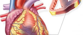
The heart is one of the most important human organs. Like any other organ, it can only function when it is sufficiently supplied with nutrients and oxygen from the blood.
Interestingly, the heart, which is filled with blood and pumps several liters of blood through it per minute, is very dependent on the relatively small arteries running along its surface.
The heart is supplied with blood by two vessels: the right and left coronary arteries
These arteries are called coronary arteries. The heart has two such vessels - the right and left coronary arteries, which supply blood, respectively, to the posterior and anterior walls.
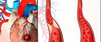
Over time, atherosclerotic plaques appear in the vascular wall of these arteries, which can block their lumen, partially or completely. This overlap leads to the development of coronary heart disease – angina pectoris and myocardial infarction.
Coronary heart disease is one of the main causes of mortality and disability throughout the world, and therefore occupies the most important place among the medical problems of our time.
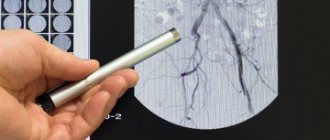
Coronary angiography is a procedure that examines the blood vessels of the heart (coronary arteries) using X-ray imaging. To do this, a radiopaque contrast agent is injected into a separate coronary artery and, in parallel, X-ray imaging is performed using an angiograph.
Consequences from the cardiovascular system
Coronary angiography. During the procedure.
Like most procedures performed on the heart and blood vessels, coronary angiography has some risks. Serious complications are rare, though. Potential risks and complications include:
- Heart attack
- Move
- Trauma arterial catheterization
- Heart rhythm disturbances (arrhythmias)
- Allergic reactions to paint or drugs used during the procedure
- Tears in your heart or artery
- Kidney damage
- Excessive bleeding
- Infection
- Blood clots
- Exposure to radiation from X-rays
see also
Like any invasive intervention, coronary angiography can have complications. Their severity ranges from minor and short-lived complications to life-threatening situations that can lead to irreversible consequences. Fortunately, thanks to improvements in equipment and increased experience of medical personnel, the incidence of complications has decreased significantly.
The risk of complications increases with the patient's advanced age, renal failure, uncontrolled diabetes mellitus, and obesity. From the cardiovascular system, the risk is influenced by the severity of coronary heart disease, features of the anatomy of the coronary arteries, the clinical situation (acute myocardial infarction, cardiogenic shock), congestive heart failure, low contractility, recent stroke or myocardial infarction, tendency to bleeding. The incidence of complications is also influenced by the experience of the medical personnel who perform coronary angiography.
However, severe complications are quite rare, occurring in less than 2% of patients; mortality rates are less than 0.08%.
see also
How do you prepare
There are other methods for diagnosing heart disease: cardiography (ECG) and echocardiography (ultrasound); MRI; X-ray examination (scintigraphy); ECG test under stress (bicycle ergometry). But only coronary angiography of the heart vessels helps the doctor to clearly see the causes of the disease - their narrowing, blockage, thinned areas of the walls (aneurysms). Angiography allows you to diagnose congenital heart defects in children and adults.
Instrumental examination of the heart vessels is prescribed less often than other diagnostic methods, since it carries more risks than non-invasive diagnostic methods. It is recommended for shortness of breath, chest pain, heart rhythm disturbances, the cause of which is unclear; and:
- when treatment with tablets and injections does not help, and symptoms intensify;
- for serious chest injuries;
- if a heart attack is suspected or in the first hours after it - to see the site of blockage of the vessel and remove the blood clot.
Coronary angiography of the heart vessels is the most reliable and most accurate way to diagnose coronary artery disease. Having identified the disease in time, the doctor decides on treatment tactics: whether stenting, coronary artery bypass grafting or angioplasty is necessary. On the eve of operations, it helps to more accurately identify the area of surgical intervention and see the condition of the heart vessels; in the postoperative period, it is carried out to evaluate the results of the intervention.
The procedure is not performed on people with severe diseases of the circulatory system, kidneys and lungs; patients with reduced blood clotting or bleeding; at elevated body temperature. It is not recommended, but in some cases it is allowed for diabetics and (with caution) elderly people.
Before coronary angiography, the patient is prescribed a urine test and blood tests - general, biochemical, coagulability, HIV and hepatitis, group and Rh factor. In addition, cardiography, cardiac ultrasound with Doppler, and chest x-ray are performed.
If you come to the examination from home, rather than wait for it during hospitalization, you will have to remember a few simple rules. They largely coincide with recommendations for preparing for any surgical interventions:
- The day before and on the day of the procedure, you should not eat or drink, otherwise nausea and vomiting may occur during the procedure.
- Take any medications you take with you, but check with your doctor before taking pills on the day of your coronary angiography. This is especially true for insulin. Since you cannot eat on the day of the procedure, you most likely will not need insulin: without eating, your blood glucose levels drop sharply, and a hypoglycemic coma is possible.
- Tell your doctor about any drug allergies you have.
Before the procedure, you need to empty your bladder. The doctor will also ask you to remove rings, earrings, chains, glasses, and contact lenses.
Before going for the procedure, it makes sense to learn not only about what coronary angiography is, but also to inquire about how it is done. The study of cardiac vessels is often carried out in a cardiology hospital, less often on an outpatient basis, in specialized multidisciplinary clinics. The procedure lasts from minutes to an hour:
- The patient's hair is shaved in the groin area (if the catheter is inserted into the femoral artery) or on the arm (when it is inserted into the radial artery) and given local anesthesia. The skin over the artery is then cut and a thin plastic tube is inserted. A very thin flexible probe with a catheter at the end is inserted through it and advanced through the vessels to the lumen of the coronary arteries.
- A special contrast agent is injected into the bloodstream through a catheter, and while it moves along the bloodstream along with the blood, the doctor takes X-rays with magnification. The results are recorded in digital format, and in the future the “picture” can be viewed on a personal computer.
- During this time, the patient's cardiac activity is monitored using electrodes attached to his chest. At the same time, his pulse and blood pressure are measured.
General anesthesia is not given during coronary angiography, but it is not necessary. There are no nerve endings inside the vessels, so you will not feel pain or experience other unpleasant sensations. From time to time, the doctor may ask you to take a deep breath, hold your breath, or change the position of your hands.
After the examination, you will have to lie down for quite a long time without bending your leg, and the doctor will forbid you to get up for hours. To quickly remove the contrast agent from the body, you need to drink more. Before discharge, you will be told when you can resume taking medications, whether dietary restrictions are necessary, and so on. For several days after the intervention, you should not engage in heavy physical work or train in the gym.
Nowadays, coronary angiography is done in two ways: through the femoral or through the radial artery. In the first case, the patient must be in the hospital; in the second, they come for the procedure in the morning, and in the evening they can return home. What is the reason for this difference?
- The peripheral (femoral) artery is one of the largest vessels in our body, lying at a depth of 2-4 cm under the skin in the groin area. Bleeding from it is very dangerous, so after the procedure you should not move much and do not rush home.
- The radial artery is a relatively small vessel that comes to the surface of the skin at the wrist. Doctors call this approach radial. Puncture of a vessel in this place very rarely causes life-threatening bleeding; the procedure itself lasts only 20 minutes and is less likely to cause complications. The patient is released home after 4-5 hours; in rare cases, he is detained until the morning.
If at home the puncture site is swollen and red, a large bruise appears in this area and sharp pain occurs, you should immediately consult a doctor. Sometimes the pressure drops at the same time, a sharp weakness is felt and shortness of breath begins - in this case, immediately call an ambulance.
Complications
Serious complications after coronary angiography are rare (on average less than 2% of cases), but they do exist, and you need to be prepared for them. First of all, these are allergic reactions to the contrast agent: skin allergies, shortness of breath, anaphylactic shock. The second group of complications is mechanical damage to the heart and blood vessels and consequences caused by the complex influence of the procedure:
- death from bleeding due to rupture of the heart or artery -
In some cases, coronary angiography is performed as an emergency. More often, however, they are planned in advance, giving you time to prepare.
Angiograms are performed in the catheterization (cathode) laboratory of the hospital. You usually go to the hospital on the morning of the procedure. Your health care team will give you specific instructions and talk with you about any medications you are taking. General recommendations include:
- Do not eat or drink after midnight the day before your angiography. Angiograms will often continue into the morning hours.
- Take all your medications to the hospital with you - in their original bottles. Ask your doctor if you should take your regular medications in the morning.
- If you have diabetes, ask your doctor whether you should take insulin or other oral medications before the angiography.
Before the angiogram procedure begins, the team's doctor will review your medical history, including allergies and medications you are taking. The team may perform a physical examination and check vital signs such as blood pressure and pulse. You will empty your bladder and change into a hospital gown. You may need to remove contact lenses, glasses, jewelry, and hair clips.
see also
Why does my arm hurt after coronary angiography?
" Health "
Question for experts: After coronary angiography of the vessels through the right arm, new bruises appeared, the arm hurts. What to do?
Sincerely, Natalya Samoilova (Shcherbatova)
Best answers
When you signed the consent for the intervention, there should have been a phrase stating that this study is invasive and complications and side effects from this medical procedure with a description are possible. The contrast leaked into superficially located vessels, which is why there are bruises, but the blood is not isolated in the heart, it penetrates into all other vessels, as well as into the coronary ones.
-answer
This video will help you figure it out
Answers from experts
Before the coronary examination, everyone is explained that the catheter is inserted through the artery. The artery is the largest blood vessel, after removing the catheter, the artery is pressed with a plastic ball. And after it was removed, you were told to lie on your back for a day and not toss and turn! You can’t get up for 24 hours! After that, don’t sit for another two days! it was necessary to follow the regime. Contact your doctor at the department where you had it done.
Make a compress with dimexide (pre-dilute 1:4) + 1 ampoule of analgin solution or semi-alcohol solution under film.
The largest arterial vessel in humans is the AORTA (located from the left ventricle of the heart in the chest and abdomen to approximately just below the navel)!! ! The femoral artery through which catheterization is performed has a diameter of about 7-10 millimeters! Dear “K”! I am a surgeon.
The matter may be more serious than you think! The catheter is about 2 mm. diameter and the femoral artery is punctured (pierced).
There are bleedings from this hole (for various reasons: non-compliance with the regime is only one of them!) in the tissue with the formation of hematomas (a cavity with blood, sometimes large in size - up to 5-8 centimeters! - it was necessary to operate) and (or) extensive bruising on the anterior surface of the thigh.
If you have a tumor (swelling) in the puncture area (upper third of the thigh), and you feel a semblance of pulsation with your hand, then quickly go to a surgeon (preferably a vascular one) - to the center where they did CAG (coronary angiography)! They will do an ultrasound.
If it is a hematoma, then it is necessary to operate - evacuate the hematoma and close the hole in the artery! If there is no swelling and you are simply worried about a huge “bruise,” then use heparin ointment (or LYOTON 1000) 2-3 times a day topically on the bruise if you are not allergic to heparin. Get better!
Research Institute named after Bakulev. I don’t know how much it costs, but coronary angiography is a mandatory procedure before installing a stent.
Stenting | Russian Cardiology Center RKNPK Ministry of Health RF Diagnosis of coronary artery disease using angiographic examination of the coronary arteries. .kardiology /stent/ — 22k — Saved in cache — Similar pages — Write
Coronary stentingCoronary stenting. ..Coronary stenting. Coronary stenting. Coronary stenting. On this site you can get information about ... .stent / - 24k - Saved in cache - Similar pages - Record
Stenting (coronary angioplasty) - Treatment in Germany ... Stenting (coronary angioplasty) is the most gentle method of surgical treatment for narrowing of blood vessels. Most often, stenting is used for ... dhaus /pages/stent/ - 22k - Saved in cache - Similar pages - Write
Stenting: Prevention and treatment of cardiovascular ... Stenting. Treatment of vascular atherosclerosis at the German Cardiology Center for patients from Russia and the CIS, including children. d-most.de/programm_stentirovanie - 20k - Coronography Vital systems · Departments · Doctors' advice · Guide. Coronography. Coronography. Coronary angiography is the most accurate and reliable... [link blocked by decision of the project administration] - 15k - Saved in cache - Similar pages - Write
Coronography Vital systems · Departments · Doctors' advice · Guide. Coronography. Coronography. Coronary angiography. Let's dwell on the description of individual stages... [link blocked by decision of the project administration] - 4k - Saved in cache - Similar pages - Write
Questions for a cardiovascular surgeon: CoronographyForum: Questions for a cardiovascular surgeon. Question: Coronography. dlinks /modules.php?op=modload&name=Forum&file=viewtopic&topic=55196&forum=103 — 38k — Saved in cache — Similar pages — Write
Questions for a cardiologist: coronography after a major heart attackForum: Questions for a cardiologist. Question: coronography after a massive heart attack. dlinks /modules.php?op=modload&name=Forum&file=viewtopic&topic=55542&forum=78 — 40k —
I don’t know about Coronography, but I have encountered stenting, although this was done to the child using an endoscope...
Coronography is, one might say, not an operation. It is an examination of the blood vessels of the heart for the presence of problem areas (narrowings) in them. Stenting is the installation of stents in areas of narrowing of these same vessels.
The operation is easy and painless under local anesthesia. About 30 minutes. You can be discharged the next day. I’m not a doctor, I just had such an operation after a heart attack about 3 years ago.
The Bakulevsky Center for Cardiovascular Surgery is a very good institution.
Don't be afraid of good health for you and your husband. The operation is expensive.
It all depends on the degree of damage to the coronary arteries, at what distance from the mouth the narrowing is located, and how many arteries are affected. It is not always possible to install a stent in a remote section of the artery. Sometimes it is enough to place a stent or two and not resort to bypass surgery.
But only a cardiac surgeon can answer this question after performing coronary angiography. Coronary angiography should be done in any case. This procedure is safe, but only it allows one to assess the situation in the coronary artery.
In some cases, especially when the circumflex artery or several vessels are affected, aorto-coronary and mammary-coronary bypass surgery has to be performed. This is done in almost all cardiac surgery centers. Here in St. Petersburg - at the Institute of Cardiology named after. V.A. Almazova.
Prices vary for different procedures and operations. You can find out more after you choose where to get treatment.
Source: https://dom-voprosov.ru/zdorove/pochemu-bolit-ruka-posle-koronarografii
What can you expect
For the procedure, you lie on your back on the x-ray table. Because the table may be tilted during the procedure, seat belts may be attached to the chest and legs. X-ray cameras can move through and around your head and chest to take pictures from different angles.
An intravenous (IV) line is inserted into a vein in the arm. You may be given a sedative through an IV to help you relax, as well as other medications and fluids. You will be awake during the procedure so you can follow the instructions. Throughout the procedure, you may be asked to take deep breaths, hold your breath, cough, or place your arms in various positions.
Electrodes on your chest monitor your heart throughout the procedure. A blood pressure cuff monitors your blood pressure and another device, a pulse oximeter, measures the amount of oxygen in your blood. You may be given drugs (anticoagulants) to help prevent your blood from clotting at the catheter and in your coronary arteries.
A small amount of hair may be shaved from the groin or arm where the catheter is to be inserted. The area is washed and disinfected, and then numbed with an injection of local anesthetic. A small cut is made at the entry site and a short plastic tube (sheath) is inserted into the artery. The catheter is inserted through the sheath into the blood vessel and carefully threaded to the heart or coronary arteries.
Inserting the catheter should not be painful, and you will not feel it moving through your body. Tell your doctor if you have any discomfort.
Dye (contrast agent) is injected through the catheter. When this happens, you may have a brief flushing or warm sensation. Don't be alarmed if you feel your heart skipping beats - this is a common occurrence during an angiography. But again, what is your doctor's command if you feel pain or discomfort.
Definition. After the procedure.
The dye is easy to see on x-rays, and as they move through the blood vessels, your doctor can observe its flow and identify any blockages or narrowing in the area. Depending on what your doctor finds during the angiography, you may have additional catheter procedures at the same time, such as balloon angioplasty or stenting, to open up the narrowed artery.
Having an angiogram takes about one hour, although it can be longer, especially when combined with other cardiac catheter procedures. You can add a few more hours to the preparation and post-care procedure.
When the angiography is complete, the catheter is removed from your arm or groin and the incision is closed with manual pressure, a clamp, or a small plug.
You will be taken to a recovery area for surveillance and monitoring. When your condition is stable, you will return to your room, where you will be monitored regularly. You will need to lie down for several hours to avoid bleeding. During this time, pressure can be applied to the incision to prevent bleeding and promote healing.
Sometimes, the plastic sheath that was first inserted into the blood vessel remains in place for several hours or even overnight if you have had angioplasty or stenting. If you received anticoagulants during the procedure, removing the sheath too soon may cause severe bleeding.
You may be able to go home the same day, or you may have to stay in the hospital for a day or longer. Drink plenty of fluids to help flush the dye from your body. If you feel for her, there is something. Ask your healthcare provider when you should resume taking your medications, bathing or showering, returning to work, and resuming normal activities.
Call your doctor if:
- You notice bleeding, new bruising, or swelling at the catheter site
- You develop increasing pain or discomfort in the catheter
- You have signs of infection such as redness, drainage, or fever
- There is a change in temperature or color of the leg or arm that was used for this procedure
- You will feel weak or weak
- You develop chest pain or shortness of breath
If the catheter site actively begins bleeding or swelling, apply pressure to the site, and contact emergency medical services.
see also
During the procedure
After the procedure
see also
Coronary angiography of the heart vessels - dangerous and non-dangerous consequences
Coronary heart disease (CHD) is the absolute leader in the world in the number of deaths. IHD is diagnosed when the blood supply to the heart is partially or completely disrupted due to damage to the coronary arteries. Most often, IHD is caused by progressive atherosclerosis, which impairs the patency of blood vessels.
Research methods
In the arsenal of modern medicine there are various methods for intravital study of the blood vessels of the human heart.
The most informative include:
- ultrasound Dopplerography of blood vessels (USDG),
- Cardiography of cardiac vessels with contrast agent,
- magnetic resonance imaging (MRI),
- angiography of the blood vessels of the heart,
- MSCT of coronary vessels (with and without contrast).
Cardiac ultrasound (US) is the basis of both Dopplerography and cardiography. MRI is a scan of blood vessels using a magnetic field and radiofrequency pulses. The essence of angiography is a contrast X-ray examination of the vessels of the heart. MSCT examination is carried out using a multislice computed tomograph.
The method is part of angiography. It got its name because it can be used to study the coronary vessels of the heart. In the medical literature you can find another name - coronary angiography.
Coronary angiography is often used in ischemic heart disease because it has earned a reputation as a reliable test of blood vessels in this disease.
In this regard, many heart patients and their relatives have a well-founded interest in how coronary angiography of the affected vessels is performed in coronary heart disease. Of interest are the possible negative consequences that such diagnostics of the arterial vessels of a diseased heart may have on human health.
Coronary angiography consists of two stages:
- preparatory,
- diagnostic procedure.
Preparation
The doctor must tell the person undergoing coronary angiography about the purposes of diagnosis, the procedure, and possible complications. The patient must inform the doctor about all diseases.
- The patient is given an electrocardiogram (ECG).
- Blood tests required:
- general,
- biochemical,
- for clotting,
- for the presence of a number of infections (HIV, hepatitis B and C, syphilis).
Be sure to do tests to determine the tolerability of the X-ray contrast agent and medications used in the procedure.
Contraindications
For a number of diseases, coronary angiography cannot be done:
- Contraindicated in people with uncontrolled hypertension, in whom stress during a coronary angiography procedure can cause a hypertensive crisis.
- Not performed after a recent stroke to avoid recurrent brain damage.
- Another prohibition is associated with decompensated diabetes mellitus, when there is serious damage to internal organs and the possibility of a heart attack cannot be ruled out.
- Internal bleeding or very low blood clotting is another reason to avoid coronary angiography.
- Kidney damage due to various diseases does not allow for coronary angiography, since a sharp deterioration in the patient’s condition is possible after the administration of an X-ray contrast agent.
- High temperature also makes coronary angiography impossible.
- Intolerance to the substance used for contrast during the procedure.
Procedure
Coronary angiography is performed on an outpatient or inpatient basis in the cardiology ward of a hospital.
- It is done on an empty stomach, before it you need to go to the toilet to empty your intestines and bladder.
- The places where the puncture of the vessel is punctured (wrist, armpit, groin, etc.) are shaved.
- In addition to the surgeon performing the operation, a resuscitator and an anesthesiologist are present in the room.
- Before the procedure, the patient takes a sedative so as not to worry too much and keep the heartbeat normal.
- During the operation, the patient lies on the operating table (on his back), his body is fixed so that damage to the vessel does not occur as a result of involuntary movement.
- After applying local anesthesia, a puncture of the vessel is made, through which access to the coronary arteries will be provided.
- An introducer plastic tube is inserted into the puncture site. It has a built-in hemostatic valve to prevent backflow of blood, an additional channel for collecting blood for analysis and administering medications.
- Through the introducer, the surgeon inserts a catheter, which is advanced into the area of the artery to be examined.
- After reaching the required position, a radiopaque agent containing iodine isotopes is injected by the catheter.
- The computer on the monitor shows a shadow image of a vessel in which a radiopaque substance is present.
- The study is done from several angles in order to obtain maximum information about the condition of the vessel or vessels of the heart.
- The results of the examination are recorded on digital media.
- After completing the procedure, the surgeon removes the catheter and introducer from the patient's circulatory system and applies a special bandage to the puncture site to stop the bleeding.
Depending on the scope of the study, the duration of the procedure ranges from 20 minutes to an hour.
Possible complications
Modern high-tech methods for studying heart vessels are quite safe. However, coronary angiography of the heart vessels can also have undesirable consequences, since the human body is complex, and it is impossible to foresee and calculate absolutely everything, even with an experienced surgeon and advanced medical equipment.
The cardiovascular system
The most severe consequences for the heart, brain and blood vessels are:
- myocardial infarction,
- stroke,
- perforation of blood vessels or heart cavity.
The probability of a heart attack is estimated at 1:1000. The risk of heart attack during or after coronary angiography is higher in patients with severe lesions of the coronary arteries.
Lower chance of stroke (7 in 10,000). It can occur in a patient if the movement of blood to the brain is blocked by a blood clot, cholesterol plaque, or air.
In 3-6 cases out of 1000, perforation or dissection of the coronary vessels or aorta is possible. The probability of injury to the iliac or femoral artery is estimated at 4:1000.
Vascular injuries are dangerous because retroperitoneal bleeding can occur, in which blood gradually accumulates in the retroperitoneal space. Moreover, blood loss occurs without visible external manifestations.
Complications that do not pose a direct threat to human life are more common.
In people suffering from diabetes mellitus with narrow lumens of blood vessels in the leg, when introducing an introducer and catheter that does not match the size of the vessel, thrombosis of the vessel of the lower extremity may develop. In this case, additional treatment will be required, either surgery to remove the blood clot or drug therapy.
If an artery and a vein are simultaneously damaged by a needle, an arteriovenous fistula can form. Probability 1:100. To eliminate it, surgery is required.
Hematomas often occur at the puncture site. If they are small in size, they will dissolve on their own.
If the hematoma is large in size, it can connect to the lumen of the artery, which leads to the appearance of a false aneurysm of the vessel. In most cases, surgery is not required.
Possible heart rhythm disturbances during diagnosis. More often the rhythm decreases (bradycardia). Less common are cases of increased heart rate (tachycardia) and uneven rhythm (arrhythmia).
Another common complication is a drop in blood pressure, which can be caused by various reasons related to the functioning of the cardiovascular system.
Allergic reactions
It is possible that an allergy may develop in response to the introduction of a radiopaque substance, sedatives, anticoagulants or antiplatelet agents, or anesthetics into the body. Therefore, preparation for coronary angiography includes careful checking of the patient's response to all drugs intended for the procedure.
If the check is not done properly, anaphylactic shock is possible, which threatens the patient's life. This happens extremely rarely, but more often a skin reaction is observed (rash, itching, redness).
Kidneys
An organ that may be damaged during coronary angiography.
In people with chronic renal failure, diabetes, or advanced age, the kidneys may not respond well to X-ray contrast agents.
Acute renal failure may develop. Severe complications require medical attention; for minor dysfunction, drinking plenty of fluids after coronary angiography is recommended.
Respiratory system
The most serious consequence is pulmonary edema. May develop due to heart failure and a severe allergic reaction. The risk of pulmonary edema is low, especially with careful preparation.
Thrombocytopenia
During coronary angiography, heparin is used, which reduces blood clotting. After a few days, thrombocytopenia may develop, triggered by heparin. Thrombocytopenia is a pathology characterized by a low number of platelets in the blood and increased bleeding.
Infections
Pathogenic pathogens enter the patient’s body at the site of puncture of the vessel.
To reduce the likelihood of infection, it is better to use an electric razor for shaving before diagnosis rather than razor blades, which can leave small scratches.
Strict compliance with hygiene requirements in the operating room is mandatory for medical staff.
After diagnosis, the puncture site should not be wetted with water for at least two days.
The easiest form of coronary angiography is considered to be when the catheter is inserted through the radial artery. If there are no complications, the person returns home within a few hours.
When access is made through the femoral artery, the patient remains in the hospital for a day.
A person who has undergone coronary angiography should monitor their well-being. If alarming symptoms appear (pain, weakness, low blood pressure, swelling at the puncture site), you should consult a doctor, and not expect that it will go away on its own, and not self-medicate.
Additional information about coronary angiography can be obtained from the video:
You can learn more about pathologies of the coronary arteries from the video:
Loading…
Source: https://dlja-pohudenija.ru/serdcze/posledstviya-koronarografii-sosudov-serdcza-metody-issledovaniya-i-vozmozhnye-oslozhneniya
results
Angiography can show doctors what's wrong with your blood vessels. He can:
- Show how many of your coronary arteries are blocked or narrowed by fatty plaques (atherosclerosis)
- Determining where blockages are located in blood vessels
- Show how much blood flow is blocked through your blood vessels
- Check the results of previous coronary artery bypass surgery
- Check blood flow through the heart and blood vessels
Knowing this information can help your doctor determine which treatment is best for you and how much of a risk your heart poses to your health. Depending on your results, your doctor may decide, for example, that you would benefit from having coronary angioplasty to help unblock blocked arteries. It is also possible that angioplasty or stenting may be done during the angiography to avoid the need for another procedure.
Risks of coronary angiography for the patient
Since diagnosing coronary blood flow involves puncturing a peripheral artery of the thigh or shoulder, inserting a catheter through it, moving it through the aorta and coronary vessels, and supplying a contrast iodine-containing substance, this may be accompanied by a negative reaction of the body.
The risk of complications increases if the patient suffers from:
Depending on the stage of coronary angiography, it can cause the following complications:
- puncture of a peripheral artery - bleeding, hematoma, false aneurysm, fistula between artery and vein, wall dissection, thrombosis, embolism, vascular spasm, infection, allergy to painkillers;
- contrast – allergy, anaphylaxis, intoxication, kidney damage;
- administration of heparin - a decrease in blood clotting ability and, as a result, bleeding;
- insertion of a catheter - arrhythmia, embolism with parts of a cholesterol plaque, dissection of the aorta or coronary vessels, heart attack, stroke.
And here is more information about bypass surgery of the heart vessels.