Types of cardiomyopathy
Cardiomyopathy is divided into three main types depending on the course of pathological processes in cardiomyocytes with corresponding changes in the structures of the heart chambers.
Stand out:
- Hypertrophic
- Dilatational
- Obstructive cardiomyopathy.
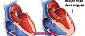
Hypertrophic cardiomyopathy
It is a thickening of the walls of the heart muscle, due to which the overall size of the heart can increase.
It can be diffuse (symmetrical), when the myocardium of the left ventricle is evenly and symmetrically thickened in all sections, including the interventricular septum. The right ventricle is extremely rarely involved in the process. This option is called non-obstructive hypertrophic cardiomyopathy, since the thickened myocardium does not affect the expulsion of blood from the left ventricle and intracardiac hemodynamics.
There is also an asymmetrical option. It occurs with and without obstruction.
Obstructive cardiomyopathy develops due to thickening of the upper wall of the interventricular septum. As a result, during heart contractions, this enlarged part blocks the lumen for blood to exit the cavity of the left ventricle. Intracardiac hemodynamics (blood circulation in the cavities) are gradually disrupted. This form is also called idiopathic hypertrophic subaortic stenosis.
Apical hypertrophic cardiomyopathy occurs when the lower part of the left ventricular myocardium thickens at the apex of the heart.
Unfortunately, the prognosis is quite pessimistic when a diagnosis such as hypertrophic cardiomyopathy is identified. The average life expectancy for this form of the disease will be about 15–17 years, which is slightly higher than with the dilated variant (does not exceed an average of 5–7 years).
However, the earlier the disease is detected, the higher the survival rate and the effectiveness of the treatment.
To exclude pathology, if suspicious symptoms occur, it is recommended to immediately contact a specialist for examination and timely diagnosis.
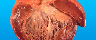
Dilated cardiomyopathy
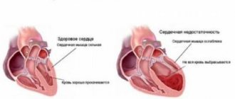
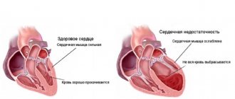
Its main manifestation is the expansion of intracardiac cavities with thinning of the walls and disruption of their contractile function.
Another name for this option is congestive cardiomyopathy: due to the development of myocardial weakness, congestion in the body is observed with the formation of persistent peripheral edema and edema of internal organs - up to the development of anasarca.
The causes of dilated cardiomyopathy are most often associated with hereditary predisposition and immune defects in the regulation of the body's defenses against infectious factors. As a result of exposure to an unknown cause, some of the myocardial cells die, followed by their replacement by fibrous tissue and a gradual “spreading” of the walls of the heart chambers in diameter. The myocardium becomes like a “rag”.
Dilated cardiomyopathy in children is the most common compared to other forms of the disease.
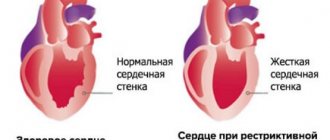
Restrictive cardiomyopathy
This form combines manifestations of fibrosis (hardening) of the myocardium and endocarditis (inflammation of valve structures). The walls of the heart become inactive and thick, and the ability to contract muscle fibers decreases. A common cause of such changes is the saturation of the myocardium with amyloid (a specific protein in amyloidosis). As a result, even in diastole, the heart does not relax (does not stretch due to blood flow), which also manifests itself in cardiovascular disorders.
There is another division of the entire pathology into forms of cardiomyopathy, the classification of which reflects the main causative factor of the disease. The disease is divided into idiopathic (primary) and secondary variants.
Idiopathic cardiomyopathy
This is most often a congenital cardiomyopathy, the causes of which are unknown.
Cardiomyopathy in newborns can develop as a result of intrauterine infections of a viral or bacterial nature. Also, such changes in the heart muscle can occur as a result of exposure to certain medications or chemicals, intoxication, or lack of nutrients. The influence of genetic factors and metabolic disorders cannot be excluded.
Cardiomyopathy in children can occur in any form of pathology, causing severe suffering and health problems, including death.
To help children suffering from cardiomyopathy and other chronic heart diseases, the national non-profit organization CCF was created. The main directions in her work are targeted assistance to sick children, as well as an active search and promotion of new methods for diagnosing and treating cardiomyopathy in children.
Secondary cardiomyopathy
This is a cardiomyopathy, the causes of which are known and can be determined in a particular patient. Among them, the main ones are the effects of alcohol, toxins, ischemia, and metabolic products.
Secondary cardiomyopathy, death from which is recorded in 15% of cases among other forms of cardiomyopathy, is divided into alcoholic, dyshormonal, ischemic and toxic.
Alcoholic cardiomyopathy
Develops as a result of prolonged exposure to alcohol (ethanol) on cardiomyocytes. More common in men suffering from alcoholism. Dystrophic processes develop in the myocardium with a gradual decrease in myocardial contractility and weakness of the heart muscle. Often accompanied by hepatitis, liver cirrhosis and pancreatitis, which also negatively affect overall metabolism.
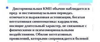
Dyshormonal cardiomyopathy
Develops with metabolic disorders in the body. The causes may be hormonal diseases such as hypo- or hyperthyroidism (diseases of the thyroid gland with decreased or increased production of thyroid hormones), menopause in women, as well as metabolic disorders of electrolytes, vitamins and microelements.
Cardiomyopathy occurs during pregnancy (myocarditis is diagnosed), when dystrophic changes are observed in the myocardium due to hormonal imbalance. One of the forms of dilated cardiomyopathy. After childbirth, in half of the cases it resolves on its own, the other half progresses to the point of serious congestion and the need for a heart transplant.
Ischemic cardiomyopathy
Develops as a final stage of coronary heart disease. It is one of the types of dilated cardiomyopathy. When collecting anamnesis, attention is paid to the course of symptoms typical of coronary artery disease, as well as signs of myocardial ischemia when conducting additional examination methods.
Toxic cardiomyopathy
May be caused by exposure to toxins, as well as physical factors (for example, ionizing radiation) on myocardial cells. Developing inflammation with transition to necrosis and fibrosis leads to the formation of secondary dilated cardiomyopathy.
Tonsillogenic cardiomyopathy
Diseases of the tonsils or adenoids can lead to metabolic disorders in the heart muscle. Chronic tonsillitis can cause the development of cardiomyopathy with degenerative processes in the myocardial stroma. Often develops in childhood.
Description of the disease
Dyshormonal cardiomyopathy is a disorder of the myocardial muscle that occurs when hormone levels change. In most cases, this is a reversible process caused by an imbalance in the endocrine system. Patients' metabolism slows down, and the blood supply to internal organs and tissues deteriorates.
In the International Classification of Diseases (ICD 10), the disease does not have a separate name; it belongs to code 142 “Cardiomyopathies”. This is one of the reasons for the appearance of internal abdominal fat covering organs, causing atherosclerosis and hypertension. With its development, the performance of the autonomic, lymphatic and circulatory systems decreases, swelling appears, and the heart works with overstrain.
Important! Among doctors, the disease is called “climacteric cardiomyopathy” due to its frequent development in women during menopause.
Degrees of compensation for cardiomyopathy
The classification presented below evaluates the general condition of patients in accordance with the severity of the disease. There are three stages:
1 – stage of compensation, clinical picture with minimal symptoms, hemodynamics at the proper level.
2 – stage of subcompensation, more pronounced symptoms of the disease appear with minor hemodynamic disturbances. In the heart muscle, dystrophic processes are moderately expressed.
3 – stage of decompensation, when ordinary physical activity causes serious hemodynamic changes, and myocardial systolic dysfunction is expressed significantly or sharply.
Why is cardiomyopathy dangerous?
Death with this pathology can occur suddenly. This is explained by the fact that acute cardiomyopathy can be complicated by severe disturbances in the rhythm and conduction of the heart that are incompatible with life. These include ventricular fibrillation and the development of complete atrioventricular block.
Acute myocardial infarction often develops with hypertrophic cardiomyopathy. When it spreads widely, acute heart failure develops.
Cardiomyopathy itself is not the main cause of death. Deadly complications develop against its background.
Pathogenesis
Under the influence of causative factors, the number of functionally functional cardiomyocytes decreases, which is accompanied by expansion of the heart chambers and a decrease in the pumping function of the myocardium. Dilatation of the heart leads to diastolic and systolic dysfunction of the ventricles and causes the development of congestive heart failure in the pulmonary and then systemic circulation. In the initial stages of dilated cardiomyopathy, compensation is achieved through the action of the Frank-Starling law, an increase in heart rate and a decrease in peripheral resistance. As the reserves of the heart are depleted, myocardial rigidity progresses, systolic dysfunction increases, minute and stroke volume decreases, and end-diastolic pressure in the left ventricle increases, which leads to even greater expansion. As a result of stretching of the ventricular cavities and valve rings, relative mitral and tricuspid insufficiency develops. Myocyte hypertrophy and the formation of replacement fibrosis lead to compensatory myocardial hypertrophy. With a decrease in coronary perfusion, subendocardial ischemia develops. Due to decreased cardiac output and decreased renal perfusion, the sympathetic nervous and renin-angiotensin systems are activated. The release of catecholamines is accompanied by tachycardia and the occurrence of arrhythmias. Developing peripheral vasoconstriction and secondary hyperaldosteronism lead to sodium ion retention, an increase in blood volume and the development of edema. In 60% of patients with dilated cardiomyopathy, parietal thrombi form in the cavities of the heart, causing the further development of thromboembolic syndrome.
The difficulties of modern medicine
Cardiac cardiomyopathy has many causative factors that directly affect the course and outcome of the disease. The large number of serious complications makes the prognosis of cardiomyopathy generally unfavorable.
The patient always has an increased risk of sudden death from thromboembolism or fatal arrhythmias.
In addition, the signs of heart failure gradually become more pronounced every year.
Of course, medicine has come a long way. And where the five-year survival rate barely reached 30%, now with early detection and adequately prescribed treatment, it is possible to extend the patient’s life for a longer period.
The transplantation industry is developing widely, giving hope to such patients. There are known cases after heart transplantation with life extension by more than one dozen years.
With the help of surgery, it is possible not only to replace the heart “pump”. Methods for eliminating obstruction of the left ventricular outflow tract in hypertrophic cardiomyopathy have positive results.
Cardiac cardiomyopathy is a direct contraindication to pregnancy in women, since maternal mortality with this pathology is very high.
It is not yet possible to prevent the development of cardiomyopathy due to the lack of knowledge about the causes of its occurrence.
Preventive measures are general in nature and are aimed at stimulating immune defense, preventing infection with infectious agents and maintaining a healthy lifestyle.
Symptoms of cardiomyopathy
Symptoms of cardiomyopathy are:
- the appearance of shortness of breath (especially during physical activity);
- increased fatigue;
- swelling of the legs;
- causeless cough;
- pale skin;
- blueness of fingers;
- the appearance of pain in the chest;
- frequent fainting;
- increased sweating;
- significant decrease in performance;
- increased heart rate;
- dizziness;
- sleep disorder and others.
Treatment of cardiomyopathy is long-term and quite complex. It depends on the form of the disease, the age of the person and the severity of his condition. Cardiomyopathy in children can be congenital. It develops due to a violation of embryogenesis. The prognosis for detecting this disease is not always comforting. Over time, the manifestation of all symptoms worsens, which leads to the development of pathologies that are incompatible with life.
Diagnostics
Dyshormonal myocardiopathy is clinically similar to diseases such as coronary heart failure, angina pectoris and heart attack. Therefore, making a diagnosis requires the use of several diagnostic methods.
The examination is performed by a cardiologist. First of all, he conducts a visual examination, listens to heart sounds, studies symptoms and medical history. Based on this, he can only make a preliminary diagnosis. The final conclusion is made after the patient undergoes the following examination methods:
- laboratory blood tests,
- electrocardiogram (ECG),
- ultrasound examination of the heart,
- magnetic resonance or computed tomography,
- chest x-ray.
Based on the results of these diagnostic methods, the attending doctor identifies the cause of hormonal imbalance, the stage of development of the pathology and, taking this into account, selects treatment tactics.
Dilated cardiomyopathy - symptoms
Dilated cardiomyopathy is most often observed in people of working age (20-40 years). But it is also possible that this disease is diagnosed in children or the elderly. Symptoms of the development of cardiomyopathy of this form are:
- the appearance of shortness of breath, which most often manifests itself during physical activity;
- increased heart rate;
- the appearance of edema;
- development of orthopnea - difficulty breathing at rest, which manifests itself in a horizontal position;
- complete intolerance to physical activity;
- attacks of suffocation that appear at night. This condition resembles that observed in bronchial asthma;
- fast fatiguability;
- upon palpation, a slight enlargement of the liver is observed;
- swelling of the veins in the neck;
- blueness of fingertips;
- increased frequency of urination in the evening and at night;
- decreased performance;
- muscle weakness;
- feeling of heaviness in the lower extremities;
- increasing the size of the abdominal cavity;
- feeling of discomfort in the right hypochondrium.
In approximately 3-12% of patients, this disease occurs without pronounced symptoms. Despite this, pathological processes in the cardiac and vascular system are worsening every day. This is especially dangerous when, when the first symptoms of cardiomyopathy appear, it cannot be diagnosed. But as the disease progresses, its signs are detected in 90-95% of cases.
Dilated cardiomyopathy can occur in a slow form. In this case, there is a slight enlargement of the heart, which is accompanied by mild shortness of breath, exercise intolerance, and swelling of the extremities. With rapidly progressing cardiomyopathy, acute heart failure, arrhythmia, suffocation and other symptoms are detected. In this case, most often the disease ends in the death of a person in approximately 2-4 years.
Hypertrophic cardiomyopathy in humans - symptoms
Signs of this type of cardiomyopathy are almost invisible in most cases, but as the disease progresses they become more pronounced. First of all, a person experiences shortness of breath. It manifests itself in some disturbance of respiratory activity, which can even turn into attacks of suffocation. At the initial stage of the disease, this symptom is observed during significant physical activity or during stress. But as hypertrophic cardiomyopathy progresses, shortness of breath develops even at rest. Due to the accumulation of blood in the vessels of the lungs, their swelling is observed. In this case, moist wheezing and cough appear. At the same time, sputum with foam is expectorated.
Also, with hypertrophic cardiomyopathy, the heart rate increases significantly. Usually, at rest, a person does not feel his heart beating, but with the development of this disease, it is felt even in the upper abdomen and at neck level. Due to the deterioration of the heart muscle, other tissues are not supplied with sufficient blood. This is accompanied by pale skin. At the same time, the limbs and nose are always cold, and the fingers may turn blue. This also causes significant muscle weakness and fatigue.
Another symptom of this disease is the formation of edema in the lower extremities, enlargement of the liver and spleen. Also, at the initial stage of hypertrophic cardiomyopathy, pain in the sternum is observed due to oxygen starvation of the heart. At the first stage, a person feels slight discomfort after physical activity. Over time, pain appears even in a calm state.
As hypertrophic cardiomyopathy progresses, oxygen starvation of the brain develops due to circulatory disorders. In this case, the sick person constantly feels dizzy. Also, with hypertrophic cardiomyopathy, frequent cases of sudden fainting are observed. This occurs when blood pressure drops sharply as the heart stops pumping blood.
Content
Definition and general information [edit]
Hypertrophic subaortic stenosis
Etiology and pathogenesis[edit]
Although the etiology of subvalvular stenosis is unknown, it is likely that the anatomical or physiological prerequisites for its development exist from birth. A discrete type of obstruction beneath the aortic valve annulus develops over time as a result of abnormal contractions or abnormal growth and hypertrophy. A multifactorial etiology of subaortic stenosis has been suggested, and possible factors include morphological features such as an excessively steep aortoseptal angle, a long and narrow LV outflow tract, excessive degree of curvature of the aortic root, and genetic predisposition. These congenital morphological features of the structure of the LV outflow tract increase hemodynamic stress on the area of the interventricular septum under the aortic valve, resulting in abnormal cell proliferation with the gradual formation of a fibromuscular ridge.
Therefore, a discrete subaortic membrane is not a congenital formation in the true sense of the word. It develops gradually throughout life due to the effect of turbulent blood flow on the abnormally formed LV outflow tract. In these patients, two-dimensional echocardiography shows that the angle between the long axis of the LV and the aorta is more acute than normal. Turbulent blood flow in the LV outflow tract causes hemodynamic injury to the endocardium, and then subsequent proliferation and fibrosis. The resulting fibromuscular membrane, in turn, increases the degree of turbulence in the blood flow leaving the LV, and due to constant hemodynamic trauma, fibrosis progresses. Turbulent flow also injures the aortic valves. Their subsequent thickening can lead to the impossibility of complete closure in diastole or to outright prolapse, which leads to aortic insufficiency. In some cases, the fibrous membrane extends directly under the aortic valve leaflets, causing their deformation and increasing regurgitation. The tunnel type of subaortic stenosis often develops secondary to previous resection of a simple subaortic membrane. Scar formation at the site of primary excision of the fibrous ridge at the junction with the abnormally formed LV outflow tract can lead to progressive fibromuscular proliferation with the formation of a tunnel in its outflow tract. The mitral valve also takes part in the development of this tunnel damage due to the location of the fibrous cord extending from the septum to the surface of the anterior MV leaflet facing it.
Rare causes of subaortic stenosis include septal attachment of the MV leaflet or excessive development of endocardial tissue attached to the anterior MV leaflet and pushing it into the LV outflow tract during systole. Both of these forms of obstruction are usually observed in combination with other defects, such as incomplete atrioventricular canal or L-transposition of TMS (cTMS).
Aortic regurgitation occurs in approximately 65% of patients with subvalvular aortic stenosis and often persists after correction of the subaortic stenosis. It places additional volume load on the LV, which is already suffering from pressure overload. Due to aortic regurgitation, coronary blood flow is reduced, which leads to increased ischemic damage to the myocardium. Increasing aortic regurgitation may require aortic valve replacement after excision of the subaortic membrane.
Clinical manifestations[edit]
Subaortic stenosis is characterized by its progression with a gradual increase in the degree of obstruction and the addition of aortic insufficiency. Subaortic stenosis rarely causes clinical symptoms until it becomes quite severe. On auscultation, a systolic ejection murmur is heard at the base of the heart, typical of obstructive defects of the LV outflow tract, and the second sound may be enhanced. With significant obstruction, intolerance to exertion and anginal pain appear.
Read also: Hypertrophic cardiomyopathy pictures
Although diagnosis may be made in infancy, clinically significant symptoms of subaortic stenosis rarely require surgical treatment before 3–6 years of age, most often occurring during adolescence. Obvious clinical symptoms of the defect include left ventricular dysfunction and exercise intolerance, and chest pain and syncope are less common.
In contrast to the discrete form of subaortic stenosis, the diffuse tunnel type of subaortic obstruction is often an integral component of complex hereditary syndromes.
Obstructive hypertrophic cardiomyopathy: Diagnosis[edit]
On a direct chest x-ray with pronounced stenosis, cardiomegaly and signs of venous congestion in the lungs appear. However, even in patients with severe subaortic obstruction, the size of the cardiac shadow is sometimes normal.
On the electrocardiogram, signs of LV hypertrophy are found in 85% of patients (primarily in cases with severe obstruction).
Doppler echocardiography clarifies the location and type of stenosis, the gradient of obstruction at the site of narrowing and the degree of hypertrophy of the LV walls. Typically, signs of discrete stenosis (membranes) under the aortic valve leaflets are visualized. Assessing the competence of the aortic valve is very important, since the occurrence of aortic regurgitation is usually an indication for surgical intervention.
Cardiac catheterization helps to assess the gradient at the site of narrowing and its level, but with sufficient echocardiographic information it is usually not required to make a decision on the type and timing of surgical treatment. Right and left sounding is recommended in cases of heart defects associated with subaortic stenosis.
Differential diagnosis[edit]
Obstructive hypertrophic cardiomyopathy: Treatment[edit]
Before surgery, β-blockers or calcium channel blockers are indicated and, if indicated, dual-chamber ventricular stimulation. Surgery is rarely used in childhood for this disease, but septal myectomy is a safe and effective procedure for relieving LV outflow tract obstruction.
Surgical treatment is indicated for symptoms resulting from stenosis or for obvious signs of progression of stenosis according to dynamic studies. You should not wait until subaortic obstruction causes pulmonary congestion as a result of increased LA pressure.
In asymptomatic patients, the gradient at the site of obstruction is greater than 30 mmHg. is an indication for surgery for a discrete type of stenosis and more than 50 mm Hg. - with tunnel type of subaortic obstruction. The appearance of aortic insufficiency, even with small gradient values, also usually indicates the need for surgical intervention. Some surgeons operate on such patients immediately after diagnosis, taking into account the progressive nature of the disease and the risk of further hemodynamic damage to the aortic valve.
Types of surgical treatment
1. Treatment of subaortic fibromuscular stenosis
Resection of subvalvular fibromuscular obstruction and septal myectomy are standard surgical treatments for this form of the disease.
2. Treatment of diffuse forms of subaortic stenosis
The surgical method is determined by the condition of the aortic valve annulus. If it is well developed, then septal ventriculoplasty is performed, the so-called Konno-Rastan operation. If the aortic valve ring is initially hypoplastic or damaged due to hemodynamic trauma during subaortic stenosis, then aortoventriculoplasty is performed, i.e. excision of obstruction of the outflow tract of both ventricles (classical Konno-Rastan procedure). In this case, the incision along the interventricular septum extends upward through the aortic ring and the aortic valve is additionally replaced with a mechanical prosthesis or the Ross-Konneau procedure is performed [Brown J. et al., 2006].
Read also: Alcoholic cardiomyopathy ICD 10
Prevention[edit]
Other [edit]
Early surgical mortality is less than 3% in most cardiac surgery centers. The survival rate after surgical treatment is 85-95% after 15 years. Late mortality is usually associated with residual (recurrent) subvalvular obstruction and repeated operations. A high rate of recurrence of subaortic obstruction (10-50% at 10-year follow-up) is associated with a high preoperative gradient (>50 mm Hg), tunnel type of obstruction, incomplete removal of the subaortic membrane, and age less than 10 years at the time of the first operation. There is evidence that the Ross-Konneau operation reduces the incidence of postoperative recurrence of stenosis in tunnel type obstruction.
Symptoms of restrictive cardiomyopathy
In restrictive cardiomyopathy, there are signs of serious heart failure, which is accompanied by the following symptoms:
- severe shortness of breath even at rest or with little physical activity;
- swelling in the lower extremities;
- accumulation of fluid in the peritoneum;
- an increase in the size of the liver, which is determined by palpation;
- fast fatiguability;
- muscle weakness;
- swelling of the veins in the neck;
- a significant decrease in appetite, and in some cases its complete absence;
- the appearance of pain in the abdominal cavity. In this case, the patient cannot determine their location;
- causeless weight loss;
- the appearance of nausea, sometimes vomiting;
- the development of a dry cough, which may be accompanied by the release of a small amount of sputum in the form of mucus;
- there is a slight itching of the skin, formations appear that are characteristic of urticaria;
- increased body temperature;
- excessive sweating, especially at night;
- dizziness, which may be accompanied by headaches.
With restrictive cardiomyopathy, general symptoms first appear that do not indicate a disturbance in the normal functioning of the heart. As the disease progresses, other signs appear that already clarify the picture of what is happening.
Signs of alcoholic cardiomyopathy
Alcoholic cardiomyopathy develops against the background of serious addiction, when a person takes a certain dose of ethanol-containing drinks every day for several years. In this case, there is a violation of respiratory activity. The patient suffers from constant shortness of breath or asthma attacks, especially after some physical activity. In parallel with this symptom, swelling of the lower extremities and tachycardia appear.
As the disease progresses, other more serious symptoms develop that indicate pathological changes in all systems:
- instability of emotional state;
- increased excitability;
- in some cases, excessive activity and talkativeness;
- increased fussiness;
- significant irritability;
- sleep disturbance, insomnia;
- inappropriate behavior;
- significant pain in the chest, where the heart is located;
- the appearance of hand tremors;
- legs and arms are always cold;
- redness and even some bluishness of the skin;
- increased sweating;
- blood pressure surges;
- severe headaches, migraines;
- dry cough that leads to chest pain.
Mechanism of disease development
With dismetabolic cardiopathy, pathological changes occur in the heart muscle. Against the background of the underlying disease, energy metabolism in cells slows down, which provokes the following processes:
- Myocardial tissue lacks oxygen and nutrients.
- Trying to compensate for the energy deficit, the body starts the process of increased cell growth. Hypertrophy appears, the ventricle or valve increases in volume.
- Chemical reactions are disrupted and toxins accumulate. Decay products provoke the death of heart muscle tissue.
Dyshormonal cardiomyopathy - symptoms
Dyshormonal cardiomyopathy is accompanied by the appearance of unpleasant nagging pain in the chest in the region of the heart. They suddenly become sharp and acquire a stabbing character. The pain is very intense, often radiating to the left shoulder blade, arm and lower jaw. This condition can last from several hours to 2-3 days. The pain can only be relieved with the help of painkillers. Its appearance does not depend on physical activity and can occur even during a period of complete rest.
Also, a sick person feels that he is getting hot. Unpleasant sensations are localized in the upper body in the face and chest. Because of this, a person sweats a lot. This condition is replaced by chills, the limbs become cold. Most often, such a person experiences a drop in blood pressure, tachycardia, dizziness, and tinnitus.
Main causes and risk factors
The main cause of dyshormonal cardiomyopathy is a slowdown in metabolism and a decrease in the level of certain hormones. They are involved in many vital processes, regulate fat and lipid balance, muscle cell regeneration, support heart function, and vascular tone.
Cardiomyopathy often occurs as a secondary manifestation or complication of the following pathologies:
- adrenal dysfunction;
- diabetes mellitus;
- prostate tumors;
- obesity;
- pathologies of the thyroid gland.
But in most cases, the disease occurs during menopause in women after 45 years of age. During menopause, the ovaries stop producing the amount of estrogen needed by the body. The adrenal glands begin to work with increased intensity, trying to correct the situation, the hormonal background changes. Adolescent girls are also at risk. Cardiomyodystrophy occurs when there is a malfunction of the reproductive system, congenital pathologies, ovarian or adrenal cysts.
Important! Inflammation of the heart muscle can occur with improper or uncontrolled use of hormonal contraceptives and medications for diabetes and bronchial asthma.
Doctors identify risk factors that increase the likelihood of developing the disease:
- late onset of the first menstruation in girls (after 15 years);
- a large number of abortions, miscarriages, missed pregnancies;
- early menopause at the age of 45–47 years;
- diseases of the female genital area (fibroids, cysts, fibroids, endometriosis).
In men, the disease is rare and is often associated with a prostate tumor, metabolic disorders and grade 2-3 obesity. Peak visits to a doctor occur between 45 and 55 years of age.
Treatment of cardiomyopathy
Cardiomyopathy is a dangerous disease that very often leads to death in patients. The causes of this disease have not been fully identified, however, it has been established that disorders in the development of the heart are often transmitted genetically. Therefore, prevention of cardiomyopathy is not always effective. It can only be carried out when planning a pregnancy in case of existing similar diseases in the family. As a preventive measure, a genetic consultation is carried out.
There are several types of cardiomyopathy, depending on which pathologies arise in which parts of the heart. There are dilated, hypertrophic and restrictive cardiomyopathy. Based on the diagnostic results, it is necessary to determine what type of cardiomyopathy the patient has in order to prescribe the correct treatment regimen for the disease.
Treatment of dilated cardiomyopathy
First of all, when treating dilated cardiomyopathy, it is necessary to eliminate heart failure, which is characteristic of this type of disease. This will help avoid further complications of the disease. When treating heart failure, beta blockers are used, which prevent stress hormones, such as adrenaline, for example, from being absorbed.
At the same time, angioprosthetic enzyme inhibitors should be prescribed, which will not allow blood pressure to increase. The most commonly prescribed drugs are Renitec or Cardopril, as they are highly effective and have fewer side effects.
In case of individual intolerance to ACE inhibitors, blockers of receptors that react to these enzymes are prescribed. This is a less effective but safer way to maintain normal blood pressure. A cardiologist who supervises the course of the disease must choose one or another drug to lower blood pressure. He will make a choice based on tests and individual patient indicators. Independent choice of medications is fraught with the development of serious complications.
Antioxidants have a positive effect on the myocardium. Therefore, they are also often used in complex therapy of dilated cardiomyopathy. If there is swelling during illness, it is very important to control the patient's urination volume in accordance with his weight. If during treatment it is determined that fluid is retained in the body, it is necessary to start taking diuretics. Within a week, with the correct dosage, the swelling should subside.
With small amounts of swelling, you can treat not only with medications, but also with folk remedies. Of course, only after consulting a doctor on this issue. Among the folk diuretics are elderberry, oregano, clover and much more. Traditional methods are no less effective than medicinal ones and have fewer contraindications. Homeopathy treatment methods are also practiced, however, studies of this treatment method have not shown positive results.
When treating dilated cardiomyopathy, blood electrolyte composition should be regularly studied, which will indicate its effectiveness.
Despite the correctly selected drug treatment, the prognosis for this disease is always negative. Most patients die within the first 5 years after the onset of the main symptoms. In addition, pregnancy is contraindicated for women suffering from dilated cardiomyopathy for several reasons. Firstly, the disease is transmitted genetically and can occur in a child. Secondly, during pregnancy the disease often begins to progress sharply. Thirdly, childbirth with such a diagnosis often ends in death for the mother, since the heart cannot withstand the excessive load.
That is why a heart transplant is necessary for effective treatment. However, today the waiting list for a donor heart is so long that patients often do not have time to live to see their operation and only a few manage to receive a new heart.
Currently, a method of treating cardiomyopathy such as stem cell treatment is practiced. These cells are universal and can be used as building material for any organ or part of the body. The stem cell donor is the patient himself after a thorough examination. These cells are extracted from bone marrow or adipose tissue and left in a special laboratory for further reproduction.
Effective treatment requires a large number of stem cells. Therefore, their reproduction takes some time. In total, at least 100 million cells are needed to start therapy. However, when the required number of cells appears, they still need to be prepared. To do this, using special chemical compounds, cells are converted into cardioblasts - the building material of the heart muscle.
Part of the finished material is frozen and stored for some time. This is necessary in case of relapse of the disease and repeated therapy. The cells are injected intravenously, after which they themselves find the affected area and restore it.
Myocardial dystrophy ICD code 10. instructions for use
Meldonium is a structural analogue of gamma-butyrobetaine, a substance that is present in every cell of the human body. Meldonium suppresses gamma-butyrobetaine hydroxygenase, reduces the synthesis of carnitine and the transport of long-chain fatty acids through cell membranes, prevents the accumulation in cells of activated forms of unoxidized fatty acids - derivatives of acylcarnitine and acyl coenzyme A. Under conditions of ischemia, it restores the balance of the processes of oxygen delivery and its consumption in cells, prevents disturbances ATP transport. At the same time, it activates glycolysis, which occurs without additional oxygen consumption. As a result of a decrease in carnitine concentration, gamma-butyrobetaine, which has vasodilating properties, is intensively synthesized.
The mechanism of action determines the variety of its pharmacological effects: increased performance, reduced symptoms of mental and physical stress, activation of tissue and humoral immunity, cardioprotective effect. In case of acute ischemic damage to the myocardium, it slows down the formation of a necrosis zone and shortens the rehabilitation period. In heart failure, it improves myocardial contractility, increases exercise tolerance, and reduces the frequency of angina attacks. In acute and chronic ischemic disorders of cerebral circulation, it improves blood circulation in the area of ischemia. Effective in case of vascular pathology of the fundus. The drug eliminates functional disorders of the nervous system in patients with chronic alcoholism during the abstinence period.
complex therapy of coronary heart disease (angina pectoris, myocardial infarction); chronic heart failure, dyshormonal cardiomyopathy;
complex therapy of acute cerebrovascular accident (ischemic stroke, cerebrovascular insufficiency);
reduced performance; physical overexertion (including among athletes);
withdrawal syndrome in chronic alcoholism (in combination with specific therapy);
hemophthalmia and hemorrhages in the retina of various etiologies, thrombosis of the central retinal vein and its branches, retinopathy of various etiologies (diabetic, hypertensive).
age under 18 years (efficacy and safety have not been established).
The safety of the drug during pregnancy has not been proven. To avoid possible adverse effects on the fetus, the drug should not be prescribed during pregnancy. It is not known whether the drug is excreted in breast milk. If it is necessary to use the drug Idrinol® during lactation, breastfeeding should be discontinued.
Can be combined with antianginal drugs, anticoagulants, antiplatelet agents, antiarrhythmic drugs, diuretics, bronchodilators.
Enhances the effect of cardiac glycosides.
Due to the possible development of moderate tachycardia and arterial hypotension, caution should be exercised when combined with nitroglycerin, nifedipine, β-blockers, antihypertensive agents and peripheral vasodilators.
JSC "Pharm"
141345, Russia, Moscow region. Sergiev Posad municipal district, rural settlement Bereznyakovskoye, village. Belikovo 11.
Tel/fax
Owner of the registration certificate: CJSC Pharm.
Consumer complaints should be sent to the manufacturer's address.
On prescription.
Synonyms of nosological groups
Heading ICD-10 Synonyms of diseases according to ICD-10I25.9 Chronic ischemic heart disease, unspecified IHD Coronary atherosclerosis in patients with IHD Coronary circulatory insufficiency I21 Acute myocardial infarction Left ventricular infarction Myocardial infarction without Q wave Myocardial infarction in the acute period Non-transmural myocardial infarction (subendocardial) Acute myocardial infarction Myocardial infarction with and without pathological Q wave transmural myocardial infarction Myocardial infarction complicated by cardiogenic shock Non-transmural myocardial infarction Acute phase of myocardial infarction Acute myocardial infarction Subacute stage of myocardial infarction Subacute period of myocardial infarction Subendocardial myocardial infarction Coronary artery thrombosis (arthrombosis) terium) Threatening myocardial infarction I20 Angina pectoris [angina pectoris] Heberden's disease Angina pectoris Attack of angina Recurrent angina pectoris Spontaneous angina pectoris Stable angina pectoris Angina syndrome X Angina pectoris Angina pectoris (attack) Angina pectoris Angina pectoris Angina pectoris rest Angina progressive Angina mixed Angina spontaneous Angina stable Chronic stable angina I50.0 Congestive heart failure Anasarca cardiac Decompensated chronic heart failure Congestive circulatory failure Congestive heart failure with high afterload Congestive chronic heart failure Changes in liver function in heart failure Cardiomyopathy with severe chronic heart failure insufficiencyCompensated chronic heart failureSwelling due to circulatory failureSwelling of cardiac originSwelling of the heartEdematous syndrome in heart diseasesEdematous syndrome with congestive heart failure Edema syndrome with heart failure Edema syndrome with heart failure or cirrhosis of the liver Right ventricular failure Congestive heart failure Congestive heart failure Heart failure with low cardiac output Chronic heart failure Cardiac edema Chronic decompensated heart failure Chronic congestive heart failure Chronic heart failure I4 3.1 Cardiomyopathy in metabolic disorders Dishormonal cardiomyopathy Climacteric myocardial dystrophy I67.9 Cerebrovascular disease unspecifiedAngioneuropathyArterial angiopathyCerebral hypoxiaDyscirculatory encephalopathyCerebral disease of a vascular and age-related natureComa due to cerebrovascular accident Lacunar statusMetabolic and vascular disorders of the brainImpaired blood supply to the brainImpaired cerebral circulationImpaired functions of the brainImpaired functions of the cerebral cortexImpaired cerebral circulationInsufficiency of cerebral circulationAcute insufficiency cerebrovascular accident Acute cerebrovascular accident Damage to cerebral vessels Progression of destructive changes in the brain Cerebrovascular accident Syndrome cerebral insufficiency Vascular cerebrovascular insufficiency Vascular encephalopathy Vascular diseases of the brain Vascular disorders of the brain Vascular lesions of the brain Functional disorders of the brain Chronic cerebral ischemia Chronic circulatory insufficiency Chronic cerebrovascular insufficiency Chronic cerebrovascular insufficiency Chronic cerebral vascular insufficiency Cerebral insufficiency Cerebral organic failure Cerebral vascular insufficiency school pathologyCerebroasthenic syndromeCerebrovascular diseaseCerebrovascular diseaseCerebrovascular disorderCerebrovascular disorderDiscirculatory encephalopathyI63 Brain infarctionIschemic strokeIschemic brain diseaseIschemic brain lesionsIschemic strokeIschemic stroke and its consequences Ischemic cerebral stroke Ischemic cerebrovascular accident Ischemic brain damage Ischemic brain damage Ischemic condition Cerebral ischemia Acute cerebral hypoxia Acute cerebral ischemia Acute ischemic cerebrovascular accident Acute cerebral infarction Acute ischemic stroke Acute period of ischemic stroke Focal cerebral ischemia Post-existing ischemic stroke Repeated stroke Syndrome Morgagni-Adams-Stokes rumChronic cerebral ischemiaCerebrovascular strokeEmbolic strokeI67 Other cerebrovascular diseasesPain syndrome with vertebrogenic lesionsVertebro-basilar insufficiencyVertebrobasilar insufficiencyCerebral circulatory disordersAcute disturbance of blood supply to the brainCerebrovascular insufficiencyZ73.6 Limitations in activity caused by decreased ability to workGeneral weaknessMental fatigueSyndrome of decreased performance and physical overstrainWeaknessDecreased motor activityDecreased performanceDecreased physical performanceReduced performance Reduced performance of the body Reduced performance when working in extreme situations Z73.0 Overfatigue Restoration of physical performance of athletes Restoration of energy body statusGeneral weaknessMental fatigueE63 Physical and mental overloadRecovery period in athletesRestoration of physical performance of athletesHigh psycho-emotional stressHigh psycho-emotional stressHigh physical and mental stressHigh physical stressProlonged overload and stressProlonged physical and mental stressProlonged physical activityIntellectual stressIntense mental and physical stressIntense physical activityStrong mental activityImpaired mental work abilities Lack of nutrition due to physical and mental overload Nervous -emotional overload Nervous and physical overstrain Nervous overstrain Nervous overfatigue Increased mental and physical stress Increased mental stress Increased physical activity Increased physical and mental stress Increased physical and psycho-emotional stress Increased physical and emotional stress Increased physical stress Increased psycho-emotional stress Increased physical stress Decreased mental work abilities Decreased physical and mental performance Reduced performance Mental and physical fatigue Psychological and physical stress Psycho-emotional overload Psycho-emotional stress Psycho-emotional disorder Syndrome physical overstrain Decreased mental performance Reduced tolerance of psychophysical stress Reduced mental performance Fatigue state Mental and psycho-emotional stress Mental and physical stress Mental and physical overload Mental and physical fatigue Mental stress Mental and physical exhaustion Mental and physical stress Mental and physical stress Mental and physical fatigue Mental overload Increased physical and mental physical loadPhysical and mental stressPhysical and mental performancePhysical stressPhysical overloadPhysical and emotional overloadPhysical overloadPhysical stressPhysical and mental stressPhysical and mental stressPhysical overstrainPhysical stressChronic fatigueChronic physical overstrainExcessive physical and mental stressExcessive physical and mental stressExcessive physical stressExcessive mental and physical stressExtreme physical stressH44.8 Other diseases of the eyeballHemophthalmosEye adhesionsH35.6 Retinal hemorrhage bleeding Hemorrhagic retinopathy Retinal hemorrhage Retinal hemorrhage Hemorrhages in the eye Hemorrhages in retina at heights Repeated hemorrhage in the retina Mouth spots F10.3 Withdrawal condition Alcohol withdrawal syndrome Withdrawal syndrome Withdrawal syndrome in alcoholism Abstinence Alcohol withdrawal Alcohol withdrawal Alcohol withdrawal condition Alcohol withdrawal syndrome Post-withdrawal disorder Post-withdrawal condition Hangover syndrome Withdrawal syndrome tions Alcohol withdrawal syndrome Alcohol withdrawal syndrome Withdrawal condition H34 Retinal vascular occlusions Arterial thrombosis of the vessels of the eye Venous thrombosis of the vessels of the eye Retinal circulation disorders Intraocular circulatory disorders Insufficiency of blood supply to the retina and choroid eyes Occlusion of the central vessels of the retina Acute obstruction of the retinal arteries Subacute and chronic circulatory failure in the retina or in the choroid of the eye Vascular diseases of the retina Vascular disorders in the retina of the eye Thrombosis of the retinal vessels Thrombosis of the central retinal vein Thrombosis of the central retinal vein and its branches Thrombosis of the central retinal vein and its branches H35.0 Background retinopathy and retinal vascular changesAtherosclerotic retinopathyRetinopathyRetinopathyRetinopathy of newbornsH36.0 Diabetic retinopathy (E10-E14+ with a common fourth sign .3)Hemorrhagic diabetic retinopathyDiabetic retinopathyRetinal dystrophy in patients with diabetes mellitusH36.8 Other retinal disorders in diseases classified elsewhereAr theriosclerotic retinopathyAtherosclerotic retinopathyHypertensive retinopathy
Treatment of hypertrophic cardiomyopathy
The entire treatment regimen for hypertrophic cardiomyopathy is aimed at improving the contractility of the left cardiac ventricle. For this purpose, a drug such as Verapamil or Diltiazem is most often prescribed. They reduce the number of heart beats, slowing the pulse, and even out the rhythm of the heartbeat. Often, the drug Disopramide is added to the classic drug treatment regimen, which enhances the effect of the main drug.
For more serious forms of hypertrophic cardiomyopathy, pacemaker implantation is prescribed, which generates electrical impulses and normalizes the heart rate. This is the most effective way to treat hypertrophic cardiomyopathy, however, it is prescribed only if drug treatment is not effective.
Additional facts
The term “myocardial dystrophy” (secondary cardiomyopathy, myocardial dystrophy) in cardiology combines a group of non-inflammatory and non-degenerative myocardial lesions, accompanied by a pronounced disorder of metabolic processes and a significant decrease in the contractility of the heart muscle. Myocardial dystrophy is always a secondary process, including dysmetabolic, electrolyte, enzyme, neurohumoral and autonomic disorders. Myocardial dystrophy is characterized by degeneration of myocytes and structures of the conduction system of the heart, which leads to disruption of the basic functions of the heart muscle - contractility, excitability, automaticity, conductivity. Myocardial dystrophy, especially in its initial stages, is usually reversible, which distinguishes it from degenerative changes in the myocardium that occur with hemochromatosis and amyloidosis of the heart.
Treatment of restrictive cardiomyopathy
Restrictive cardiomyopathy is the most difficult to treat. Because the disease manifests itself too late. Compared to other stages, treatment is most often no longer effective. By the time the disease is finally diagnosed, the patient becomes unable to work and, as a rule, does not live more than 5 years.
Therefore, doctors do not know how to treat cardiomyopathy in this case, since all treatment can only be aimed at relieving symptoms and improving the patient’s quality of life and slightly prolonging its life.
At the moment, no reliable methods have been found to completely stop the pathological process. The fact is that even a placed implant may not save the situation, since relapses of the disease often occur on the implanted heart. When lining up for heart implantation, doctors take into account the specifics of this type of disease, and patients with other less complex types of diseases remain at an advantage. Therefore, surgery for this type of cardiomyopathy is a very rare case in medical practice.
Thus, the effectiveness and method of treatment depends on the type of disease, and the prognosis of treatment may also vary. Overall, cardiomyopathy is a dangerous disease that can be fatal even with proper treatment.