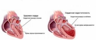
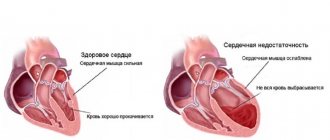
Heart failure is associated with decreased heart function. The heart muscle cannot produce the energy needed to pump the required amount of blood throughout the body.
In Russia alone, about 7 million people suffer from heart failure. In people over 70, one in four people are affected, with men typically affected at a much younger age than women. The risk for men is approximately one and a half times higher than for women. In Russia, diseases of the circulatory system are the most common cause of death.
- What is heart failure?
- What causes heart failure?
- What are the types of heart failure and what are their symptoms?
- What classes is heart failure divided into?
- How is heart failure diagnosed?
- How is heart failure treated?
- What are the chances of recovery from heart failure?
What is heart failure?
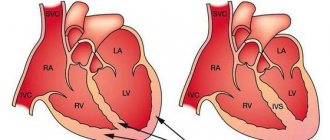
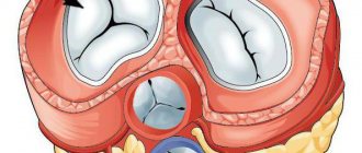
With a healthy heart, oxygen-rich blood from the left ventricle is pumped through the body to the organs, providing them with oxygen and nutrients. After supplying the organs, low-oxygen blood returns from the body to the right side of the heart, from where it is transported to the lungs. In the lungs, the blood is enriched with oxygen so that it can again be pumped throughout the body through the left ventricle.
Heart failure is a weakening of the pumping function of the heart. Typically, either the right side of the heart is affected (right-sided heart failure) or the left side of the heart (left-sided heart failure). With advanced heart failure, both sides of the heart can be affected (global heart failure). Heart failure can also be chronic or acute in nature. Chronic heart failure is more common than acute heart failure, which occurs suddenly and unexpectedly. Acute heart failure can occur suddenly against the background of an acute cardiovascular accident and/or decompensated heart failure.
What causes heart failure?
Heart failure is caused by diseases that affect or damage the heart muscle. The most common cause of chronic heart failure is coronary artery disease.
Coronary artery disease (CAD) is caused by narrowing of the coronary arteries (coronary arteries), most often due to atherosclerosis. Coronary arteries are vessels that supply the heart with oxygen and other important nutrients. Progressive narrowing (also called stenosis) of the arteries leads to poor circulation of the heart muscle. IHD is often diagnosed when angina (chest pain and tension) is present, but otherwise goes undetected.
A heart attack occurs due to decreased circulation of oxygen-rich blood to the heart muscle, leading to irreversible tissue death. This damage affects the pumping function of the heart, leading to heart failure. A large proportion of patients also suffer from high blood pressure, which further aggravates the situation.
High blood pressure (hypertension) is the single cause of heart failure in nearly 20% of people, making it the second most common cause of the disease. High blood pressure causes the heart to constantly work harder. The heart cannot work under additional stress for a long period of time, and therefore deteriorates.
This effect may be caused by a problem with the heart valve. When the aortic valves are narrowed or leaky, the heart must work harder or beat faster, which also increases the workload.
Bradycardia, a heart rhythm disorder in which the heart rate is reduced, can also cause heart failure because too little blood is circulating. A heartbeat that is too fast (tachycardia) is associated with a decrease in stroke volume and can therefore also lead to heart failure.
Inherited heart disease, pregnancy, autoimmune disorders, alcohol, drugs or medication abuse, an overactive thyroid gland and metabolic disorders (diabetes mellitus) can be causes of heart failure.
What does an electrocardiogram show?
An ECG is used not only to detect heart failure, but also other possible heart problems. It is carried out as follows: the patient takes a lying position, after which electrodes are connected to the arms, legs and chest, transmitting information to the machine, which prints out a cardiogram. These electrodes pick up any electrical activity of the heart, even the weakest. ECG shows:
- heart rate;
- condition of the myocardium (heart muscles);
- general condition of the heart.
Cardiography most accurately reflects the rhythm of heart contractions. Based on it, the cardiologist determines whether the patient has tachycardia (rapid heartbeat), or, conversely, the rhythm is too slow or there are any malfunctions.
Accordingly, a set of further clarifying diagnostics and treatment is determined:
- supraventricular arrhythmia - laboratory tests and a review of existing treatment are prescribed;
- ventricular arrhythmia - angiogram, exercise tests, study of the blood supply to the heart muscle, sometimes a special device is implanted - ICD (implantable cardioverter-defibrillator);
- ischemia or previous heart attack - echocardiography, angiography, coronary bypass surgery are prescribed to restore blood supply to the myocardium;
- left ventricular hypertrophy - Doppler ultrasound and echocardiogram are recommended.
The health of the heart muscle is considered a slightly less accurate indicator that can be extracted from an ECG. The fact is that dead tissue reacts in a special way to electrical signals passed through it. In this way, the doctor can determine whether the patient is predisposed to a heart attack, whether there are scars on the myocardial muscle tissue, dead areas of muscle, and so on.
However, it is impossible to rely entirely on the cardiogram readings regarding the condition of the heart muscle. If any abnormalities are detected, the doctor must refer the patient for further, more accurate diagnosis.
As for the general condition of the heart, a cardiogram allows you to see any problems only if the patient is observed during intense physical activity.
What are the types of heart failure and what are their symptoms?
Heart failure is divided into the following types:
- Left-sided heart failure
- Right-sided heart failure
- Global heart failure
- Systolic heart failure
- Diastolic heart failure
- Chronic heart failure
- Acute heart failure
Each type of heart failure has different symptoms, and the symptoms can vary in intensity. However, the main symptom of heart failure is difficulty breathing during exercise or at rest. Warning signs may include sweating with mild exercise, an inability to lie flat, tightness in the chest, or swelling in the legs.
Left-sided heart failure
The left side of the heart is responsible for pumping oxygen-rich blood throughout the body to the organs. In left-sided heart failure, the pumping function of the left ventricle is limited, resulting in insufficient oxygenated blood to be pumped throughout the body. Instead, the blood remains in the pulmonary circulation, which can lead to fluid in the lungs (pulmonary edema), difficulty breathing, throat irritation, a rattling sound when breathing, weakness, or dizziness.
It is most often caused by coronary artery disease (CHD), high blood pressure or a heart attack, and less commonly by problems with the heart muscle or heart valves.
Left-sided heart failure may present acutely or develop over time. It is usually first noticed by shortness of breath from physical activity. When severe, it can even lead to hypotension (low blood pressure) at rest.
Right-sided heart failure
The right side of the heart is responsible for returning low-oxygen blood back to the lungs. In right-sided heart failure, the right ventricle does not work properly. This causes increased pressure in the veins, forcing fluid into the surrounding tissue. This leads to swelling, especially in the feet, toes, ankles and legs. This can also lead to an urgent need to urinate at night when the kidneys receive better blood circulation.
The cause is most often an acute or chronic increase in pulmonary circulatory resistance. Caused by pulmonary diseases such as pulmonary embolism, asthma, severe emphysema, chronic obstructive pulmonary disease (COPD, most commonly due to tobacco use), or left-sided heart failure. Rare causes include heart valve problems or heart muscle disease.
Global heart failure
When both the left and right sides of the heart are affected, it is called global heart failure. There are symptoms of left and right heart failure.
Systolic and diastolic heart failure
Systolic heart failure is associated with loss of normal functioning of heart muscle cells or external impairment of pumping function. Blood enters the lungs, and the organs do not receive enough oxygen.
In diastolic heart failure, the elasticity of the ventricle is lost, causing it to not relax and fill appropriately. One of the most common causes of diastolic dysfunction is high blood pressure. Due to increased resistance in the arteries, the heart must work harder. The elasticity of the heart muscle decreases, and less blood can be pumped from the ventricles to the body between contractions. This results in the body not receiving enough blood and nutrients.
Heart valve disease can also cause the heart muscle to thicken. The heart muscles become stiffer and less elastic due to the accumulation of proteins. Symptoms range from cough to shortness of breath.
Chronic and acute heart failure
Chronic heart failure is a progressive disease that takes months or years to develop and is more common than acute heart failure. In chronic heart failure, symptoms are often not taken seriously because the body is able to compensate over a long period of time or symptoms are associated with increasing age. Symptoms reflect either left- or right-sided heart failure.
Acute heart failure occurs suddenly, minutes or hours after a heart attack, when the body can no longer compensate. Some symptoms include:
- severe difficulty breathing and/or coughing;
- Gurgling sound when breathing;
- Heart rhythm disturbances;
- Pallor;
- Cold sweat.
How to decipher the electrocardiogram of the heart in children and adults?
An ECG, also known as the electrocardiography procedure, is one of the most common ways to detect many diseases of the cardiovascular system nowadays. You can find out what the cardiogram of the heart shows after decoding the doctor, who interprets the information received by the device and makes a conclusion regarding the patient’s health.
The electrocardiography method is a simple and painless way to non-invasively diagnose the functioning of internal organs, which does not cause discomfort and does not directly affect the body. However, it is also an extremely informative method of examination, which is what makes it so popular for a long time.
Only, unlike ultrasound examinations, a cardiogram does not emit any waves, but only reads information, therefore, to find out what the ECG actually shows, you need to refer to the operating principle of the device itself. An electrocardiograph has a system of sensors that are attached to certain places on the patient’s body and record the information received from there.
All these highly sensitive mechanisms are capable of capturing the signals of electrical impulses produced by the work of the heart and converting them into a curve, each tooth of which has its own special meaning. Thanks to this, doctors have the opportunity to quickly and easily identify various possible pathologies and abnormalities in the functioning of the heart and cardiovascular system and even find out exactly what diseases led to this.
Despite the fact that this examination procedure has been used to diagnose cardiovascular diseases for many years, it remains relevant to this day due to its accessibility for patients and its effectiveness. The results obtained during the examination are an accurate reflection of the process occurring inside the human myocardium.
The cardiogram reflects the rhythm of the heart and its impulses that are produced during operation, and also records the pulse, conductivity and the time that the organ needs to fill with blood. All this allows us to create a fairly complete clinical picture of the electrical activity of the myocardium and the general condition of the heart.
All information transmitted from the sensors is recorded on tape and compared with the results that should be normal for a person. If pathologies are present, they will certainly be reflected on the cardiogram in the form of deviations of the main teeth of the curve. Based on what kind of teeth they are and how exactly they differ from the norm, the doctor can make a conclusion about the patient’s diagnosis, since each pathology is characterized by a certain set of deviations.
Thus, an electrocardiogram allows you to determine the speed at which the ventricles of the heart fill, identify myocardial problems and notice disturbances in the heart rhythm and frequency of its contractions. The method makes it possible to learn about the state of muscle tissue due to the fact that the injured myocardium transmits impulses differently than healthy muscles. These changes can be detected by highly sensitive sensors on the patient's skin.
Often, in addition to the presence of pathology, the doctor can determine the type of damage and its location on the heart. A qualified cardiologist is able to identify deviations from the norm by the angles of inclination of the teeth of the cardiogram, without confusing them with variants of the norm, and make a diagnosis.
It would be a good idea to take the results of previous electrocardiographic studies with you to your appointment with a cardiologist so that the doctor can determine the dynamics of the state of the heart and cardiovascular system, as well as track rhythm changes, calculate whether the heart rate has increased, and whether any pathologies have appeared.
An electrocardiogram of the heart is the main diagnostic study that allows us to draw conclusions about the functioning of the organ, the presence or absence of pathologies and their severity. The interpretation of the ECG of the heart is carried out by a cardiologist who sees not only the curves on paper, but can also visually assess the patient’s condition and analyze his complaints.
The indicators, collected all together, help make the correct diagnosis. Without making an accurate diagnosis, it is impossible to prescribe effective treatment, so doctors especially carefully study the patient’s ECG results.
We suggest you familiarize yourself with: In case of heart failure, it is necessary to prescribe -
Electrocardiography studies the electrical currents generated by the human heart. This method is quite simple and accessible - these are the main advantages of the diagnostic procedure, which has been carried out by doctors for quite a long time and doctors have accumulated sufficient practical experience in interpreting the results.
The heart cardiogram was developed and introduced in its modern form at the beginning of the twentieth century by the Dutch scientist Einthoven. The terminology developed by the physiologist is still used to this day. This once again proves that ECG is a relevant and in-demand study, the indicators of which are extremely important for diagnosing heart pathologies.
An electrocardiogram is extremely important, since its correct reading makes it possible to detect serious pathologies, on the timely diagnosis of which the patient’s life depends. A cardiogram is performed in both adults and children.
Upon receipt of the results, the cardiologist can evaluate the frequency of heart contractions, the presence of arrhythmia, metabolic pathologies in the myocardium, disturbances in electrical conductivity, myocardial pathologies, localization of the electrical axis, and the physiological state of the main human organ. In some cases, a cardiogram can confirm other somatic pathologies that are indirectly related to cardiac activity.
Important! Doctors recommend doing a cardiogram if the patient experiences obvious changes in heart rhythm, suffers from sudden shortness of breath, weakness, or faints. It is necessary to do a cardiogram in case of primary pain in the heart, as well as in those patients who have already been diagnosed with abnormalities in the functioning of the organ and experience murmurs.
An electrocardiogram is a standard procedure when undergoing a medical examination, in athletes during medical examination, in pregnant women, and before surgical interventions. An ECG with and without exercise has diagnostic value. A cardiogram is done for pathologies of the endocrine and nervous systems, and for increased lipid levels.
The results of the study will be absolutely incomprehensible to dummies, so you cannot read a heart cardiogram yourself. The doctor receives from the electrocardiograph a long graph paper with curves printed on it. Each graph reflects an electrode attached to the patient's body at a certain point.
In addition to graphs, devices can provide other information, for example, basic parameters, the norm of one or another indicator. A preliminary diagnosis is generated automatically, so the doctor needs to independently study the results and only take into account what the device gives in terms of a possible disease. Data can be recorded not only on paper, but also on electronic media, as well as in the device’s memory.
Interesting! A type of ECG is Holter monitoring. If the cardiogram is taken in the clinic in a few minutes with the patient lying down, then with Holter monitoring the patient receives a portable sensor, which he attaches to his body. The sensor must be worn for a full day, after which the doctor reads the results.
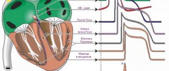
Curves on graph paper are represented by an isoline - a straight line, which means there are no impulses at the moment. Deviations up or down from the isoline are called teeth. In one complete cycle of cardiac contraction there are six teeth, which are assigned standard letters of the Latin alphabet.
Each tooth is not just a picture with a letter; behind it lies a certain phase of the heart’s work. You can decipher a cardiogram if you know which teeth mean what. For example, the P wave demonstrates the moment when the atria are relaxed, R indicates the excitation of the ventricles, and T indicates their relaxation.
For example, if the distance between the R waves is unequal, doctors talk about extrasystole, atrial fibrillation, and weakness of the sinus node. If the P wave is elevated and thickened, this indicates thickening of the walls of the atria. An extended PQ interval indicates artrioventicular block, and an expanded QRS suggests ventricular hypertrophy and His bundle block.
To interpret the ECG, there is a table containing the normal values. Based on it, doctors can see deviations. As a rule, in the process of long-term work with cardiac patients, doctors no longer use a table at hand; in adults, they have learned the norm by heart.
In addition to the table values, doctors also consider other parameters of the heart:
- rhythm of heart contractions - in the presence of arrhythmia, i.e. disruptions in the rhythm of contractions of the heart muscle, the difference between the indices of the teeth will be more than ten percent. People with a healthy heart have a normosystole, but pathological data make the doctor wary and look for abnormalities. The exception is sinus arrhythmia in combination with sinus rhythm, as often happens in adolescence, but in adults, sinus rhythm with deviations indicates the onset of the development of pathology. A striking example of deviations is extrasystole, which manifests itself in the presence of additional contractions. It occurs with cardiac malformations, myocardial inflammation, ischemia;
- Heart rate is the most accessible parameter; it can be assessed independently. Normally, in one minute there should be from 60 to 80 complete cycles of the heart. With an accelerated cycle, more than 80 beats indicate tachycardia, but less than 60 is bradycardia. The indicator is more illustrative, since not all severe pathologies give rise to bradycardia or tachycardia, and in isolated cases, the ECG of a healthy person will also show such phenomena if he is nervous during electrocardiography.
What classes is heart failure divided into?
There are several classifications of heart failure:
- Classification according to V. Kh. Vasilenko, N. D. Strazhesko, G. F. Lang;
- Classification of acute heart failure according to the Killip scale;
- And the most common is the New York Heart Association classification.
According to the New York Heart Association (NYHA) functional classification, heart failure is classified into classes I-IV depending on the severity of symptoms and limitation of physical activity.
Heart failure is divided into four classes depending on the severity of symptoms:
- NYHA I: heart disease without any restrictions on physical activity. Normal activity does not cause increased fatigue, palpitations, or difficulty breathing.
- NYHA II: Heart disease causing moderate limitation in daily activities. No symptoms at rest.
- NYHA III: Heart disease causing marked limitation in daily activities. Simple activities such as brushing your teeth, eating, or talking cause fatigue, palpitations, or difficulty breathing. There are no symptoms at rest.
- NYHA IV: Heart disease that causes symptoms at rest (and with any degree of light physical activity).
Heart failure significantly reduces quality of life. Patients often experience great frustration with physical limitations and tend to withdraw from social life. For this reason, psychological disorders such as depression are often present in addition to the expected physical symptoms.
How is heart failure treated?
For chronic heart failure, medications (such as ACE inhibitors, beta blockers and diuretics) are used. Medicines are used to prevent complications and improve quality of life. ACE inhibitors and beta blockers can prolong life, but they must be taken regularly to achieve a beneficial effect.
In addition, rhythm therapy (to treat cardiac arrhythmias) and implantation of a three-chamber pacemaker are used. The latter ensures timely activation of the atria and both ventricles. A defibrillator is also often implanted as part of a pacemaker to counteract dangerous heart rhythm disturbances in the setting of severe heart failure. This treatment is also known as resynchronization therapy. An important part of successful treatment is physical therapy.
How does heart failure manifest?
Depending on which part of the heart is affected, the symptoms of heart failure may vary. Most often, the patient begins to feel the following:
- dyspnea;
- heart rhythm disturbance;
- dizziness, darkening of the eyes;
- veins protrude from the neck;
- the skin turns pale;
- My legs swell in the evenings and start to hurt.
Later, so-called dropsy may occur (fluid accumulates in the abdominal cavity). The patient cannot perform any physical work, after which coughing attacks occur, which many attribute to other reasons, for example, smoking.
However, not all of the above symptoms are unambiguous confirmation of the presence of heart failure in a person. For example, swelling of the legs can be varicose veins; it is also common in older people, as is fatigue. Shortness of breath may be a consequence of pneumonia.
Symptoms can manifest with varying intensity, and there is even the possibility of death. Therefore, if one, or even more so several phenomena occur, you should immediately seek medical help. First of all, you need to do an electrocardiogram. Whether heart failure is visible on an ECG remains an open question, but it may help detect other conditions that will contribute to the development of heart failure and require treatment on their own.