The main muscle of the human body is the heart. This is a complex mechanism, the components of which are valves. The tricuspid valve is located on the right side. It separates the atrium and ventricle. When it malfunctions, the blood flow in that part of the muscle is disrupted. The result is a life-threatening condition. In medical practice, it is also known as tricuspid valve insufficiency. Today's article will discuss the main causes, symptoms and treatments for this disease.
Anatomical certificate
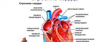
The human heart is a muscular organ. Its cavity is divided into 2 atria and 2 ventricles. Communication between these structures occurs through valves. They are responsible for blood flow in one direction.
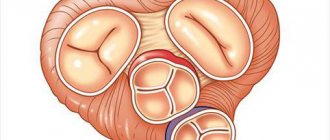
On the right side of the heart, the chambers are connected through the tricuspid valve. It consists of the following anatomical formations:
- three valves (septal, anterior and posterior);
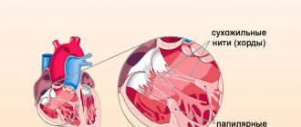
- tendon tract;
- papillary muscles;
- fibrous ring.
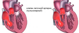
During contraction of one atrium, the tricuspid valve opens. Gradually, blood fills the ventricle. After this, the myocardium begins to alternately contract, and the valve leaflets slam shut under strong pressure. Such a well-functioning mechanism prevents the reverse flow (regurgitation) of blood. Under the influence of certain factors, sometimes its operation fails. In this case, they say that they are experiencing tricuspid valve insufficiency. However, he can no longer fully perform his functions.
With this pathology, blood from the right ventricle returns to the atrium. A slight increase in fluid volumes has virtually no effect on the functioning of the main muscle of the body. With severe insufficiency, the chambers of the heart are subject to deformation, and their pressure changes sharply. It can increase up to 8 times compared to natural values.
How does the aortic valve work?
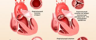
The tricuspid structure of the aortic valve is distinguished from the bicuspid mitral valve by the absence of papillary muscles and tendon chords. Therefore, it opens and closes only under the influence of the difference in pressure in the cavity of the left ventricle and the aorta.
During opening, elastin fibers from the ventricle press the valves against the walls of the aorta, opening the hole for blood flow. At the same time, the aortic root (the initial part) contracts and pulls them towards itself. If the pressure in the ventricular cavity exceeds the pressure in the aorta, then blood flows into the vessel.
The valves close with swirling currents in the area of the sinuses. They move the valve away from the walls of the aorta towards the center. Elastic flaps close tightly. The closing sound is heard with a stethoscope.
Brief description of the disease
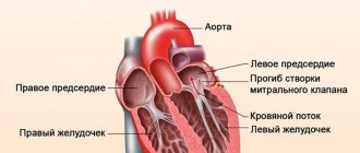
Tricuspid insufficiency is a heart defect that develops due to inadequate closure of the valve leaflets, backflow of blood from the right ventricle into the atrium. Against the background of constant regurgitation, diastolic volume and pressure in this heart structure increase. This entails hypertrophy and dilatation of its walls. As a result of disruption of the compensatory mechanisms, the patient develops stagnation in the body.
Tricuspid regurgitation is often accompanied by other congenital heart defects. For example, an open foramen ovale or atrial septal defect.
Set of diagnostic measures
Diagnosis of PTC is an important step in the process of treating the disease. It must be comprehensive and timely, which will increase the patient’s chances of recovery. A doctor can assume the development of a disease in a patient based on complaints, as well as objective examination data: palpation, percussion, auscultation of the heart.
The following instrumental research methods can confirm the diagnosis:
- electrocardiography, which allows you to determine dilatation of the right ventricle;
- echocardiography, which makes it possible to diagnose the presence of flexion of the tricuspid valve leaflets and regurgitation;
- catheterization of the right heart, which can be used to determine the increase in pressure in the heart chambers and lungs;
- X-ray of the chest cavity can confirm the diagnosis of pulmonary hypertension;
- computed tomography and MRI to detect enlargement of the right heart.
Echocardiogram of the heart with tricuspid valve prolapse
Main causes and forms of pathology
Tricuspid valve insufficiency always has a different course. To make an accurate diagnosis, it is first necessary to determine the form of the pathological process. For this purpose, various criteria are used in medical practice: period of occurrence, localization of the damaged area of the valve, severity of regurgitation.
Depending on the time of development, the disease in question is of two types. As a rule, in all cases it is not acquired, but occurs after an inflammatory process. The congenital defect is diagnosed extremely rarely and develops in the womb. Its appearance is influenced by the effect of negative factors on the pregnant woman’s body. This could be radiation, an infectious disease, or x-ray exposure.
Based on the part of the tricuspid valve that is damaged, insufficiency can be of two forms. The organic version is accompanied by a physiological change in the valves. They gradually become deformed and become covered with plaque. In the functional form of the disease, the valve does not close completely.
The development of acquired organic tricuspid insufficiency occurs under the influence of the following reasons:
- Rheumatism is an inflammation that spreads to the internal organ systems.
- Carcinoid syndrome is damage to various organs caused by a small intestinal tumor. Pathogenic elements from the neoplasm enter the blood and begin to attack the endocardium. They then penetrate the pulmonary vessels.
- Infectious endocarditis is an inflammatory lesion of the lining of the heart.
- Mitral commissurotomy. This is an operation that is used to treat stenosis.
Acquired functional (relative) insufficiency of the tricuspid valve develops due to damage to the papillary muscles or expansion of the fibrous ring. In the first case, the structures presented are responsible for maintaining the motor ability of the valves. During myocardial infarction, the papillary muscles are damaged. This leads to disruption of the functionality of the tricuspid valve.
Pathologies of the annulus fibrosus can cause various disorders. These include myocarditis, chordal ruptures, tumors and neoplasms that prevent blood from flowing out of the ventricle.
Causes
Acquired disorders can occur due to diseases:
- rheumatism,
- infectious inflammation of the inner lining of the heart,
- carcinoid tumors,
- diseases that impair blood circulation and provoke an enlargement of the right ventricle;
- traumatic injuries to the heart area,
- complications caused by the consequences of mitral valve surgery.
Valve malfunctions can be caused by:
- an anomaly in the structure of the valve or a disorder in its structure due to disease,
- damage to some parts of the mechanism involved in controlling the operation of the valve.
Stages of development of deficiency
Based on the severity of the regurgitation process, it is customary to distinguish several stages during the disease:
- Degree 1. Reverse blood flow remains minimal and is not accompanied by hemodynamic disorder.
- Degree 2. At this stage, the pathology is accompanied by the advancement of a reverse blood stream over a distance that does not exceed 2 cm.
- Grade 3. Blood flow moves into the atrium more than 2 cm from the valve surface.
- Degree 4. At the time of regurgitation, blood penetrates into the upper zone of the atrium, passing throughout the chamber.
The symptoms that accompany each stage of the disease will be discussed below.
Degrees
Valve insufficiency can be expressed to varying degrees. A measure to determine the depth of the problem is the amount of blood returning (regurgitation) to the atrium.
- 1st The reverse flow of blood towards the atrium touches the valve leaflets. The first degree of the disease is considered harmless and treatment is not prescribed.
- 2nd Pathology of the second degree expresses itself by advancing the reverse jet beyond the valve to a distance of up to two centimeters.
- 3rd. If the backflow moves into the atrium more than two centimeters from the surface of the valve, then this violation is classified as third degree.
- 4th. The entry of blood into the upper zone of the atrium at the time of regurgitation, that is, the stream passes throughout the entire chamber, indicates the fourth degree of the disease.
Signs of tricuspid valve insufficiency
At the initial stage, heart disease practically does not manifest itself. Sometimes after physical activity, patients note the appearance of strong pulsation of the veins in the neck. The pathological process can only be detected using Doppler ultrasound. A regular ECG does not show significant changes in the functioning of the heart.
Tricuspid valve insufficiency of the 2nd degree is accompanied by an increase in the volume of reverse blood flow. Right ventricular disorder leads to an increase in heart size. As a result, the clinical picture is supplemented by the following symptoms:
- loss of strength, decreased physical and mental activity;
- frequent urination at night;
- severe swelling of the lower extremities;
- dyspnea;
- feeling of heaviness under the ribs;
- dyspeptic disorders.
When listening to the heart, pansystolic murmurs and atrial fibrillation are detected.
As the pathology progresses, the number of symptoms displayed increases. It is not recommended to ignore their occurrence. It is better to seek qualified medical help at the initial stage of development of the disease. Tricuspid valve insufficiency of 1-2 degrees can still be treated with medication. The transition of the disease to the next stage always requires surgical intervention. Even after the operation, complications cannot be ruled out.
Clinical picture
In the early stages of the formation of TB prolapse or with the development of a pathological process of 1st degree of severity, the patient, as a rule, does not have any complaints from the heart, and the disease can only be suspected during a medical examination. The first symptoms of the disease appear when the volume of regurgitation increases. In severe forms of tricuspid valve insufficiency, patients experience the following clinical manifestations of the pathological process:
- feeling of weakness, tiredness and fatigue;
- swelling of the distal parts of the lower extremities and the anterior abdominal wall;
- visible pulsation of the neck veins;
- decreased diuresis;
- palpitations with rhythm disturbances (tachycardia);
- pain in the heart area;
- dyspnea;
- in severe cases - hemoptysis.
When visiting a doctor, patients with degrees 2, 3 and 4 of tricuspid valve prolapse complain of a feeling of weakness, loss of performance and heaviness in the abdomen and legs. Often in such patients, complaints related to a decrease in the volume of urine produced come to the fore, which they mistakenly perceive as kidney pathology.
Symptoms of PTC from the heart:
- hypertrophy of the right heart;
- symptoms of damage to other valves;
- pansystolic murmur, which increases with inspiration;
- On auscultation, the sounds of a “flapping sail” are heard above the heart
In addition to changes in the heart, tricuspid valve prolapse is also indicated by an increase in the size of the liver, dyspeptic disorders, heaviness in the right hypochondrium and portal hypertension.
According to statistics, prolapse with tricuspid valve insufficiency is most often diagnosed in patients who regularly use drugs.
This is due to the fact that taking intoxicating drugs in unsanitary conditions is one of the main causes of infectious or toxic endocarditis, as a result of which the valve leaflets lose their elasticity and ability to completely close in diastole.
Features of the disease in children
Newborn babies with severe regurgitation and the presence of concomitant cardiac pathologies are susceptible to the rapid development of right ventricular failure. Very often it ends in death.
The acquired form of the disease develops against the background of complications of other diseases. Most often we are talking about streptococcal infection, which entails rheumatic lesions.
Other causes of deficiency in childhood include:
- acute form of hypertension;
- myocardial diseases;
- traumatic injuries;
- malignant neoplasms.
At the initial stage, tricuspid valve insufficiency in children is considered as an anatomical feature. The first degree of the pathological process does not require treatment. With age, the disease usually goes away on its own.
Bicuspid valve condition
Bicuspid aortic valve is a common congenital disease of the cardiovascular system. The disease is not independent, but only a consequence of stenosis. This occurs because the valves open only partially, allowing reverse blood flow to enter the ventricle.
Most often, the symptoms do not bother children in the first years of life, but the disease continues to develop and makes itself felt in adolescence. The bicuspid aortic valve often occurs against the background of another disease. Various complications can stimulate this defect. The diagnostic echocardiographic method allows for a timely diagnosis, which allows reversing the process.
Diagnostic methods
Early detection of the disease plays a direct role in the success of treatment and also affects the patient’s life expectancy. Since tricuspid valve insufficiency of the 1st degree is practically asymptomatic, pathology is detected only after its transition to the next stage of development.
Diagnosis begins with interviewing the patient, studying his medical history and physical examination. By asking various questions to the patient, the doctor determines the clinical picture and the time of onset of the first symptoms. He also needs to know what diseases preceded the deficiency. An external examination usually reveals cyanosis of the skin and swelling, and upon auscultation, a disturbance in the heart rhythm.
As part of the diagnosis, several laboratory tests are required. Among them, the most informative is a blood test and a study of the patient’s immunological status. Based on the results of laboratory tests, one can judge the presence of inflammatory processes in the body and parallel illnesses.
To clarify the diagnosis, the doctor can use hardware examination methods. These include:
- ECG. It is carried out to detect an increase in atrium volumes.
- Phonocardiogram. Demonstrates the presence of systolic murmurs.
- Spiral CT. Provides an informative image of the main muscle of the body.
- X-ray. Gives an idea of the size of the heart and reveals congestion.
- Coronocardiography. Used before surgery and allows you to assess blood flow.
- EchoCG. Shows deformation of the valves, the presence of new formations on them.
Based on the results of a complete examination, the doctor gets an idea of the condition of the tricuspid valve. Insufficiency of this structure responds well to treatment only at the initial stage of development. Treatment options for this disease will be discussed in more detail below.
What pathologies of the right atrioventricular valve most often occur?
Dysfunction of the right atrioventricular orifice most often takes the form of stenosis or insufficiency. Pathological changes in the valvular apparatus of the heart significantly impair blood circulation, which is manifested by certain clinical symptoms.
Tricuspid valve stenosis
- It is associated with certain diseases of an infectious nature, infection with streptococcal, enterococcal or treponemal infections.
- It is more often detected in patients with rheumatism or syphilis.
- Represents a narrowing and reduction in the diameter of the AV opening (stenosis), which significantly impedes the flow of blood through the valve.
- In 60% it is combined with damage to other valves, mitral or aortic.
- Circulating in the bloodstream, the infection settles in all parts of the heart, affecting the elements of the valve apparatus.
- Due to the progressive inflammatory process, the tricuspid valve becomes sclerotic. The valves, annulus fibrosus, muscular elements and chords fuse, reducing the lumen of the AV foramen.
- Normally, the size of the valve is 3-4 centimeters; with stenosis, the diameter decreases from 3-1.5 cm.
- As a result of changes in hemodynamics, not the entire volume of blood flows from the atrium to the ventricle, and therefore stagnation develops in the pulmonary circulation.
- The examination is characterized by pathological reflux - swelling of the neck veins when pressing on the abdominal area where the liver is located.
- Auscultation of the heart reveals diffuse pulsation and enlargement of the cardiac boundaries, loud pathological murmur in the diastole phase.
- It manifests itself as portal hypertension (increased pressure in large hepatic vessels) with subsequent stagnation of blood in the spleen, intestinal vessels, and stomach.
- Characteristic symptoms are severe weakness, shortness of breath, swelling, cyanosis of the hands and face, irregular heartbeat, increased blood pressure, hemoptysis, swelling of the abdomen and fatty tissue, swelling of the veins around the navel.
- Without treatment, it leads to fatal heart failure and death.
- Drug treatment is ineffective; surgical intervention is indicated to replace the affected valve and save the patient’s life.
Right atrioventricular valve insufficiency
- Consequences of tricuspid valve dysfunction
The prognosis for progressive stenosis or insufficiency of the right atrioventricular valve is extremely unfavorable. According to statistics, most patients develop complications of cardiac pathology within 5-10 years. The most dangerous of them: thromboembolism of the pulmonary vessels with a thrombus from the AV valve or fatal atrial fibrillation (arrhythmia). Cardiac arrest can be triggered by complete blockade or secondary infection in the presence of a defect.Progressive pathology of the tricuspid valve is complicated by congestive heart failure with damage to the liver and blood vessels of the internal organs. This increases the risk of developing gastrointestinal bleeding from portal hypertension from the esophageal veins.
Principles of treatment
Tricuspid valve insufficiency of the 1st degree does not require therapy. If the disease progresses to the next stage of development, the patient is prescribed treatment. It can be either medicinal or surgical. The help of the latter is resorted to in especially serious cases, when the use of tablets and injections does not produce results.
For the entire period of treatment, the patient must adhere to the following rules:
- Quit smoking completely.
- Avoid hypothermia and stressful situations.
- Follow a diet to reduce stress on the heart.
- Reduce the intensity and amount of physical activity.
Compliance with these recommendations increases the effectiveness of the therapy and is also a kind of prevention of the development of complications.
Disease prevention
The main prevention of PTC comes down to the treatment of rheumatism and the prevention of its relapses, the same applies to other diseases that can cause the appearance of pathology.
- A person at risk must be constantly monitored by a cardiologist and cardiac surgeon.
- It is also important to maintain a healthy lifestyle in terms of nutrition and exercise.
Now let's talk about a special case of the disease: primary prolapse of the tricuspid (grade 1) and mitral valves.
Drug treatment
Moderate tricuspid valve insufficiency is not considered an indication for surgery. In this case, treatment is carried out with the help of medications. The standard treatment regimen involves the use of the following medications:
- Diuretics (Britomar, Hydrochlorothiazide). Eliminate congestion in the body, accelerate the process of fluid removal.
- Potassium preparations (Panangin, Asparkam). Help the body not accumulate excess fluid.
- Venous dilators (Corvaton, Nitrosorbide). Reduce the load on the heart through blood deposition.
- Anticoagulants (Warfarex, Warfarin).
- Cardiac glycosides (Digoxin, Korglykon). Helps in the fight against arrhythmia.
- Beta-blockers (Diltiazem, Carvedilol). Reduce the frequency of contractions of the left ventricle.
The regimen and dosage of drugs are determined individually, taking into account the severity of the disease.
Main functions
Throughout a person’s life, the heart provides a closed path of blood flow, delivery of oxygenated blood to organs and tissues, and venous outflow of carbon dioxide and breakdown products. The cardiovascular system consists of blood circulation circles. The large one originates in the left ventricle and ends in the right atrium, the small one starts in the right ventricle and goes to the left atrium.
The tricuspid valve is actually an element of a small circle that performs the following functions:
- During heart contractions, it prevents reverse regurgitation (blood flow from the lower ventricle into the atrium).
- Directly involved in blood circulation, ensures the delivery of venous blood to the vessels of the lungs.
- Through which the process of gas exchange in the alveoli of the lung tissue and heat transfer takes place.
Surgery
Tricuspid valve insufficiency grade 3 is considered the main indication for surgical intervention. Surgery is also recommended for patients with serious deformations of the valves or severe malfunctions in their operation.
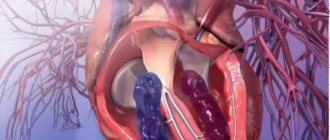
If the valve apparatus is preserved, annuloplasty is used. During the operation, the doctor places U-shaped sutures on the damaged area.
If the intervention turns out to be ineffective, there are obvious structural changes, the patient is given an artificial cap. After implantation of the prosthesis, anticoagulant therapy is required. When implanting a biological prosthesis, it is short-term. If it is made of medical alloys, treatment of tricuspid valve insufficiency takes a little longer.
Possible complications and prognosis for recovery
Lack of timely treatment can lead to progression of the disease. The pathological process in this case affects not only the tricuspid valve. Insufficiency gradually leads to the fact that the body begins to look for new compensatory mechanisms. In this way he tries to cope with existing violations. Depending on the severity of the disease, the following disorders may appear:
- pneumonia;
- increased liver size, cirrhosis;
- pulmonary embolism;
- ascites.
According to statistics, the listed complications develop in 90% of cases.
As for the prognosis for recovery, it depends solely on the degree of development of the disease. Tricuspid valve insufficiency of the 2nd degree responds well to drug treatment, and the likelihood of complications is almost zero. With the third degree of the pathological process, the five-year survival rate is approximately 60-70%. Such figures are typical for patients who have already undergone surgery. In decompensated forms of insufficiency accompanied by chronic pulmonary diseases, the prognosis is disappointing.
Types of tricuspid regurgitation
There are 2 main classifications of this pathology - according to the time of appearance and the reasons for its occurrence.
- By time of appearance: congenital and acquired.
Congenital is registered during the intrauterine development of the child or in the first months after birth. In this case, heart valve function may return to normal over time. In addition, it is much easier to keep a congenital anomaly under control if you take care of the heart and follow measures to prevent cardiovascular diseases.
Acquired disease appears in adults already during life. This pathology almost never occurs in isolation; it can be caused by a variety of diseases - from dilatation (expansion) of the ventricle to obstruction of the pulmonary arteries.
- For reasons: primary and secondary.
Primary tricuspid anomaly is diagnosed against the background of heart disease. The patient does not have any problems with the respiratory system with this diagnosis. The main cause of secondary disruption of blood flow in the tricuspid valve is pulmonary hypertension, that is, too high pressure in the pulmonary artery system.
Regurgitation of the tricuspid valve traditionally goes hand in hand with insufficiency of the valve itself. Therefore, some classifications divide the types of reverse blood flow based on the form of tricuspid insufficiency, that is, the valve disease itself:
- Organic (absolute) failure, when the cause is damage to the valve leaflets due to a congenital disease.
- Functional (relative), when the valve is stretched due to problems with the pulmonary vessels or diffuse damage to the cardiac tissue.