Why does sinus rhythm appear on the cardiogram?
Quite often the patient is told that sinus rhythm is diagnosed on the cardiogram. How scary and harmful is this to the heart? To answer the question, you need to understand the very meaning of “sinus rhythm.”
Sinus rhythm is called oscillations that first arise in a special node and then spread to other points. These points are the ventricles of the heart and the atria.
Important! For the cardiogram result to be truthful and informative, the patient needs to relax. He should not feel nervous or afraid.
If you follow medical terminology, the sinus rhythm of the heart represents the elevation of the P wave in the QRS complex. Pulse in the range of 60-80 beats per minute. In sinus rhythm, the distance between the PP and RR teeth is identical. To diagnose such a rhythm, the doctor takes into account:
- the size of the P waves in each individual lead,
- duration of the segment PQ along the entire perimeter,
- armholes P before each QRS complex,
- in the second lead, the P notch is necessarily present.
By analyzing these parameters, the doctor makes a conclusion about the condition and rhythm of the heart, the presence or absence of sinus rhythm. If signs of sinus rhythm are fully present, then the heart is excited in the right direction from top to bottom. If these features are absent, the doctor diagnoses the presence of non-sinus rhythm. The source of excitation in such cases is located in the atria, atrioventricular node or ventricles. The causes of sinus rhythm are further diagnosed.
How does the analysis take place?
The norm for the first stage of analysis is to check the direction of the electronic axis. When analyzing the vector, the physician takes into account the weight of the ventricles and their conductivity, location features, etc. During the analysis, he must take into account the horizontal or vertical vector.
Carrying out an ECG
At the first stage of the analysis, the doctor sets the standard indicators and then compares them with the result obtained. The normal heart rate is 60-90 beats per minute, the normal QT interval is 390-450 ms. If the interval is not normal, then the doctor diagnoses ischemic heart disease, rheumatism or atherosclerosis. If the specified interval is less than normal, then the likelihood of hypercalcemia is high.
If the QRS complex that forms in the ventricles is more than 120 ms, then this is a deviation from the norm. The displacement of this complex indicates heart block, hypertrophy of the right or left ventricle.
If a child has a bad cardiogram, this is not yet a reason to panic. Cardiologists at the children's medical center will help you understand the results, make an accurate diagnosis and prescribe treatment. The clinic is located in the North-Eastern Administrative District of Moscow, not far from the Otradnoe, Bibirevo, and Vladykino metro stations.
Bad cardiogram, or how does it manifest itself?
A bad ECG most often indicates that the heart is experiencing a blockage. The blockade occurs due to improper transmission of impulses from the central nervous system to the heart. An accelerated pulse is present due to the accelerated vibrations of the organ.
A change in tact results from an inappropriate sequence and frequency of contractions of the heart muscle. To understand that the cardiogram is bad, you need to consider the following factors:
- The distance between the P waves. Normally they are the same.
- The appearance of non-sinus rhythm (probability of blockade).
- Heart rate. To count, take into account the number of cells between the R-waves. The norm is 60-90 beats per minute.
- The source that causes contraction of the heart and sinus rhythm. Normally it is in the sinus node. During diagnosis, the doctor analyzes the parameters of the R waves. The presence of a source between the ventricles and atria indicates a deviation from the norm.
- Duration of segments and teeth. The parameter allows you to see the quality of heart conduction in the ventricles, the presence of blockade in the atria, etc.
- Placement of the electrical axis of the heart. The displacement of the axis is a deviation from the norm, which indicates pathologies and blockages.
The doctor examines each parameter of the cardiogram in a comprehensive manner to see the real condition of the patient at the time of diagnosis.
Can an ECG across the Sky completely replace a classic cardiogram?
Bad heart cardiogram - what to do and how to restore heart health?
Marina Dmitrievna
25 911
Symptoms
When there is pain in the heart area, most people are wary, because a person’s full life depends on the work of this organ. Many people go to the hospital to see a cardiologist. However, electrocardiography does not always answer all their questions. Can a person’s heart hurt if the cardiogram is good? Why does my heart hurt for more than one month?
What does an ECG of the heart show, interpretation in adults
Cardiovascular diseases are one of the leading causes of death among people worldwide. Over the past decades, this figure has decreased significantly due to the advent of more modern methods of examination, treatment, and, of course, new drugs.
Electrocardiography (ECG) is a method of recording the electrical activity of the heart, one of the first research methods, which for a long time remained practically the only one in this field of medicine.
About a century ago, in 1924, Willem Einthoven received the Nobel Prize in Medicine; he designed the apparatus with which the ECG was recorded, named its waves, and identified the electrocardiographic signs of certain heart diseases.
Many research methods are losing their relevance with the advent of more modern developments, but this does not apply to electrocardiography. Even with the advent of imaging techniques (echocardiography, CT, MRI, etc.)
) For decades, the ECG continues to be the most common, very informative, and in some places the only available method for studying the heart.
Moreover, over the century of its existence, neither the device itself nor the method of its use have changed significantly.
What do moderate changes in the myocardium mean?
Very often, doctors detect moderate changes in the myocardium on an ECG or when examining a patient’s heart using ultrasound equipment.
Such a diagnosis practically does not mean serious problems for a person, since it only shows changes in the heart muscle that can occur under the influence of various factors, such as diseases, age, and inflammation.
In children and adolescents, changes in the myocardium can occur due to incomplete formation of processes during the development of the cardiovascular system, and in patients over the age of 50 years, the aging of the body plays a major role.
How dangerous are transformations?
Minor changes in the left ventricular myocardium may not give any signs of disease. They occur relatively frequently in every person, so they are considered harmless. Most often they are identified when examining a patient for another reason, during preventive examinations or during medical examination. If a person does not complain about heart problems, then there is no need to worry.
Our readers successfully use Normaten to treat HYPERTENSION. Seeing how popular this product is, we decided to bring it to your attention. Read more here...
But if manifestations of cardiac dysfunction are detected, especially from the left ventricle, then it is urgent to undergo an examination, make a diagnosis, find the cause of the pathology, and carry out a course of treatment. Reasons for concern may include the following:
- The patient developed shortness of breath.
- Heart muscle discomfort or pain.
- Severe interruptions in the functioning of the heart. Often the left ventricle is to blame.
- The patient was diagnosed with a rhythm disorder.
In case of such violations, you should urgently seek medical help.
What changes are happening?
Depending on the factors influencing the person and the course of the changes occurring, doctors distinguish dystrophic, diffuse and metabolic lesions. Such tissue changes most often begin to cover increasingly large areas of the heart, although in some cases they are limited to some cardiac region.
Most often, doctors find a diffuse type of changes in the myocardium in patients. This type occurs when all parts of the heart are uniformly affected, usually due to inflammatory processes in the myocardium.
Most often, the cause of such changes is improper water-salt metabolism between individual parts of the body. But in some cases this is possible under the influence of medications.
Changes in the metabolic plan occur due to a shift in metabolic processes in the myocardium. Most often, this development of events can be expected due to poor nutrition of the heart muscle. At the same time, the contractile performance of the heart drops sharply. But, as practice shows, such changes can be reversed. Typically, negative transformations can occur for the following reasons:
- The person is hypothermic or has a severe cold.
- Too much nervous or physical stress.
- Overweight.
- Chronic diseases that affect the myocardium.
Often moderate changes in the heart occur due to sensitivity to various irritants or stresses, after the removal of which the disease quickly disappears. If the lesions occur sequentially, the heart will not be able to recover. Then the patient may experience irreversible changes in the heart muscle, which will negatively affect the functioning of the organ.
If the energy entering the myocardium is less than it gives out, then dystrophic changes subsequently begin. If they are moderate, then there will be no symptoms of the disease. But most often the disease manifests itself in the form of severe fatigue and shortness of breath, even with little physical activity.
If this happens, it is recommended to consult a doctor and undergo an examination.
Movements of a non-specific nature
Sometimes, when taking an electrocardiogram, doctors find nonspecific lesions. In most cases they occur outside the heart, so they are not of particular concern. Most often on the electrocardiogram, this type of change is found in the ventricles of the heart muscle.
Most often this occurs due to the process of repolarization, when the myocardium recovers after the passage of control nerve impulses. In most cases, such changes do not pose any danger and are completely reversible.
They appear due to the effects of various diseases, hormonal system failures, poor nutrition, and metabolic disorders in the human body.
For what reasons does the cardiogram worsen?
A person is not always able to calm down before the procedure. There is “white coat syndrome” when the patient is afraid of doctors. Due to fear, the heart rate increases, which affects the final result.
The main reasons that worsen ECG readings are:
- excessive alcohol consumption, smoking, drug addiction,
- hard physical work before taking data,
- stress,
- blockades of different locations,
- cardiac hypertrophy,
- arrhythmia,
- bradycardia,
- scarring of tissue in the myocardium,
- myocardial infarction,
- inflammation of the membranes, ventricles, atria, etc.
To make a diagnosis, one electrocardiogram is not enough. Cardiologists prescribe diagnostics using echocardiography, Holter monitoring, ultrasound, general blood test, biochemical blood test, and sometimes thyroid hormone testing.
What to do if a child has a bad cardiogram?
If the doctor tells you that the ECG has revealed signs of pathological changes, you need to urgently contact a pediatric cardiologist to decipher the data. You should not try to interpret the changes yourself: the data should be interpreted comprehensively, taking into account age characteristics, concomitant diseases, and other symptoms present.
In most cases, one cardiogram is not enough to make a serious diagnosis. Other examinations will be required: echocardiography, cardiac ultrasound, MRI, Doppler monitoring, blood tests. The ECG will also need to be repeated to verify the correctness of the results obtained, as well as to evaluate the dynamics of changes. Only after this is a final diagnosis made and treatment prescribed.
What diseases are diagnosed using an ECG?
The ECG records any deviations from the norm even at those stages when the patient does not feel pain in the heart area. Doctors often use this method to diagnose:
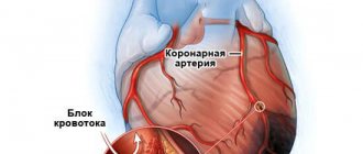
- Hypertrophy of the atria and ventricles. Occurs due to overload of the heart chambers, as blood is transported through the vessels incorrectly. The atria and ventricles increase in size. On the ECG it is manifested by the growth of R waves, deviation of the electrical axis of the organ, and increased excitability vector.
- Angina pectoris. When sick, the patient may not feel any deterioration in health for a long time. On the ECG, angina is displayed as: ST segments below the isoline, the T wave changes display.
- Arrhythmia. Manifests itself in the form of chest pain and rapid heart rate. On the ECG it appears as: the presence of fluctuations in the PQ and QT intervals, the R waves deviate from the norm, the PQ interval decreases.
- Bradycardia. The interval between the R waves increases, the area between the QT waves increases, the direction of the waves changes, and the heart rate slows down.
- Pericarditis. With this heart disease, inflammation of the pericardial sac begins, which affects the rhythm of the organ, the functioning of the ventricles and atria.
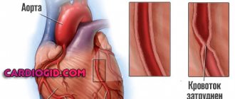
In addition to these diseases, ECG diagnoses myocardial infarction, myocarditis, coronary heart disease, and heart failure.
Poor ECG results, causes, mechanism of formation, signs and treatment
Electrocardiography is a technique for examining the heart based on recording electrical potentials. An ECG allows you to identify signs of heart disease (rhythm disturbances (presence of extrasystoles, tachycardia, bradycardia, blockades), heart defects, heart attack, hypertrophy of organ tissue). A bad cardiogram allows you to identify the disease and begin treatment on time.
Normal ECG signs in children and adults
Twelve ECG leads create a three-dimensional picture of the heart's electrical activity
In adults and children, cardiogram indicators are slightly different. Due to the immaturity of the cardiovascular system, as well as the large mass of the heart, children under 1 year of age and older may have their own ECG characteristics.
The cardiogram is recorded using an electrocardiograph. It registers the electrical impulses of the heart and records them on a special film. The recording is made in the form of a curve. The results are recorded on 3 leads: standard (I, II, III), enhanced (avF, avR, avL), chest (v1-6).
Differences in children's cardiogram:
- right ventricle (in a one-year-old child, the right ventricle is larger than the left);
- the intervals in the graphic image are small;
- there is a high P wave, since the atria are larger relative to the mass of the entire body than in an adult patient;
- respiratory arrhythmia (when inhaling, an increase in rhythm is observed);
- heart rate is 80 or more per minute;
- P-QRS-T interval is 120-200 ms;
- incomplete blockade of the right bundle branch.
Normal values
| Teeth, intervals | Duration, focus | |
| adult | child | |
| QRS | Positive, 0.06-0.1 s | 0.06-0.1 s |
| P | Positive, 0.07-0.11 s | Less than 0.1 s |
| PQ | 0.12-0.20 s | 0.2 s |
| T | 0.12-0.28 s, positive | QT up to 0.04 s |
| Q | 0.03 s, negative | |
| Rhythm frequency (beats per minute) | 60-80 | up to 3 years old - 100-110, 3-5 years old - no more than 100, 6-8 years old - 90-100, 9-12 years old - 75-85. |
An ECG is done in any clinic and hospital. It can also be carried out by ambulance workers. To make an ECG, the patient is placed on a couch, electrodes are placed on the chest (suction cups, 6 pieces), on the wrists and limbs (clamps).
The patient should lie still and breathe evenly. Any movement or crying of the child may distort the results. Next, an electrocardiogram is taken using an electrocardiograph. After taking an ECG, the doctor interprets the results.
No preparation is required for the examination. To increase the reliability of the results, the doctor may conduct stress tests (squat exercises, treadmill, exercise bike). Load can provoke the appearance of pathology that is not visible at rest.
Indications for prescribing an ECG:
- murmurs during auscultation of the heart;
- shortness of breath on exertion;
- a history of severe infectious diseases;
- family history of heart disease;
- arrhythmias;
- chest pain;
- preparatory stage before surgery.
Diseases manifested on ECG
The causes of a poor cardiac cardiogram can be various heart diseases, as well as hormonal and electrolyte disorders.
In children, changes in the ECG appear due to heart defects, cardiomyopathies, infectious myocarditis, and arrhythmias.
In adults, rhythm disturbances, ischemic damage to myocardial tissue, hypertension, atrioventricular block, His block, atrial fibrillation and others are possible.
Bundle branch block is not considered an independent disease
A poor ECG often develops due to arrhythmias.
These include tachyarrhythmias, bradyarrhythmias, His block, atrioventricular block, and extrasystoles. With tachycardia, the patient feels a rapid heartbeat and shortness of breath at the height of the arrhythmia.
In such patients, an ECG can reveal a heart rate of up to 140 beats per minute or more.
Bradyarrhythmias are manifested by a decrease in rhythm of less than 60 beats per minute. Such patients complain of weakness, drowsiness, and increased fatigue during physical activity or during training.
In athletes, bradyarrhythmia is considered a normal condition, since the heart is accustomed to stress in anaerobic mode. It may be a little exaggerated.
On an ECG, bradycardia is recorded as a decrease in heart rate of less than 60 beats per minute.
Autonomic dystonia in adolescents is often accompanied by tachycardia or bradyarrhythmia, depending on the type of pathology. An attack of rhythm disturbance can be triggered after strong experiences. These conditions may include fainting (bradyarrhythmia) or palpitations (tachyarrhythmia).
In adolescents, extrasystoles are very often detected on ECG, especially on Holter (24-hour) ECG monitoring. They are described by the patient as a sensation of turning over in the chest or a strong single contraction, during which the patient takes his breath away.
Extrasystoles can be single, double (bigeminy), triple (trigeminy), group (more than 3 in a row). The more extrasystoles in the group, the stronger the hypoxia in the heart muscle after an attack of arrhythmia. Extrasystoles appear on the ECG in the form of an extraordinary complex.
They are ventricular and atrial.
Extrasystoles are divided according to the place of origin
Why else can pathologies appear on the electrocardiogram? A change in the ECG may show 1st, 2nd, 3rd degree atrioventricular block (decreased conduction).
The first degree of AV block is the mildest and is often asymptomatic. It looks like a prolongation of the P-Q interval. Second degree AV block may be symptomatic.
On the electrocardiogram, 2nd degree blockade appears as disappearance of QRS complexes.
The third degree is more severe. The patient often faints. The atria and ventricles contract spontaneously (each in its own rhythm). In some cases, the P wave may overlap the QRS. Intraventricular blockades are possible. These include bundle blockades.
Complete atrioventricular block is considered very dangerous. It is accompanied by fainting states and collapse.
Children often develop endocarditis due to weak immunity. The disease is provoked by tonsillitis, as well as other diseases caused by streptococcus and staphylococcus. Endocarditis can also be caused by fungal infections.
Inflammation of the inner lining of the heart can cause impaired repolarization (relaxation) of the heart. In this case, the heart gradually begins to experience hypoxia. A bad cardiogram during exercise is detected faster.
There may be arrhythmias and blockades.
A poor ECG occurs with rheumatic endocarditis in adults. This pathology is rare in children. The disease is considered a complication of rheumatoid arthritis, as well as other rheumatic diseases. Electrocardiography shows rhythm disturbances and possible blockades.
A routine ECG in patients over 40 years of age can reveal a poor cardiac cardiogram with signs of angina pectoris. Angina is a disease in which ischemic (oxygen and nutrient deficiency) damage to the heart muscle develops.
Ischemia can develop due to increased thrombus formation (factors: thrombophlebitis of large veins in the legs, blood thickening, taking contraceptives, frequent pregnancies).
Angina pectoris on the electrocardiogram looks like a decrease in the ST segment, the appearance of a negative T wave.
Acute infarction on the electrocardiogram is characterized by an increase in the ST segment. The T wave merges with the ST segment. This formation has been compared to the arched back of a cat.
After an acute infarction, a subacute infarction develops. Both stages are accompanied by the appearance of a pathologically deep Q wave. It goes away as it recovers.
During the recovery period, the opposite T wave remains for 14 days. The ST segment returns to baseline.
How to improve ECG readings?
Bad heart cardiogram, what to do to improve the result? When a person notices a change in heart rate in everyday life due to stress, then he needs to psychologically prepare for the procedure. Don't worry or be nervous.
Advice! Before the ECG procedure, do not take heart medications so that the result is as reliable and informative as possible.
There are several simple but effective recommendations that can affect the result of the cardiogram:
- If the diagnosis is preventive, then get at least 8 hours of sleep before the ECG.
- Give up morning exercise to keep your heart in a calm state.
- When the procedure is in the morning, it is easy to have breakfast or refuse food. Eat food two hours before the procedure.
- If a person drinks a lot of water, then the day before the cardiogram, reduce the amount of fluid you drink.
- Avoid consuming alcoholic drinks, energy drinks and caffeinated drinks.
- To better attach the electrodes, men with increased chest hair growth can shave the area where the electrodes will be attached.
- To prepare psychologically and be less nervous, it is better to come to the procedure in advance.
These are simple guidelines, but they are important for the correct collection of information.