The electrical axis of the heart (EOS) is a clinical parameter that is used in cardiology and is reflected in the electrocardiogram. Allows you to evaluate the electrical processes that move the heart muscle and are responsible for its correct functioning.
From the point of view of cardiologists, the chest is a three-dimensional coordinate system in which the heart is enclosed. Each contraction is accompanied by a number of bioelectrical changes, which determine the direction of the cardiac axis.
Normal values and causes of violation
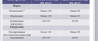
The direction of this indicator depends on various physiological and anatomical factors. Position +590 is considered the average norm. But the normogram options fall into a wide range from +200 to +1000 .
In a state of health, the electrical axis shifts to the left under the following conditions :
- at the moment of deep exhalation;
- when the body position changes to horizontal, the internal organs put pressure on the diaphragm;
- with a high-standing diaphragm - observed in hypersthenics (short, strong people).
A shift of the indicator to the right in the absence of pathology is observed in the following situations :
- at the end of a deep breath;
- when changing body position to vertical;
- For asthenics (tall, thin people), the norm is the vertical position of the EOS.
The location of the electrical axis is determined by the fact that the mass of the left ventricle under normal conditions is greater than the mass of the right half of the heart muscle. Due to this, electrical processes occur more intensely in it, so the vector is directed towards it.
Mathematics of the heart muscle: sinus rhythm, EOS deviation to the left
Proper heart performance is a guarantee of long human life. And the deciphered sinus rhythm and deviation of the EOS to the left are an indicator of the condition of the heart muscle. Thanks to the electrical axis, it is possible to diagnose and treat it at an early stage, prolonging the normal state of the body and the life of the sick person.
What is the electrical axis of the heart
By deviation of EOS, the diagnosis of heart disease can be determined
EOS - electrical axis of the heart - is a cardiological concept that means the electrodynamic strength of the organ, the level of its electrical activity. Based on its position, the specialist deciphers the state of the processes occurring in the main organ every minute.
This parameter represents the total amount of bioelectrical changes in the muscle. Using an electrocardiogram, in which electrodes record certain excitation points, it is possible to mathematically calculate the location of the electrical axis relative to the heart.
The conduction system of the heart and why it is important for determining EOS
The part of muscle tissue formed from atypical fibers that regulate the synchronization of contractions of the organ is called the conduction system of the heart.
The contractile property of the myocardium consists of a sequence of stages:
- Organization of an electrical impulse in the sinus node
- The signal enters the ventricular node of the atrium.
- From there it is distributed along the bundle of His, located in the interventricular septum and divided into 2 branches
- The activated bundle moves the left and right ventricles
- With normal signal transmission, both ventricles contract synchronously
The cardiac conduction system is a kind of energy supplier for the functioning of the body. It is here that electrical changes initially occur, provoking contraction of muscle fibers.
When the wiring system is dysfunctional, the electrical axis changes its location. This moment is easily determined by an electrocardiogram.
What is sinus rhythm on an ECG
Sinus rhythm on an electrocardiogram shows that the signal of an electrical nature is produced only in the sinus node. This area is located in the right atrium under the membrane and is directly supplied with arterial blood.
The cells of this organ are spindle-shaped and collected in small bundles. The low level of ability to contract is compensated by the production of electrical impulses, the analogues of which are nerve signals.
The sinus node produces low-frequency signals, but is capable of delivering them to muscle fibers at high speed. A run-up of 60-90 shocks in 60 seconds is considered an indicator of the quality functioning of the organ.
Semi-vertical and semi-horizontal position of the EOS is more common
The normal state corresponds to the predominance of the left-sided ventricle over the right-sided ventricle. Thanks to this, the processes of the electrical nature of the first are stronger in total, and the EOS will be directed specifically at it.
Source: https://organserdce.com/diagnostika/ekg/otklonenie-eos-vlevo.html
Diagnosis using ECG
An electrocardiogram is the main tool for determining EOS. To identify changes in the location of the axis, two equivalent methods . The first method is more often used by diagnosticians, the second method is more common among cardiologists and therapists.
Alpha angle offset detection
The value of the alpha angle directly shows the displacement of the EOS in one direction or another. To calculate this angle, find the algebraic sum of the Q, R and S waves in the first and third standard leads . To do this, measure the height of the teeth in millimeters, and when adding, take into account whether a particular tooth has a positive or negative value.
Next, a special table is used - according to Died . By substituting the obtained values into it, the exact value of the alpha angle or the displacement of the electrical axis is calculated.
The value of the sum of teeth from the first lead is found on the horizontal axis, and from the third - on the vertical axis. The intersection of the resulting lines determines the alpha angle.
The use of this determination method is suitable for those specialists who have the appropriate table at hand.
Visual definition
A simpler and more visual way to determine EOS is to compare the R and S waves in the first and third standard leads . If the absolute value of the R wave within one lead is greater than the value of the S wave, then we speak of an R-type ventricular complex. If on the contrary, then the ventricular complex is classified as S-type.
When the EOS deviates to the left, a picture of RI - SIII is observed , which means the R-type of the ventricular complex in the first lead and the S-type in the third. If the EOS is deviated to the right, then SI - RIII is determined on the electrocardiogram.
What is the electrical axis of the heart and what can be the consequences of deviations from the norm?
The electrical axis of the heart (EOS) is a concept that implies the activity of conducting nerve excitations synthesized and executed in the heart.
This indicator is characterized by the sum of the conduction of electrical signals through the cavities of the heart that occurs during any contraction of the heart tissue.
The electrical axis of the heart is one of the characteristics determined on an ECG. To make a diagnosis, additional hardware tests are necessary.
During an electrocardiogram study, the device records nerve excitations emitted by different parts of the heart by applying electrocardiograph sensors to different parts of the chest.
To calculate the direction of the EOS, doctors use a coordinate system, comparing the location of the heart with it. Due to the projection of electrodes onto it, the EOS angle is calculated.
In places where the zone of the heart muscle in which the electrode is installed emits stronger nerve excitations, that is where the EOS angle is located.
Why is the normal conduction of electrical excitations of the heart so important?
The fibers that make up the heart perfectly conduct nervous excitations, and with their multitude they create the cardiac system, where they conduct these nervous excitations.
The initial functioning of the heart muscle begins in the sinus node, with the appearance of nervous excitation. Next, the nerve signal is transported to the ventricular node, which transmits the signal to the His bundle, through which the signal propagates further.
Electrical axis of the heart
The location of the latter is localized in the septum separating the two ventricles, where it branches into the anterior and posterior legs.
The nerve conduction system is very important for the healthy functioning of the heart, since, thanks to electrical impulses, it sets the normal rhythm of heart contractions, which determines the healthy functioning of the body.
If deviations appear in the signal conduction structure, then significant deviations in the position of the EOS are possible.
How is the electrical axis of the heart determined?
It is up to the attending physician to identify the location of the EOS, deciphering the ECG, using diagrams and tables, and finding the alpha angle.
This angle is formed from two straight lines. One of them is the 1st lead axis, and the second is the vector line of the electrical axis of the heart.
Location features include:
| Normal | If the angle is within plus thirty - plus sixty nine, then this indicates normal indicators of the electrical axis of the heart |
| Vertical EOS | Registered when determining the axis within seventy to ninety degrees |
| Horizontal | When the angle is between zero and thirty degrees |
| Shift left | The position of the ventricle is located within the angle from zero to minus ninety degrees |
| Offset right | It is registered when the ventricular position indicators range from ninety-one to one hundred and eighty. |
Another way to identify the electrical axis of the heart is to compare QRS complexes, the main task of which is the synthesis of nerve excitations and contraction of the ventricles.
The definition indicators are given below:
| Normal | With such indicators of the electrical axis, the R-wave of the second lead is larger than the R-wave in the first lead, and the similar wave of the third branch is smaller than in the first. (R2>R1>R3) |
| Left deviation | If the normal position of the electrical axis to the left is violated, the R-wave of the first compartment is recorded - the largest, and the second and third, respectively, smaller. (R1>R2>R3) |
| Right deviation | Violation of the electrical axis of the heart to the right side is characterized by the largest third R-wave, and a corresponding decrease in the second and first. (R1 |
Source: https://moyakrov.info/heart/elektricheskaya-os-serdtsa
Establishing diagnosis
What does it mean if the electrical axis of the heart is deviated to the left? EOS displacement is not an independent disease. This is a sign of changes in the heart muscle or its conduction system that lead to the development of the disease. Deviation of the electrical axis to the left indicates the following violations:
- an increase in the size of the left ventricle - hypertrophy (LVH);
- malfunction of the left ventricular valves, which causes the ventricle to be overloaded with blood volume;
- cardiac blockades, for example, blockade of the left bundle branch (on the ECG this looks like this, which you can learn about from another article);
- disturbances in electrical conductivity inside the left ventricle.
All of these factors lead to the fact that the left ventricle does not work correctly, and the conduction of impulses through the myocardium is impaired. As a result, the electrical axis deviates to the left.
The electrical axis of the heart (EOS) is deviated to the left: what does this mean?
If the electrical axis of the heart (EOS) is deviated to the left or right, this may indicate problems with the functioning of this organ. Let's look at why this can happen, when it is dangerous and when it is not, and how this condition is treated.
How is offset determined?
The position of this axis is determined by electrocardiography, after analyzing an electrocardiogram from several leads.
To detect changes in the normal location of the axis, 2 methods can be used.
Alpha angle deviation
This technique is most often used by diagnosticians. Normally, the EOS completely coincides with the anatomical axis (the heart is located semi-vertically, and the lower end deviates down and slightly to the left). Its location is determined by the alpha angle formed from 2 straight lines (1 abduction axis and the EOS vector line).
To identify the angle, the sum of the S, R and Q waves in 3 and 1 standard leads is calculated. The positive and negative value of each tooth must be taken into account.
https://www.youtube.com/watch?v=df4W0wnXCVs
Diede's table is then used. Having entered the result, the doctor determines the criteria for the alpha angle.
Here's what it looks like:
Click on the picture to enlarge
Normally, this angle should be from – 29° to +89°. A significant left-sided displacement of the axis is a sign of pathological disorders. When it changes to -30°, we are talking about a left-sided deviation, and with values from +90° to +180° - a right-sided deviation.
Left-sided deviation of the angle from – 30° to – 44° is insignificant, at – 45° to – 90° it is considered significant and in most cases accompanies cardiac pathologies.
Additional diagnostic methods
After an ECG is performed, its results are carefully studied to identify the cause of the pathological condition. In most cases, a repeat cardiogram is prescribed, which is necessary to eliminate technical errors (incorrect application of electrodes, device malfunction, etc.).
In addition, the following studies are recommended:
- Holter monitoring - if the doctor diagnoses a conduction disorder or arrhythmia using an ECG, then 24-hour monitoring of cardiac activity (24-hour ECG) is performed, which makes it possible to more accurately determine the area of the heart with a conduction disorder.
- Ultrasound of the heart - this study is aimed at obtaining more information about cardiac output, blood flow, and the condition of the heart chambers. If indicated, ultrasound can be supplemented with Doppler ultrasound.
- ABPM (daily blood pressure monitoring) is prescribed for a sharp increase in blood pressure against the background of left ventricular hypertrophy with deviation of the cardiac axis. This examination allows you to find out the stage of hypertension and determine the most appropriate treatment.
- Cardiac surgery consultation is prescribed for any pathologies of the heart and, especially for defects with a tendency to progress.
It must be borne in mind that deviation of the EOS to the left is just an ECG sign indicating diffuse changes in various pathologies, therefore a comprehensive diagnosis must be prescribed.
Reasons for displacement
Changes in heart activity on the electrocardiogram are provoked by many factors.
Let's consider each case in more detail.
Heart diseases
The main reason for the shift to the left of the heart axis is left ventricular hypertrophy. Changes can be provoked by: ischemia (including heart attacks and post-infarction cardiosclerosis), aortic and mitral valve disease, cardiomyopathy, myocardial dystrophy and other diseases.
Changes in the cardiogram are possible with atrial fibrillation, heart defects (acquired and congenital), and left bundle branch block.
Physiological conditions
A slight deviation of EOS on electrocardiography often occurs in completely healthy people, for example, athletes, thin and tall patients.
The electrical axis can shift to the left during deep exhalation, when the diaphragm is high, and when the body position changes (from vertical to horizontal), which is caused by compression of the diaphragm by internal organs. Such shifts are considered quite normal.
In what cases is EOS rejected in children?
In children, EOS can change according to age. For example, newborns are characterized by a right-sided deviation and this is not a pathology. In adolescence, the EOS angle has stable indicators.
Most often in children, left-sided axis deviation (up to –90°) is caused by congenital defects that can be complicated by concomitant cardiovascular anomalies.
This is possible with an open ductus arteriosus, in case of high loads on the left ventricle, which happens with mitral heart defects or coarctation of the aorta.
This picture in a child is possible with a defect of the interventricular septa or with a high position of the diaphragmatic dome.
A shift of the axis to the left (from 0 to –20°) is also possible due to a change in the position of the ventricles. Congenital heart disease with incomplete atrioventricular communication, as well as atrial septal defects, are also accompanied by a change in the axis from –20° to –60°.
Clinical manifestations
EOS displacement is not a disease, so it is not characterized by certain clinical signs. In addition, the pathologies that cause it can also occur with mild symptoms. In this case, deviations of the electrical axis of the heart to the left are often detected only when deciphering the electrocardiogram.
https://www.youtube.com/watch?v=ENJwXd5KYQk
There are certain symptoms specific to certain diseases. For example, with hypoxia of the left ventricle, they are expressed by paroxysmal pain in the chest and surges in blood pressure. Tachycardia and severe headache may occur. With left bundle branch block, fainting and bradycardia are possible.
Diseases that are accompanied by levogram
If a patient has a deviation in EOS, this may be a consequence of diseases such as:
- coronary heart disease (CHD);
- cardiopathy of various origins;
- chronic heart failure (CHF) of the left ventricular type;
- congenital heart defects;
- myocardial infarction;
- infectious damage to the myocardium.
In addition to diseases, blockage of the conduction system of the heart can result from taking certain medications.
Additional Research
The detection of a deviation of the EOS to the left side on the cardiogram is not in itself the basis for the doctor’s final conclusion. In order to determine what specific changes occur in the heart muscle, additional instrumental studies are required.
- Bicycle ergometry (electrocardiogram while walking on a treadmill or on an exercise bike). Test to detect ischemia of the heart muscle.
- Ultrasound . Using ultrasound, the degree of ventricular hypertrophy and disturbances in their contractile function are assessed.
- 24-hour Holter ECG monitoring. The cardiogram is taken within 24 hours. Prescribed in cases of rhythm disturbance, which is accompanied by deviation of the EOS.
- X-ray examination of the chest. With significant hypertrophy of myocardial tissue, an increase in the cardiac shadow in the image is observed.
- Coronary artery angiography (CAG) . Allows you to determine the degree of damage to the coronary arteries with diagnosed ischemic disease.
- Echocardioscopy . Allows targeted determination of the condition of the patient’s ventricles and atria.
EOS: what is it, the norm of the electrical axis of the heart, deviations
Electrical axis of the heart - those words that appear first when deciphering an electrocardiogram. When they write that her position is normal, the patient is satisfied and happy.
However, in conclusions they often write about the horizontal, vertical axis, and its deviations.
In order not to experience unnecessary anxiety, it is worth having an understanding of EOS: what it is, and what the dangers are if its position is different from the normal one.
General idea of EOS - what is it
It is known that the heart, during its tireless work, generates electrical impulses.
They originate in a certain area - in the sinus node, then normally the electrical excitation passes to the atria and ventricles, spreading along the conducting nerve bundle, called the bundle of His, along its branches and fibers.
In total, this is expressed as an electric vector, which has a direction. EOS is the projection of this vector onto the anterior vertical plane.
Doctors calculate the position of the EOS by plotting the amplitudes of the ECG waves on the axis of the Einthoven triangle formed by standard ECG leads from the limbs:
- the amplitude of the R wave minus the amplitude of the S wave of the first lead is plotted on the L1 axis;
- a similar magnitude of the amplitude of the teeth of the third lead is deposited on the L3 axis;
- from these points, perpendiculars are set towards each other until they intersect;
- the line from the center of the triangle to the intersection point is the graphic expression of the EOS.
Its position is calculated by dividing the circle describing the Einthoven triangle into degrees. Typically, the direction of the EOS roughly reflects the location of the heart in the chest.
The normal position of the EOS - what is it?
Determine the position of the EOS
- speed and quality of passage of the electrical signal through the structural divisions of the conduction system of the heart,
- the ability of the myocardium to contract,
- changes in internal organs that can affect the functioning of the heart, and in particular the conduction system.
In a person who does not have serious health problems, the electrical axis can occupy a normal, intermediate, vertical or horizontal position.
It is considered normal when the EOS is located in the range from 0 to +90 degrees, depending on constitutional features. Most often, normal EOS is located between +30 and +70 degrees. Anatomically, it is directed down and to the left.
The intermediate position is between +15 and +60 degrees.
On the ECG, positive waves are higher in the second, aVL, aVF leads.
- R2>R1>R3 (R2=R1+R3),
- R3>S3,
- R aVL=S aVL.
Vertical position of the EOS
When verticalized, the electrical axis is located between +70 and +90 degrees.
It occurs in people with a narrow chest, tall and thin. Anatomically, the heart literally “hangs” in their chest.
On the ECG, the highest positive waves are recorded in aVF. Deep negative – in aVL.
- R2=R3>R1;
- R1=S1;
- R aVF>R2,3.
Horizontal position of the EOS
The horizontal position of the EOS is between +15 and -30 degrees.
It is typical for healthy people with a hypersthenic physique - wide chest, short stature, increased weight. The heart of such people “lies” on the diaphragm.
On the ECG, the highest positive waves are recorded in aVL, and the deepest negative ones in aVF.
- R1>R2>R3;
- R aVF=S aVF
- R2>S2;
- S3=R3.
Deviation of the electrical axis of the heart to the left - what does it mean?
The deviation of the EOS to the left is its location in the range from 0 to -90 degrees. Up to -30 degrees can still be considered a variant of the norm, but a more significant deviation indicates a serious pathology or a significant change in the location of the heart. for example, during pregnancy. Also observed with maximally deep exhalation.
https://www.youtube.com/watch?v=aNio-BKHAaE
Pathological conditions accompanied by deviation of the EOS to the left:
- hypertrophy of the left ventricle of the heart is a companion and consequence of prolonged arterial hypertension;
- violation, blockade of conduction along the left leg and fibers of the His bundle;
- left ventricular myocardial infarction;
- heart defects and their consequences that change the conduction system of the heart;
- cardiomyopathy, which impairs the contractility of the heart muscle;
- myocarditis - inflammation also impairs the contractility of muscle structures and the conduction of nerve fibers;
- cardiosclerosis;
- myocardial dystrophy;
- calcium deposits in the heart muscle, preventing it from contracting normally and disrupting innervation.
These and similar diseases and conditions lead to an increase in the cavity or mass of the left ventricle. As a result, the excitation vector travels longer on the left side and the axis deviates to the left.
The ECG in the second and third leads is characterized by deep S waves.
- R1>R2>R2;
- R2>S2;
- S3>R3;
- S aVF>R aVF.
Treatment
Deviation of the electrical axis of the heart to the left from the normal position is not in itself a disease. This is a sign determined using instrumental research, which allows us to identify disturbances in the functioning of the heart muscle.
The doctor makes a final diagnosis only after additional research. Treatment tactics are aimed at eliminating the underlying disease.
Ischemia, heart failure and some cardiopathy are treated with medications. Additional adherence to diet and a healthy lifestyle leads to normalization of the patient’s condition.
In severe cases, surgical intervention is required , for example, with congenital or acquired heart defects. In case of severe disruption of the conduction system, it may be necessary to transplant a pacemaker, which will send signals directly to the myocardium and cause its contraction.
Most often, deviation is not a threatening symptom. But if the axis changes its position abruptly and reaches values of more than 900, then this may indicate a blockade of the Hiss bundle branches and threatens cardiac arrest. Such a patient requires urgent hospitalization in the intensive care unit. A sharp and pronounced deviation of the electrical axis of the heart to the left looks like this:
Detection of a displacement of the electrical axis of the heart is not a cause for concern. But if this symptom is detected, you should immediately consult a doctor for further examination and identify the cause of this condition. Annual planned electrocardiography allows for timely detection of cardiac dysfunction and immediate initiation of therapy.
What does it mean if the EOS (electrical axis of the heart) is deviated to the left
The first method of instrumental diagnosis of any heart disease is an electrocardiogram (ECG). Changes on it may not allow identifying the exact variant of the disease, but will help determine the direction of further diagnostic search. A shift of the electrical axis of the heart to the left is one of the ECG signs that indicate the presence of pathology and require additional diagnostics.
What is EOS
This is one of the electrocardiographic indicators that demonstrates the direction of propagation of electric current through the heart muscle.
It allows you to accurately determine the position of the heart in the chest and changes in its size.
To do this, the direction of the EOS must be correlated with a conventional horizontal line - their intersection forms an alpha angle. It is by its value that the presence/absence of pathology is assessed.
Electrical axis of the heart
The electrical axis of the heart is determined by a doctor using an electrocardiogram. There are several ways to calculate the EOS direction and alpha angle in degrees. For a person without specialized education, the simplest method is the tabular one. To use it, you need to know the lead on the ECG in which the sum of the teeth is:
- Q, R, S is zero;
- R and S have the greatest positive value;
- R and S have the greatest negative value.
The sizes of the teeth are calculated manually from the cells of the graph paper on which the cardiogram is printed. The Q and S waves are negative, and the R waves are positive. The lead appears on the ECG as a separate recording of a curved line and is signed with an alphabetic/numeric designation:
- standard – I, II, III; ECG in different leads
- reinforced - aVR, aVL, aVF.
Table for determining the alpha angle:
| Alpha angle | Lead in which the sum of Q+R+S is zero | The lead in which the sum of R+S has the greatest positive value | The lead in which the sum of R+S has the greatest negative value |
| -90 | I | aVL, aVR | aVF |
| -60 | I, II | aVL | III |
| -30 | II | aVL | III |
| 0 | aVF | I | aVR |
| +30 | III | I, II | aVR |
| +60 | aVL | II | aVR |
| +90 | I | aVF | aVL, aVR |
| +120 | aVR | III | aVL |
| +150 | II | III | aVL |
| +180 | aVF | aVL, aVR | I |
A normal indicator of the alpha angle is the interval from +30 to +60 degrees - in this case, the EOS is considered normal and indicates the correct location of the organ in the chest. In medical documentation, the norm is designated as “RII
Deviation of the electrical axis to the left is manifested by a decrease in the value of the alpha angle to less than 0 degrees (from 0 to -90). The EOS shift to the left is recorded as follows - “RI
Reasons for EOS deviation to the left
1. An increase in the size of the left half of the heart in the following diseases/conditions:
- High pressure in the arterial vessels (arterial hypertension). Its development is possible when the kidney tissue is damaged, the hormonal system is disrupted, when hypertension occurs (when there is no specific cause for high blood pressure) and a number of other pathologies.
- Cardiomyopathies are a group of heart diseases in which the normal structure of the organ is disrupted. In this case, an increase in the thickness of the heart wall or the size of its chambers (ventricles and atria) may occur.
- Heart defects - incomplete opening/closing of the valves leads to disruption of blood flow between the atria, ventricles and large vessels (aorta and pulmonary trunk). Retention of blood in the chambers of the heart can cause an increase in their size or excessive growth of the heart muscle.
- Coronary heart disease - an increase in the volume of the organ occurs due to an increase in the number of connective tissue fibers in its wall. This group of diseases develops due to insufficient blood flow to the heart tissues, most often when the vessels become overgrown with fatty plaques.
2. Development of some rhythm disturbances - sharply to the left the EOS can be deviated when a bundle branch block occurs.
Symptoms
EOS deviation to the left - what is it? This is an ECG symptom that is detected only when an electrocardiogram is taken. A person cannot feel a change in the distribution of his electrical potentials throughout the heart. However, the diseases that led to this can manifest themselves with various symptoms.
| Disease | Characteristic clinical manifestations |
| Coronary heart disease (abbreviated as IHD) | This group of pathologies is united by one symptom – chest pain. It has the following typical characteristics:
Most often, IHD develops in people after 40 years of age. However, some forms, such as vasospastic angina, can occur at a younger age. |
| Cardiomyopathy | With this pathology, symptoms of cardiac dysfunction come to the fore:
|
| Heart disease | There are various types of valve defects - and each of them has its own characteristics of clinical manifestations. However, almost all valve pathologies are characterized by signs of heart failure (described above). Also, with mild defects, a long asymptomatic course is possible with the gradual formation of EOS deviation and the development of irreversible consequences. |
| Symptomatic arterial hypertension | High blood pressure is the main symptom of these pathologies. The patient may also note the presence of periodic headaches, mainly in the occipital region, and a deterioration in general well-being. The main difference between essential and symptomatic hypertension is the cause of their development. In the second option, high blood pressure occurs due to damage to internal organs. With the essential variant, the exact cause is unclear. |
| Hypertension (essential hypertension) |
Additional diagnostics
A sharp deviation of the electrical axis of the heart to the left cannot be considered normal and the reason for this must be found out. To do this, you need to conduct a comprehensive examination, which necessarily includes:
- Anamnesis collection . The goal is to detect characteristic complaints about any pathology of the heart/large vessels, including pain, a periodically appearing feeling of “palpitations” or “heart sinking,” the development of evening edema, the presence of shortness of breath, and others.
- Blood pressure measurement . Identifying hypertension and finding out its cause is the first task of a doctor who examines a patient. If the electrical axis of the heart is deviated to the left, then what this means for the patient is important. Most often, this is a sign that hypertension has existed for a long time and has not been treated.
- Listening to the heart with a phonendoscope (auscultation) is the easiest way to identify valve defects, some types of arrhythmias (including fast/slow sinus rhythm, blockades, etc.) and signs of heart failure, in the form of weakened contractions of the heart muscle.
- Cardiac ultrasound (EchoCG) is the “gold standard” for diagnosing cardiomyopathies. Echocardiography allows you to accurately assess the size of the organ, the strength and uniformity of myocardial contractions, the condition of the cavities of the atria and ventricles, and the thickness of the walls (to identify hypertrophy). Another advantage of ultrasound is its safety - this study can be performed on patients of any age, including pregnant women.
- Dopplerography of the heart is one of the options for ultrasound, which allows you to further evaluate the movement of blood through the chambers. Doppler ultrasound is necessarily prescribed if there is a suspicion of damage to the valves, pathology of the aorta, pulmonary trunk and other vessels.
The EOS is deviated to the left - what this means will be determined by the above examinations. If necessary, the list can be supplemented with other methods designed to identify specific diseases and plan treatment tactics.
https://www.youtube.com/watch?v=36gVa-o_VAA
If a child develops a clinical picture characteristic of the development of heart failure, an examination should be started in a timely manner. Symptoms suggesting deviation of the EOS to the left include:
- rapid fatigue when screaming, active turning or other movements in a newborn;
- formation of edema of dense consistency;
- increased breathing, especially during exercise;
- lag in weight gain and growth deficiency - this symptom is clearly noticeable in adolescence and early childhood.
The most common causes of axis deviation are congenital defects - the ductus ductus, patent septum, or narrowing of the junction of the right ventricle and the pulmonary artery. With their timely treatment, negative consequences can be avoided. However, if a small patient has already developed a pronounced deviation of the EOS to the left, the changes in the heart are most likely irreversible.
In this case, after correcting the defect, the necessary therapy is determined and a number of working conditions are excluded, including military service.
Treatment
It is impossible to restore the normal position of the electrical axis with an increase in the size of the heart, since these changes are irreversible. The goal of treatment is to slow the progression of the disease, eliminate symptoms and increase life expectancy.
Therapy differs depending on the cause that caused the pathological changes. But almost any regimen includes one/several of the following drugs:
| Group of drugs | Examples of drugs | What are they doing? |
| Angiotensin-converting factor inhibitors (abbreviated as ACE) | "Enalapril", "Lisinopril", "Pirindopril". | Increases the life expectancy of patients with cardiovascular pathologies. Reduce and control blood pressure. Sartans are additionally capable of reducing the amount of uric acid in the blood. |
| AT2 receptor blockers (abbreviated as ARBs, sartans) | "Valsartan", "Irbersartan", "Losartan". | |
| Beta blockers | "Bisoprolol", "Metoprolol", "Nebivolol". | Increases the life expectancy of patients with cardiovascular pathologies. They eliminate a number of arrhythmias, help control the frequency of contractions of the heart muscle (lead to normal or cause slight bradycardia), and reduce pressure in the bloodstream. |
There is also the possibility of surgical treatment for some diseases. For example, performing coronary artery bypass surgery for coronary artery disease or vulvoplasty for valve defects.
Source: https://davlenienorm.com/bolezni-serdtsa/obsledovanie/elektricheskaya-os-serdtsa-otklonena-vlevo.html