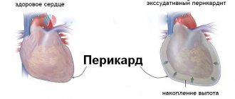
Development of the syndrome
Cor pulmonale is a life-threatening pathological process, as it often leads to death without timely assistance.
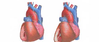
Determining the mechanism of the disease will help you understand what it is. The development of cor pulmonale is caused by a decrease in the vascular network in the pulmonary circulation due to damage to the outer layer of the alveoli. They are spherical formations in the lungs that serve for gas exchange. The alveoli are intertwined with a huge number of capillaries, so their damage leads to poor circulation. The patient will be able to experience for himself what cor pulmonale is when a tenth of small vessels are damaged, as symptoms will begin to manifest themselves clearly. Hypertrophy of the right ventricle of the heart begins with the death of 1/5 of the capillaries of the alveoli - these processes are very closely interrelated. If the blood supply decreases by more than a third, then the decompensation phase begins.
The load on the right cardiac ventricle increases already at an early stage of development due to increased pulmonary blood pressure due to the death of capillaries.
Constant overload leads to hypertrophy of heart tissue and the development of heart failure.
The functional mechanisms that arise as a result of the development of the pathological process will help the patient understand what the cor pulmonale is:
- An increase in the volume of blood transported in the pulmonary circulation. The death of capillaries and narrowing of blood vessels due to high pressure cause hypoxemia (lack of oxygen in the blood). Its deficiency is eliminated by activation of the compensatory mechanism. The body tries to restore balance by increasing the volume of blood pumped by the heart.
- Euler-Lillestrand reflex. Oxygen deficiency causes capillaries to narrow. After normalizing its amount in the blood, they expand.
- Increased blood pulmonary pressure. The extinction of capillaries and severe coughing, characteristic of lung diseases, lead to vasoconstriction. The patient's intrathoracic pressure increases and a squeezing painful sensation occurs in the area of the heart.
- Constriction of blood vessels due to the influence of substances produced by the body. With the development of hypoxemia, the body begins to produce special compounds (serotonin, thromboxane, endotolin, lactic acid) that contribute to the development of spasm of the vascular walls. Their impact also provokes an increase in pulmonary pressure.
- Blood viscosity. Due to the lack of oxygen, microaggregates are produced that slow down blood flow. The blood becomes thicker and affects the development of pulmonary hypertension.
- Infectious diseases provoke the development of cor pulmonale and aggravate the course of the pathological process. They have a negative impact due to deterioration of lung ventilation. Against this background, pressure increases and oxygen deficiency increases. Gradual inhibition of the work of the heart muscle provokes the development of myocardial dystrophy.
Development of the problem during pregnancy
During the period of bearing a child, a woman’s body undergoes physiological and hormonal changes. This significantly increases the load on the heart and blood vessels. Therefore, before conceiving, you should conduct an examination and assess how much the pregnancy will affect the organs and whether the woman can safely and without harm to her own health bear a child.
During pregnancy, the development of hypertrophy is not considered a separate disease. It occurs if a woman has a history of congenital and acquired diseases.
If a pregnant woman has heart problems, she must be admitted to the hospital three times for examination over the course of nine months.
The first time hospitalization is carried out in order to examine the defect, determine the severity of the disorders and the efficiency of the circulatory system. In case of serious violations, the option of terminating the pregnancy may be considered.
The expectant mother is re-admitted to a medical facility when physiological stress reaches its peak. The hospital will take measures to keep the heart in good condition.
The last hospitalization is needed to determine the method of delivery.
Characteristics of the disease
Based on the generally accepted classification, you can figure out what cor pulmonale is. According to the speed of development, pathology comes in the following forms:
- Acute cor pulmonale manifests itself with lightning speed. The clinical picture is getting worse every minute.
- Subacute cor pulmonale develops within 2 days to 2-3 weeks.
- Chronic cor pulmonale develops over years.
Acute syndrome often occurs due to the growth of blood clots in the artery supplying the lungs. They are a consequence of atherosclerosis, ischemia, rheumatism and other vascular diseases. In recent years, the acute form of the pathology has become increasingly common.
The subacute variety is not so dangerous, but without treatment it can be fatal. The clinical picture develops gradually, so there will be time to undergo an examination to identify the cause and eliminate it.
The chronic form of the disease develops over 2-3 years. Patients do not strive to find out what cor pulmonale is in order to become familiar with treatment methods, because it does not have any special manifestations at the beginning of its development.
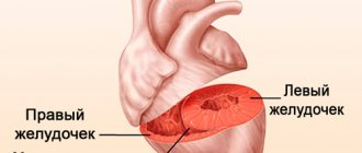
Over time, hypertrophy of cardiac tissue occurs due to lack of oxygen in the blood. It leads to dilatation of the ventricle and atrium on the right side, that is, to their expansion. As the disease worsens, blood flow is disrupted and internal organs malfunction. Chronic hypertrophy can develop even faster due to exposure to pathologies of the bronchopulmonary system (bronchitis, pneumonia).
The factor influencing development is no less important for a person who wants to know what cor pulmonale is. A total of 3 etiological groups are classified:
- The vascular group consists of the vascular causes of cor pulmonale.
- The bronchopulmonary category includes diseases of the lungs and bronchus.
- The thoradiaphragmatic class includes diseases that affect ventilation of the lungs.
To understand how to treat pathology, it is important for a specialist to find out its stage of development:
- The preclinical stage is characterized by overstrain of the right cardiac ventricle and rare attacks of increased pulmonary blood pressure.
- The subcompensated stage is manifested by an increase in the size of the right ventricle and persistent pulmonary hypertension. There are no symptoms of circulatory failure.
- The decompensated stage is determined if the patient’s symptoms of the subcompensated stage of development are accompanied by signs of insufficiency of the functions of the heart and lungs.
Characteristic signs
As a result of such changes in the heart, the patient’s condition worsens. The patient begins to feel pain in the chest on the left side, breathing becomes impaired, and even light exertion leads to severe fatigue. If you treat the underlying disease that promotes hypertrophy, your health will improve and the symptoms of the disease may completely disappear.
The clinical picture of the disease consists of signs of pulmonary disorders and venous blood stagnation.
The fact that hypertrophic processes are developing in the right side of the heart can be understood by:
- coughing attacks, shortness of breath, respiratory dysfunction;
- the appearance of edema;
- pallor and blue discoloration of the skin;
- attention disorders;
- feelings of tingling and discomfort in the heart;
- disruptions in the rhythm of heart contractions.
Most often, the development of hypertrophy does not manifest itself in any way. Deterioration in well-being occurs when the disease reaches an advanced stage. Therefore, people suffering from tachycardia, dizziness and swelling of the legs should visit a doctor to identify the causes of this condition.
Reasons for appearance
The causes of the development of the disease have certain differences, depending on its form. Acute cor pulmonale manifests itself due to the influence of the following factors:
- formation of blood clots in the pulmonary artery and its branches;
- pneumomediastinum (accumulation of air in the mediastinum);
- severe pneumonia;
- frequent attacks of bronchial asthma;
- asthmatic status.
The subacute form of the disease develops due to the following factors:
- the appearance of embolism of microscopic size in the pulmonary circulation;
- inflammation of the walls of the pulmonary vessels (vasculitis);
- persistent increase in blood pressure in the pulmonary artery of unknown origin (primary);
- diffuse inflammatory infiltration of the alveoli;
- development of neoplasms in the mediastinum;
- advanced bronchial asthma;
- hyperventilation of the lungs against the background of botulism, polio and other diseases.
A chronic disease is formed under the influence of the following pathological processes:
- persistent primary increase in blood pressure in the pulmonary artery;
- inflammatory processes in the pulmonary artery;
- pulmonary emboli of a recurrent nature;
- complications after amputation of an entire lung or part of it;
- obstructive pathologies of the bronchopulmonary system: bronchial asthma;
- pneumosclerosis;
- inflammation of the bronchial tubes of a chronic type;
- emphysema.
Causes
There are about 100-120 factors for the development of cor pulmonale, possibly more with an accurate count. All moments are divided into several groups.
Vascular factors
Vascular are relatively rare, accounting for up to 30% of recorded clinical cases.
- Vasculitis. Inflammatory disease of the endothelium, the inner lining of blood vessels. The lesion is infectious (viral) or autoimmune in nature.
When the small circle arteries are involved in the process, their gradual stenosis occurs. In case of insufficient treatment, scarring and fusion may occur. Blood flow weakens.
Then everything goes according to the scheme described above. The nature of hemodynamics in the right parts of the heart changes, overexertion leads to the growth of the muscle layer, then complications occur.
- Atherosclerosis of the pulmonary artery. It is a blockage, or less often a narrowing (stenosis) of the lumen of the corresponding vessel.
Basically, the pathological process is provoked by changes in the level of lipids in the blood. Fatty compounds, in particular cholesterol, are deposited radially on the walls, and there is a deterioration in blood flow at the local level.
The consequences are catastrophic. The heart increases in mass, blood pressure rises, and the frequency of contractions increases. Severe symptoms from the respiratory system are observed.
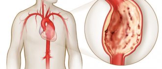
- Aortic aneurysm with pulmonary artery compression. It occurs in a relatively late period of development of a dangerous protrusion of the wall, also with a clear position of the formation. After correcting the condition with surgical methods, there is a good chance of recovery.
- Pulmonary embolism. If the blockage is complete, death occurs within minutes. It’s another matter when a blood clot does not completely close the lumen of the vessel. The effect is the same as with advanced atherosclerosis. Only the symptoms develop many times faster and the process is more aggressive.
Urgent medical correction is required in a hospital setting. Human survival depends on it. Thromboembolism usually results in acute cor pulmonale.
Extravascular factors
Occurs in 70% of situations or more. Estimates vary by region of the planet.
- Pneumonia. Inflammation of the lungs of a septic nature. It is caused by bacteria in most cases. Viruses also occur. Fungi are much less common. Even a one-sided form can provoke a significant circulatory disorder in the right atrium and ventricle.
Acute types lead to emergency conditions. Often everything is limited to a single episode. After drug correction, the situation returns to normal.
Another thing is chronic, often recurrent pneumonia. Against the background of stable respiratory failure, CHL is formed, a constantly progressive type.
- Bronchial asthma. Predominantly allergic disease. In the early stages, when DN is not yet expressed, the likelihood of disorders such as cor pulmonale is minimal. Remissions are of high quality. Advanced forms result in a chronic process; over time it progresses and enters the decompensated phase.
- Emphysema. The formation of voids in the pulmonary structures as a result of rupture of the alveoli. Usually occurs in experienced smokers. The process is not always visible on radiography, which is due to the imperfection and relative inaccuracy of the technique. Treatment as such is impossible, but there is a chance to partially compensate for pathological changes. In this situation, it turns out to slow down the development of the pulmonary heart for years.
- Tuberculosis. Acute or chronic infectious disease. It is provoked by mycobacterium or Koch's bacillus. Causes pronounced destruction of the lungs over a period of several years. Accompanied by disintegration and melting of tissues. At the end they are a curdled mass. The result is gross scarring of the structures. It is not known from which the patient will die faster: from bleeding or critical cardiac dysfunction. Without treatment, both options are likely.
- Bronchiectasis. Similar to the previous described condition. But the point is different. The alveoli are filled with purulent exudate. The process leads to the gradual disintegration of tissues, their melting and evacuation from the body. In place of functional cells, rough voids remain, similar to tuberculous cavities.
The third group of reasons is associated with defects in the musculoskeletal system. As an option, ankylosing spondylitis and kyphoscoliosis are diagnosed.
The essence of pathological processes is a violation of the formation of the chest. A stable compression of the lungs occurs with a weakening of gas exchange and an increase in pressure in the corresponding artery.
Finally, the last group of factors is intoxication, bacterial and viral pathologies.
- Botulism. Provoked by a most dangerous agent. The microorganism secretes a toxic substance that primarily affects the nervous system. The respiratory structures are also affected.
The result is a disruption of normal gas exchange. An increase in the number of movements per minute leads to severe hyperventilation. Acute cor pulmonale appears as a botulism emergency as one of the possible options.
- Polio.
Reasons are being eliminated gradually. Diagnosis is carried out urgently.
Symptoms
The clinical picture of the disease differs depending on its form. Acute cor pulmonale has the following symptoms:
- acute chest pain;
- constant shortness of breath;
- development of cyanosis;
- an increase in the size of the veins in the neck;
- severe arterial hypotension;
- attacks of tachycardia (increased heart rate);
- pain in the liver area;
- nausea to the point of vomiting.
The subacute form of the pathological process has symptoms similar to the acute type of the disease. It differs only in the speed of development.
The chronic form of the disease can manifest itself for years as symptoms of the underlying pathological process. After the onset of the decompensation phase, patients develop the following clinical picture:
- rapid pulse;
- rapid fatigue with virtually no load;
- increasing shortness of breath;
- pain in the chest, relieved by oxygen inhalation;
- spread of cyanosis;
- gradual swelling of the veins in the neck;
- development of swelling in the legs;
- an increase in the size of the abdomen due to the accumulation of fluid in the abdominal cavity (ascites).
The clinical picture of chronic pulmonary heart disease actually has no direct connection with physical activity. A painful attack cannot be relieved by taking Nitroglycerin.
Treatment
During therapy, it will be important to detect the focus, and therefore establish the disease or failure that caused the enlarged heart. It is necessary to do fluorography to get a complete picture. After carrying out the necessary diagnostics, based on the image, therapy is prescribed, which is aimed at eliminating this pathological process.
After an x-ray is taken, medications are prescribed as an additional treatment to remove obstacles to the outflow of blood and relieve the intensive functioning of the ventricles. This will make it possible to prevent the occurrence of adverse consequences in the form of myocardial infarction, angina pectoris, shortness of breath and cardiac arrhythmias.
If therapeutic actions do not give the desired result, the specialist prescribes surgery to improve blood flow. But it should be used only in extreme situations.
Diagnostics
Diagnosis and treatment are interconnected, since without an accurate determination of the cause of cor pulmonale it will not be possible to draw up a correct treatment regimen. A doctor may suspect the presence of a disease in a patient by conducting an examination and identifying the following external manifestations:
- dilation of blood vessels on the cheeks and conjunctiva (the appearance of blush and “rabbit eyes”);
- spread of cyanosis (blue discoloration of the lips, nose, ears and tongue);
- an increase in the size of the veins in the neck;
- expansion of the nail phalanges.
While listening to the heart (auscultation) with a stethoscope, the cardiologist will hear the following abnormalities:
- pronounced pathological changes in tones above the pulmonary artery;
- a variety of wheezing associated with impaired respiratory function.
- noises indicating insufficiency of the right valve (at the stage of decompensation).
The following changes are visible on the x-ray:
- swelling of the pulmonary artery;
- enlargement of lymphatic vessels;
- displacement of the heart shadow to the right side (at the stage of decompensation).
With an ultrasound scan of the chest cavity (echocardiography), the doctor will evaluate the intensity and strength of contractions of the right heart ventricle, determine the degree of dilatation and overload. During the examination, you can also notice the interventricular septum sagging to the left.
The state of respiratory function is determined using a spirograph. Focusing on the volume of inspiration, its speed, as well as the ratio of gas and oxygen, the doctor will assess the severity of pathological abnormalities.
Measurement of pulmonary pressure is required in severe cases of acute disease. Pathology is diagnosed with the following indicators:
- above 25 mm Hg. Art. in a calm state;
- above 35 mm Hg. Art. after physical activity.
During the examination of the patient, the doctor must determine the functional class of the pathology:
- The first class is assigned to patients whose clinical picture is based on symptoms of diseases of the bronchopulmonary system. High pressure in the pulmonary circulation is unstable.
- The second class is characteristic of people who have a combination of symptoms of bronchopulmonary diseases and respiratory failure.
- The third class is characterized by all the above mentioned signs in combination with manifestations of heart failure. High pressure in the pulmonary circulation is persistent.
- The fourth class is assigned to people in the stage of decompensation. The symptoms are pronounced and against its background stagnant processes develop. Heart and respiratory failure at level 3 danger level.
Pulmonary heart: mechanism of development
Pulmonary hypertension plays a leading role in the formation of cor pulmonale in patients. At the initial stage, it is closely related to increased cardiac output at the reflex level; this reaction is a response to increased respiratory function and tissue hypoxia that develops against the background of respiratory failure.
The vascular form of cor pulmonale is accompanied by resistance to blood flow in the arteries corresponding to the pulmonary circulation, which occurs primarily due to the organic form of narrowing of the lumen in the pulmonary vessels against the background of blockage by emboli (when considering the connection with thromboembolism), as well as against the background of a tumor or inflammatory form of infiltration of the walls when the lumen in them is closed (when considering the connection with systemic vasculitis).
Bronchopulmonary and thoracodiaphragmatic forms of manifestation of cor pulmonale are accompanied by a narrowing of the lumen within the pulmonary vessels, which occurs due to microthrombosis and fusion through connective tissue that is relevant to them, or due to actual compression occurring in the area of a tumor, inflammatory or sclerosing process. In addition, narrowing of the lumen of the pulmonary vessels can occur against the background of weakening of the lungs in terms of the ability of their vessels to stretch and collapse due to changes that have arisen in the pulmonary segments. Meanwhile, in the overwhelming majority of cases, the main role is given to functional mechanisms, against the background of which the already noted pulmonary arterial hypertension develops; these mechanisms are directly related to disorders that arise in the respiratory function, with ventilation and with the development of hypoxia.
This factor, arterial hypertension itself, causes overload of the heart, and in particular its right sections. The gradual development of the disease leads to changes in the acid-base balance (initially it can be defined as compensated, but subsequently reach a state of decompensation of disorders). In frequent cases, small vessels are damaged by numerous blood clots, and the heart muscle gradually reaches a state of dystrophy in combination with necrotic processes.
The chronic process of pulmonary heart disease determines the following classification of stages:
- preclinical stage - characterized by the manifestation of the pulmonary form of hypertension in combination with signs indicating strain in the functioning of the right ventricle; identifying this stage is possible only through instrumental research;
- compensated stage - its course is characterized by right ventricular hypertrophy in combination with pulmonary hypertension in a stable form of manifestation without reaching a state of circulatory failure;
- decompensated stage (cardiopulmonary failure) - symptoms appear in a form relevant to right ventricular failure.
First aid measures
If an acute type of cor pulmonale develops, it is necessary to urgently call an ambulance. Until she arrives, a person should lie on the floor and not move. It is advisable that the room is well ventilated.
In a hospital setting, the patient will receive thromboembolic therapy. It must be started as early as possible to increase the chances of restoring pulmonary artery patency and preventing death.
Along with thromboembolic therapy, doctors can use the following measures to stabilize the patient's condition:
- artificial ventilation;
- injection of analgesics and drugs for cardiac and respiratory failure;
- surgery to remove a blood clot.
Decoding the results
The heart on x-ray is assessed according to the following parameters: location, size, shape. Having assessed these indicators, the specialist draws a conclusion about the health status of the subject.
Digital code
Heart shadow
In the absence of pathologies, the organ is located in the anterior lower region of the left half of the sternum. When a person moves, the heart moves, but no more than 2 cm. An x-ray can demonstrate the following options for the organ’s shadow:
- localization on the right side;
- shift due to effusion into the pleural cavity;
- displacement under the influence of a diaphragmatic hernia or neoplasm;
- change in localization in case of lung shrinkage.
Cardiac configuration in pathologies
The cardiac configuration on x-ray indicates a certain type of damage to the valve apparatus and blood vessels of the heart:
| Cardiac configuration | Explanation |
| Mitral | The waist is smoothed, the angle between the right atrium and the vascular network is moved upward, the radius of the ventricular arch on the left side is increased, the pulmonary artery has elongated arches. A clinical picture is observed with pathologies in the structure of the mitral canal, a decrease in the lumen of the pulmonary trunk |
| Aortic | The waist is visualized, the size of the ventricle on the left is increased, and the presence of a wide aorta. Symptoms are observed in the case of Fallot's disease, aortic stenosis, arterial hypertension |
| Globular (trapezoidal) | The waist is defined, the arches of the atrium on the right side and the ventricle on the left are enlarged, the contour is smoothed. Signs are visualized in the inflammatory process of the myocardium, an increase in the lumen of the chambers, an abnormal structure of the septum |
Increase in organ size
The enlargement of the cavities of the heart and some vessels entails a change in the size of the organ. The development of a certain pathological process is judged by the location of such changes:
| Change area | Diagnostic explanation |
| Left atrium | The esophagus is moved back to the side, the appendage is enlarged, and an additional arch is visualized. Typical clinical picture of mitral valve disease |
| Ventricle on the left | The apical part has a rounded shape, the arc is enlarged. Symptoms of heart valve insufficiency, arterial hypertension |
| Aortic arch | The expansion of the first arch on the right is observed. Characteristic in case of development of hypertension, aneurysm |
| Right atrium | Ambiguous signs of arch lengthening are visualized, and the vena cava is dilated. Indicates the development of pulmonary hypertension |
| Ventricle on the right | Lengthening and expansion of the pulmonary trunk, the upper part is rounded, “looks” upward, the cardiac diameter has an increased size, the space behind the sternum is not defined. Clinical picture of congenital defects of the septum and cor pulmonale |
Drug therapy
A drug treatment regimen for cor pulmonale is drawn up based on the patient’s condition and the underlying pathological process. Basically, it includes the following groups of drugs:
- Broad-spectrum antibiotics. They are recommended if the patient has diseases of the bronchopulmonary system caused by a bacterial infection.
- Drugs with a bronchodilator effect, which are used to relieve attacks of bronchial asthma and obstructive bronchitis.
- The use of antiplatelet agents and anticoagulants is due to blood thickening and the presence of thromboembolism.
- Diuretics (diuretics) are used to eliminate edema and in the development of heart failure.
- Drugs with antiarrhythmic effects and cardiac glycosides are prescribed to normalize heart function in case of right ventricular failure and arrhythmia.
- Glucocorticosteroids are used if the disease develops against the background of an autoimmune failure.
- Nitrates serve to normalize blood circulation.
- Drugs with expectorant action and mucolytics are used to remove sputum in bronchopulmonary diseases.
- Potassium-containing medications serve to saturate the body with potassium during hypokalemia. It is responsible for saturating tissues with oxygen, maintaining water and acid-base balance and other important processes.
- A solution of sodium bicarbonate is administered by drip in case of severe acidosis.
Why do you need a heart x-ray?
Fluoroscopy
Examination of the organ is carried out using radiography and fluoroscopy. In the first case, the doctor receives an image; in alternative diagnostics, he evaluates the functioning of the organ at the current time.
In some clinical cases, radiography of the heart in three projections is prescribed or the borders are contrasted.
Examination in three projections
To obtain an image in a direct projection, the patient is placed in a position in front of the screen. This type of procedure is provided for diagnosing diseases in the area:
- ascending aortic zone;
- left ventricle;
- atrial fusion;
- two angles between the shadow of the organ and the diaphragm;
- pulmonary artery.
If a picture of the heart is taken in an oblique projection on the right, the patient stands with the appropriate shoulder forward (45˚ angle) to the device. The equipment tube is placed behind the person. Research is carried out using this method:
- areas behind the sternum;
- conus arteriosus;
- contour line of the heart and all its components;
- pulmonary fields.
The principle of x-ray examination of the heart in a beveled position on the left is identical to that described above. Only in this case the patient stands with his left shoulder to the screen.
With the described type of examination, a specialist can assess the condition of the areas of the aorta, the posterior side of the ventricle on the left, and the trachea.
X-ray with contrast injection into the esophagus
Changes in the right cardiophrenic angle.
Pericardial cyst. The location of the esophagus is behind the heart. When the size of a certain cardiac chamber increases, the esophagus is pushed towards the spinal trunk along arcs of varying radii. In the process of diagnosing the local area, the size of this arc is determined.
If movement along a small radius arc is visualized in an oblique position on the right, a decrease in the size of the mitral orifice is suspected. We talk about valve insufficiency if movement of the esophagus is observed along an arc of a large radius.
This type of examination helps to identify the right placement of the aortic arch, an increase in the width of the pulmonary trunk and the lungs in particular.
Individuals who were examined complain of the following unpleasant symptoms:
- squeezing pain in the chest;
- enlarged liver;
- shortness of breath;
- prolonged cough;
- fever, etc.
Pathologies in which the need for an x-ray is obvious: cor pulmonale, hypertension, heart defects in adults, heart failure, vascular diseases, pericardial cyst (around the heart), etc.
ethnoscience
Folk remedies are used as a complement to many treatment regimens, but not in all cases. Acute cor pulmonale requires immediate treatment, so you should immediately call an ambulance rather than selecting remedies on your own. Subacute and chronic forms do not have such restrictions. After examination and prescription of a course of basic treatment, the following folk recipes are allowed:
- Calendula infusion should be drunk 1 tbsp. l. 3 times a day for at least 2-3 months. To prepare, you will need to pour 500 ml of alcohol into 80 g of plant flowers. Then let it sit for 7 days.
- Garlic-lemon drink with honey, take 1 tbsp. l. per day before bedtime. The duration of therapy is 1 month. To prepare the product, you need to chop 3 small heads of garlic and mix them with freshly squeezed juice from 3 lemons. Add 250 ml of honey to the resulting mixture and mix well.
- The collection, consisting of hawthorn, knotweed, horsetail and tricolor violet, should be consumed as a decoction 3 times a day, 100 ml for 1 month. To prepare you will need to take 1 tbsp. l. mixture and pour it into a glass of boiling water. The product needs to sit for at least 10-15 minutes.
Types of heart dilatation
Dilatation of the chambers of the heart can be:
- Tonogenic, caused by increased pressure in the cavity. Occurs when the valve openings are narrowed or there is increased pressure in the aorta or pulmonary artery. May precede myocardial hypertrophy. The tone of the heart muscle and contractility are preserved; it becomes the myogenic type as it progresses.
- Myogenic. The cause is a decrease in the contractility of the heart muscle due to dystrophic disorders. A persistent and irreversible process, the myocardial fibers are stretched and elongated. Occurs in myocarditis and atherosclerosis.
Recommendations
There are certain recommendations that increase the chances of recovery or relief of the general condition if strictly followed. Their list:
- consumption of potassium-containing products;
- use of oxygen therapy;
- chest massage;
- breathing exercises;
- full sleep (at least 8 hours);
- avoidance of stressful situations;
- reduction of physical and mental overload;
- giving up bad habits (drinking alcohol, smoking);
- avoiding areas with poor ecology;
- reducing the amount of salt and animal fats in the diet;
- preventing exposure to allergens.
There is a narrower range of recommendations created for certain situations:
- Chronic diseases of the bronchopulmonary system require the patient to be able to perform positional drainage of the bronchi.
- In case of chronic obstructive pulmonary disease, it is advisable to carry out bloodletting procedures with the administration of Reopoliglucin.
- Severe cases of cor pulmonale will require a heart muscle or lung transplant.
As a preventative measure, it is recommended to follow the following rules:
- Identify and begin to treat pathological processes associated with the bronchopulmonary system and cardiac muscle in the early stages of development. Annual preventive examinations can help with this.
- Do not self-medicate and consult your doctor about any changes in your condition.
- Try to prevent exacerbations of pathologies of the bronchopulmonary system to prevent the development of respiratory system failure.
- Do moderate physical therapy and breathing exercises.
- Follow the rules of a healthy lifestyle and follow all doctor’s recommendations.
- Completely eliminate foci of infection when they occur in the body in order to maintain a strong immune system.
How to avoid the problem
Right atrial hypertrophy is a serious problem. Therefore, measures should be taken to prevent such violations. First of all, doctors recommend:
- avoid bad habits;
- create the right daily routine;
- balance your diet.
It is important to create an optimal training regimen when playing sports.
People who are not involved in sports are advised to avoid enormous loads and grueling exercises. To keep your mood good and your body in good shape, it is useful to walk, swim, ride a bike or jog in the morning at a moderate pace every day. Doctors say that it is difficult for the body to tolerate increased loads, which increase pressure in the circulatory system and develop hypertrophic processes.
Emotional stress has a negative impact on the functioning of the heart muscle. Therefore, they should be avoided so that the heart does not wear out prematurely. To strengthen the nervous system, it is recommended to resort to relaxation techniques, meditation, and yoga. These classes are especially necessary for residents of large cities, since they are more susceptible to stress than others.
It is also important to visit a doctor on time if you have health problems. This will help detect diseases that can cause complications in the form of hypertrophy of the heart chambers and other disorders. If there are congenital defects, maintenance therapy should be carried out periodically.
Forecast
What prognosis a doctor will give to a patient with cor pulmonale depends on the form of the pathology. In the acute version, death can overtake a person within a matter of minutes. If it was avoided, the condition improves within 10 days.
The subacute form of the disease leads to death within 1-2 weeks if the patient is not helped. If the pathology is successfully relieved, a negative prognosis remains only for future employment. It is associated with long-term treatment of the underlying pathological process, which led to the development of cor pulmonale.
The chronic course of the pathology is considered quite insidious due to the mildly expressed symptoms at the beginning of development. With each passing month, the chances of eliminating the consequences of the pathology will be less and less. With effective treatment carried out in the initial stage of the disease, the patient can live over 10 years. Therapy started already at the stage of decompensation prolongs life only by 2-3 years. On average, patients with the chronic form of the pathology live about 5 years. A lung transplant prolongs life by 2 or more years in 60% of patients.
Cor pulmonale is a complication of diseases of the cardiovascular and bronchopulmonary systems. It is divided into several stages along its course. The most dangerous of them can be fatal in a few hours. To prevent this, experts advise people who are at risk to learn what cor pulmonale is and be examined annually. If the development of a pathological process is detected, you will need to reconsider your lifestyle and follow all the doctor’s instructions. Self-medication will only worsen the situation and increase the chances of death.
Treatment methods
If hypertrophy of the right atrium of the heart occurs, the doctor must decide how to treat it.
Also read: How does adhesive pericarditis manifest?
This disease is classified as a secondary disorder. Therefore, in order for the atrium to return to its normal size, normal heart function and blood circulation to improve, the main cause of the disorder must be identified and eliminated.
Treatment of the disease is complex, aimed at treating the main pathology. Doctors correct the patient’s condition using medications and recommend introducing changes to the usual lifestyle. If the patient does not follow the advice of specialists, no therapy will give the expected results. Therefore it is important:
- Avoid bad habits such as smoking and alcohol abuse.
- Limit the presence of salt in your daily diet, do not drink a lot of liquid, and avoid foods containing high amounts of cholesterol.
- If you are overweight, solve this problem, but physical activity should be moderate.
If the patient strictly follows these rules, then the process of returning the heart to a normal state will go faster and the likelihood of recurrence of the disease will decrease.
If the problem arose as a result of impaired functioning of the lungs, then they try to compensate for the functioning of the organ with means to relieve the inflammatory process, drugs that dilate the bronchi and other medications.
In the presence of valve defects, surgical intervention must be resorted to. For diseases of the heart muscle, antiarrhythmic drugs are used, treatment is carried out with drugs to stimulate metabolism in muscle structures, and cardiac glucosides.
If you detect the development of hypertrophy in the right atrium in time, you can completely get rid of the problem and live a long life.
Diagnostic methods
When making a diagnosis, the history of the occurrence of signs of circulatory failure is taken into account - shortness of breath, tachycardia, wheezing in the lungs, swelling of the extremities, expansion of the boundaries of the heart during percussion. To clarify the diagnosis, instrumental examination methods are required:
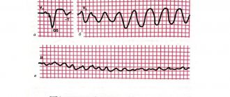
- ECG - signs of overload of one or more chambers of the heart, rhythm disturbances in the form of atrial fibrillation, conduction blockades.
- Ultrasound of the heart is the main way to detect dilatation of the atria or ventricles, as well as the presence of intracardiac hemodynamic disorders - reverse blood flow, pressure overload.
- X-ray – the size of the heart is increased due to one of the cavities.
- Scintigraphy to assess changes in the myocardium.
- MRI and CT scan to detect the cause of cardiac dilatation.
Treatment of dilatation
Therapy for enlargement of the heart chambers involves correction of manifestations of circulatory failure and arrhythmia.
In case of decompensation, patients are placed on bed rest and the intake of water and table salt is reduced. The following groups of drugs are used:
- ACE inhibitors – Enap, Capoten;
- diuretics - Veroshpiron, Triampur;
- antiarrhythmic - Bisoprolol, Carvedilol;
- long-acting nitrates (reduce blood flow to the right side of the heart) - Isoket, Nitrogranulong.
Cardiac glycosides are prescribed with caution. In extremely severe cases, a heart transplant is indicated.
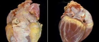
Decompensated cor pulmonale, diagnosis and search
Cor pulmonale is a deviation of the right side of the heart, which is characterized by hypertrophy (enlargement) and dilatation (expansion) of the right atrium ventricle. It is also circulatory failure, which develops with hypertension in the pulmonary hemodynamics (circulation).
Decompensated cor pulmonale (DCP) is determined by noticeable insufficiency of hemodynamics and external respiration. Diagnosis of DLS is not difficult; it is carried out only if there are obvious signs of right ventricular failure. At the very initial stages, it is difficult to recognize heart failure in cor pulmonale, because the very first symptom is shortness of breath. In this case, it is not a help. In any case, complaints and symptoms make it possible to detect the first signs of decompensation of the pulmonary heart.
The diagnostic search for DLS is divided into three stages
At the first stage, changes in the nature of shortness of breath are noticed. How to identify this? Shortness of breath becomes more and more constant and ceases to depend on the weather. The frequency of inhalations increases, but exhalation does not lengthen. The duration and intensity of shortness of breath increases after coughing, but after taking bronchodilators it does not decrease. At the same time, stage 3 pulmonary insufficiency progresses—shortness of breath at rest. Working capacity decreases and fatigue progresses. Ultimately, with hypoxia and hypercapnia, constant headaches and drowsiness occur.
There may also be complaints of pain in the cardiac region, unfortunately, the nature of the pain is uncertain. It is quite difficult to explain the root cause of pain; its appearance is influenced by many factors. For example: hemodynamic overload of the heart in pulmonary hypertension, metabolic disorders in the myocardium, insufficient disruption of collaterals in hypertrophied myocardium.
Also, heart pain can be felt along with suffocation, severe cyanosis or agitation. This is precisely what is typical for hypertensive crises in the pulmonary artery. Irritation of the baroreceptors of the right atrium is expressed by an increase in pressure in the pulmonary artery. This also causes an increase in pressure in the right ventricle.
There are several factors that help to suspect decompensated cor pulmonale: heaviness in the right hypochondrium, edema, abdominal enlargement with a pulmonary history.
At the 2nd stage of the search, a symptom of swollen neck veins is detected. When two insufficiencies combine: pulmonary and cardiac, the jugular veins become distended at both the inlet and outlet. With pulmonary insufficiency, acrocyanosis forms, the hands and fingers become cold to the touch. There is also swelling of the lower extremities and pastosity of the legs.
Tachycardia becomes constant, most of all it is expressed at rest than during exercise. Atrioventricular valve insufficiency also develops when the right ventricle dilates. All this predetermines the manifestation of systolic murmur at the xiphoid process of the sternum. There is a possibility of an increase in blood pressure during hypoxia, and when heart failure develops, its sounds become more muffled.
Also, one should not forget that when early circulatory failure is detected, the liver enlarges. It may protrude from under the costal arch. This is also present in patients with emphysema, regardless of whether they have heart failure. First of all, the left lobe of the liver enlarges, its palpation is painful and sensitive. The more symptoms become, the greater the likelihood of identifying a positive Plesha symptom.
When coronary artery disease is combined with drugs or hypertension stages 2-3, hydrothorax and ascites can be observed, but this is extremely rare.
The third stage of the diagnostic search is not so significant when diagnosing DLS.
X-ray can reveal an enlargement of the right chambers of the heart and also identify pathology of the pulmonary artery:
1. Dilatation of the descending right branch of the pulmonary artery
2. Enhanced image of pulmonary vessels
3. Increased pulsation in the lung area and its weakening in the peripheral parts.
Other ways to identify and diagnose cor pulmonale decompensation may include an ECG, hemodynamic studies, and blood tests.
general characteristics
The development of the syndrome involves three stages. This:
- acute cor pulmonale;
- subacute stage;
- a process that is chronic.
In the first case, the pathology develops very quickly (maximum - a few days). In contrast, the chronic form can be asymptomatic for two to three months or even years. Patients suffering from pulmonary diseases are also at risk of developing the syndrome, and this occurs in approximately 3% of cases.
Cor pulmonale is often a complication of cardialgia. The disease is in fourth place in terms of the frequency of deaths occurring among all diseases of the cardiovascular system.
Classification and pathogenesis
Chronic cor pulmonale is not an independent disease; it is caused by other lung pathologies. Thus, CLS can be called a complication that occurs as a result of damage to the respiratory system. According to WHO statistics, the diagnosis of CHL is most often a consequence of the following pathologies:
- bronchial asthma;
- obstructive bronchitis;
- lung abscess, pleurisy;
- interstitial pneumonia;
- pneumosclerosis;
- tuberculosis;
- bronchiolitis;
- frequent sore throats;
- bronchiectasis;
- emphysema and polycystic lung disease;
- pneumoconiosis.
The name of the disease is formulated on the basis of the relationship between two body systems, where pathological changes in one lead to dysfunction of the other. There is the following classification of forms of chronic pulmonary heart disease:
- Bronchopulmonary. In this form, the presence of chronic inflammatory lung diseases is observed.
- Vascular. Occurs with prolonged pulmonary hypertension.
- Thoradiaphragmatic. It is provoked by various disorders of the spinal column and deformation of the chest due to injury or congenital malformation.
In medicine, this disease is also classified according to the degree of compensation for heart failure:
- compensated CHL;
- decompensated CHL;
- subcompensated CHL.
In the compensated form, all body systems adjust to the unfavorable conditions created. In this condition, the patient experiences symptoms of the underlying disease, including chronic pulmonary hypertension.
In the decompensated form, the body's adaptive mechanisms are disrupted, which leads to the progression of the disease, which is accompanied by chronic heart failure.
The subcompensated form is characterized by a rapid increase in symptoms of the disease and suppression of the body's protective functions. CHF may occur, against the background of which the patient begins to have systematic heart attacks and a rapid deterioration in his general health.