The human head and brain are penetrated by a network of blood ducts. All of them are of exceptional value; their health affects not only a person’s well-being, but also his life.
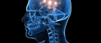
Magnetic resonance imaging (MRI) has been helping us study the complex interweaving of blood structures in the brain for some time.
Let's figure out how this device works and what MRI of brain vessels shows.
Basics of the method
MRI of the head and cerebral vessels makes it possible to visualize and examine soft tissues. The brightness and detail of its results is higher than that of radiography or CT.
The device provides tools for assessing the anatomical integrity, conductivity, and performance of the vascular system of the head and cervical region using angiography.
The method can be described as reading the electromagnetic reflection of a central atomic part, usually hydrogen, from the soft components of the human body located inside a magnetic field.
The process of magnetic resonance of nuclei is completely safe for the human body.
The tomograph produces electromagnetic radiation and reads the time and speed indicators of the response signals of elementary particles.
The vibrations resonate with the body and draw a three-dimensional model of the patient on the screen with all the information about his structures, parts and body systems.
As a rule, the power of a standard device is 1.5 Tesla. A large diameter tunnel pipe is surrounded by a magnet. The patient lies motionless on a table, which slides into the tunnel during the examination.
The length of the tunnel varies depending on the purpose. There are non-fully enclosed devices for very obese patients or for those who cannot tolerate closed spaces.
Nearby there is an office where the resulting image is processed on a computer.
To identify many serious disorders and failures, MRI of the brain and blood vessels is simply necessary. For a couple of decades now, this method has been widely used by vascular neurosurgeons and neurologists.
The device and its maintenance are quite expensive, require trained specialists and are available only in large medical centers. The service can cost from two to five thousand rubles. Certain segments of the population can get tested at a discount.
Differences from X-ray examination
X-rays work using radioactive x-rays, under the influence of which only hard bone or cartilage structures of the body are clearly visible on film. It will not be possible to see organs or vessels on it.
A small dose of radiation during an X-ray for a healthy person is unacceptable for some patients: children, women expecting a baby or breastfeeding mothers, patients who have already been exposed to a certain dose of radioactivity.
Because of all these features, tomography of cerebral vessels is ahead of x-rays in terms of reliability and objectivity. But in general, this method can be used in conjunction with a vascular program.
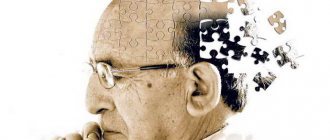
What brain pathologies does MRI show?
The distribution of white and gray matter and pathological processes within the brain are illustrated in detail by MRI. Magnetic scanning is one of the high-precision non-invasive methods of instrumental diagnostics for neurosurgeons, neurologists, and, less often, psychiatrists. Analysis of changes in the two main components of the brain - gray and white matter - is important for clinical diagnosis and therapy of a huge number of diseases: epilepsy and episyndrome, stroke, Alzheimer's disease, malignant and benign neoplasms, multiple sclerosis, infectious and inflammatory processes, post-traumatic injuries, etc.
Distribution of gray and white matter in the brain
Gray matter
The gray matter of the brain is responsible for most of the functions of higher nervous activity and is represented by neuron bodies, glial cells, a cluster of dendrites, thin tiny blood vessels - capillaries - and unmyelinated axons. The main histological structures are centers, each of which controls some action: the act of urination, defecation, heartbeat, etc. Neurosurgeons consider the gray color to be conventional; rather, this substance has an earthy tint. The composition of the main structures of the brain is clearly distinguishable, mainly due to differences in water and protein content. This allows one zone to be differentiated from another on tomograms. Pathological processes localized in the gray matter lead to disturbances in perception, speech, emotions, memory, sensory sensitivity, will, muscle movements, etc.
White matter
The white color is caused by bundles of nerve fibers covered with a myelin sheath. The main purpose of this brain structure is the transmission of impulses from the main centers to the periphery (lower links of the nervous system).
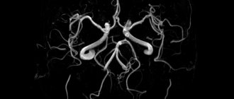
MR angiography
This is a radiation scan to identify problems specifically in the vascular network of the head. It allows you to non-invasively obtain a clear and comprehensive picture of blood movement, the condition of arteries and even the smallest capillaries.
The lymphatic network of the brain is also well visualized.
Thanks to such information content, it is possible to detect the disease at the initial stage of its development and plan surgical intervention with high accuracy.
And the possibility and likelihood of curing many pathologies depends on early diagnosis of cerebral vessels on MRI.
A three-dimensional picture of the desired area is obtained by a combination of angiographic examination and computed tomography, even without the introduction of contrast. It is used only if cancer is suspected.
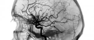
Arteries of the brain
MRI of the cerebral arteries is necessary if you suspect:
| Disease | Description |
| Ischemic and hemorrhagic stroke | Weakness in the limbs on half the body, impaired speech, consciousness, facial asymmetry, severe headache with nausea and vomiting |
| Transient ischemic attacks | Temporary disturbance of cerebral circulation - all signs of a stroke, but they disappear within 24 hours |
| Encephalopathy | Headache, dizziness, memory loss, tinnitus, loss of performance, depressed mood, irritability, tearfulness, poor coordination of movements, and at a later stage dementia occurs (dementia) |
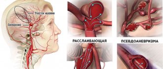
| Aneurysm | Impaired vision, eye movements, severe headache that does not go away after painkillers with nausea and loss of consciousness |
| Binswanger's disease | in young people, due to damage to small arteries, insomnia occurs, sudden changes in blood pressure, memory and gait deteriorate, and involuntary discharge of urine and feces occurs |
| My-My disease | In children, speech, vision, movements and sensitivity in the arm and/or leg are impaired, epilepsy attacks occur, in adults severe migraine-type headaches appear, movements lose coordination |
| Cerebral vasculitis | Against the background of infection, rheumatism or poisoning, a severe headache begins, limbs weaken, behavior and speech change, vision and hearing suffer, fainting and convulsions occur. |
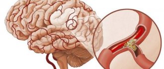
MRI reveals the presence of a cholesterol plaque or blood clot that blocks the flow of blood, changes in the walls of the arteries with their thickening, spasm, or expansion due to an aneurysm. In chronic processes, you can see a slowdown in blood flow and signs of lack of nutrition in the area of the damaged vessel.
Method capabilities
MR imaging has a wide range of diagnostic applications. It makes it possible to fully and harmlessly study the circulatory system of the head in all details, in general and in specific areas, to evaluate the biophysics and biochemistry of the brain.
Angiography is effective in detecting early symptoms of apoplexy, its monitoring and prognosis.
MRI of the vessels of the head clearly shows any structural defects in the blood vessels, the formation of blood clots, and manifestations of disseminated atherosclerosis.
Cancerous tumors, various pineal cysts and their position, size and distribution along the vascular pathways are also well visualized.
At the same time, the consequences of all kinds of injuries and concussions of the base of the skull and brain, hemorrhage, and intracranial aneurysms are also visible.
This makes it easier to detect the source of the problem and helps surgeons prepare effectively for surgery on such an important and fragile organ.
What does computer research show?
Distinguished by its particular accuracy in diagnosing ailments, radiation therapy allows medical professionals to determine the characteristics of many of the most important processes in the brain, namely:
- Changes in the vascular system associated with head trauma, such as concussion.
- Aneurysms of any size.
- Alzheimer's disease.
- Atherosclerosis in any manifestation.
- Cysts (bubble-shaped cavities of a pathological nature).
- The initial stage of ischemic disease.
- Suspicious neoplasms.
- Pick's disease (necrotic damage to neurons).
- Necrosis of certain segments of the cerebral cortex.
- Blood clots in blood vessels.
- Hydrocephalus.
- Atrophy of the cerebral cortex.
- Vascular dementia.
- Early manifestations of stroke.
- Signs confirming the presence of concussion.
- Diseases associated with destabilization of the pituitary gland (meningioma, prolactinoma, etc.).
- Meningitis.
- The extent of tumor damage to various brain vessels, as well as the size and exact location of the formation.
- Narrowed areas of blood vessels and their exact length.
Since some ailments can have an extremely negative effect on the body, often contributing to the death of the patient, it is necessary to promptly consult a doctor when suspicious symptoms are detected.
Before entering the tomography room, you must remove all types of jewelry (earrings, rings, cuffs and piercings are no exception)
Areas of application
In neurology, MRI of the brain and its vessels is done to determine brain abnormalities, problems with the locomotor system, anomalies and defects of various organs and systems.
A layer-by-layer three-dimensional image of the body without the use of harmful rays helps with this.
It helps to examine fibrocartilaginous tissue, connective and nerve fibers. The versatility of the method lies in its coverage of all types of tissues and systems of the body up and down, in its safety and high accuracy, three-dimensional result.
Why tomography is done, indications.
There must be a compelling reason to perform a magnetic resonance imaging scan of the brain. The doctor will not prescribe an expensive procedure without reason. A referral for an MRI of the head is usually issued when other diagnostic methods have not provided sufficient information to make a diagnosis, or when a serious disease that is life-threatening to the patient is suspected.
Indications for MRI of the brain include:
- Cerebrovascular accidents;
- Vascular diseases of the brain;
- Having had a stroke or heart attack;
- Brain dystrophy;
- Hearing and speech impairments;
- Traumatic brain injury;
- Inflammatory diseases of the brain;
- Neurodegenerative diseases;
- Suspicion of a tumor.
MRI is often prescribed for headaches of unknown origin. Sometimes only with the help of this diagnostic method it is possible to identify diseases whose clinical picture is erased or unclear.
Purpose of MRI
A person with suspected neurological problems of the cerebral vessels, such as hematomas, acute disruptions in blood flow in the brain of various nature, or autoimmune diseases, can receive an appointment for MRI.
Tomography is indicated for patients with injuries and damage to the skull and neck.
In oncology, MRI is also usually used to clarify the diagnosis. In this way, neoplasms of the chiasmatic-sellar zone and pituitary gland are examined.
A scan is carried out if a person simultaneously experiences significant hearing loss and speech defects, vestibular disorders, fainting, stuffy ears, or a long-term intense headache.
MRI is also done if there is a history of neurological infections, purulent processes, central nervous system disorders, epileptic seizures, pathological dilations or narrowings of the bloodstream, in the postoperative period.
Such ailments as multiple sclerosis, prolonged inflammation of the sinuses, arteritis, hypertension, congenital structural heart defect, nasal bleeding, diseases that destroy brain cells necessarily require tomography. It happens that the method is used to diagnose athletes whose daily loads and training are associated with head injuries and physical overexertion.
MRI angiography of cerebral vessels
MRI angiography of cerebral vessels is divided into arteriography and venography. This is important, since in arterial diseases there is often a need to assess the condition of the intracardial vascular networks located inside the skull and extracranial, that is, cervical.
Venography examines the venous branches and sinuses between the membranes of the brain. If, after a preliminary examination, the doctor has doubts about the diagnosis, then arteriography together with venography or MRI of the brain with angiography may be prescribed.
MRI in children and pregnancy
Carrying a child is not a reason to refuse the examination completely. But still, during this important period, MRI of cerebral vessels can be done only for serious medical reasons and after the first trimester.
In any case, expectant mothers should not be injected with a contrast agent.
Children, if they have a doctor's prescription, can undergo this procedure completely safely. The baby's ears must be protected with headphones.
Parents and doctors should emotionally prepare the child for what will happen, tell and explain in a playful way why it is so important to lie still.
After all, this is a prerequisite for successful results. While in the tomograph, the baby can hear the encouragement of his parents and immediately report his inconveniences or unpleasant sensations.
Before the age of five, babies undergo an MRI under the influence of anesthesia, which is carefully selected for a particular child by an anesthesiologist.
The idea of doing an MRI of the brain vessels of your baby may come up with the following problems:
- the baby regularly loses consciousness;
- headache often;
- involuntary muscle contractions occur;
- hearing and vision indicators fall;
- the baby develops late in terms of speech and motor skills;
- the baby has become dramatically different in everyday life;
- the child is engaged in active and traumatic sports;
- There are suspicions of epilepsy.
Types of changes
Focal changes in the brain substance often have a post-ischemic etiology, which is characterized by circulatory disorders in one of the hemispheres.
This process has several types, involving three stages:
- at the first stage, MRI signs do not have pronounced manifestations, since the formation of lesions occurs, accompanied by mild weakness, apathy, and dizziness may occur;
- at the second stage, changes in the brain intensify, migraine attacks may occur, a noticeable decrease in intelligence or mental abilities, ringing in the ears, unmotivated mood swings, and lack of coordination are observed;
- at the third stage, focal changes in the white matter of the brain enter the stage of irreversible processes, signs of dementia (dementia) appear, and loss of sensitivity and control of movements occurs.
Changes in the GM substance can occur during aging in people over 60 years of age, and the type of changes is of a single focal nature.
Another type has a dystrophic nature of lesions that arise due to lack of nutrition of brain tissue. The main reasons for the appearance are indicated above.
MRI picture
A competent specialist will glean a lot of information from an MRI image of the cerebral vessels or a three-dimensional image. In a healthy person, all brain tissue is intact and does not contain neoplasms or darkening.
This method provides the doctor with a layer-by-layer image of the walls of blood vessels, a three-dimensional model of the entire circulatory system of the head.
In this case, you can use different angles of view on individual zones or highways, and detect the slightest blockage of blood flow in the deep veins of the neck.
Any vascular defects, cancerous degenerations, aneurysms and narrowings, congenital anomalies, inflammation of the brain parenchyma, damage by pathological microorganisms, hydrocephalus are easily detected.
In the vascular mode of the brain
MRI in the vascular mode of the brain can be performed only for internal arteries (intracranial) or simultaneously with the study of the supply vessels of the neck and venous network.
Internal arteries of the brain with a vascular program
When performing an MRI of the intracranial arteries of the brain with a vascular program, the following vessels are scanned:
- basilar;
- paired internal carotid;
- midbrain, anterior and posterior;
- connecting;
- vertebrates.
They may show signs of atherosclerosis, inflammation, tumors, abnormal structure (aneurysm, malformation), damage or compression by a neoplasm. In the latter case, contrast is often used to clarify the diagnosis.
Watch the video about what an MRI of cerebral vessels shows:
MRI of neck arteries
The following paired vessels (right and left) are subject to MRI examination of the neck arteries: subclavian, vertebral and common carotid. Tomography shows the condition of the brachiocephalic trunk, external and internal branches of the carotid arteries.
As a result of the diagnosis they find:
- sclerotic changes;
- compression of a vessel in the spine or a neoplasm;
- aneurysms;
- vascular malformations;
- severe tortuosity;
- changes in anatomical structure.
MRI of neck vessels
Indicated for neck trauma, atherosclerosis, inflammatory processes, acute and chronic disorders of cerebral blood flow.
MRI of cerebral veins
The objects of study during MRI of cerebral veins are:
- venous sinuses;
- cerebral veins;
- branches in the cerebellum;
- blood outflow pathways in the trunk area.
Indicated for signs of venous stagnation:
- morning headache;
- swelling of the face;
- increased intracranial pressure – distension, pressure on the eyes;
- loss of consciousness during a coughing attack;
- staggering when walking;
- intolerance to bright light and loud sounds;
- fatigue when atmospheric pressure decreases.
Arteriovenous malformations
MRI shows expansion of the venous network, areas of local narrowing of the lumen of blood vessels, blockage of the veins and sinuses of the brain with blood clots, and arteriovenous malformations. In most cases, tomography of the brain substance and arterial branches is performed simultaneously. The procedure is performed without contrast or with additional contrast.
Preparing to Scan
As before many types of diagnostic procedures, before MRI of the brain vessels you will be given an agreement to sign.
Before signing it, make sure that you tell the doctor about all the important features of your health: chronic illnesses, allergies, claustrophobia or pregnancy. If it is difficult for you not to move for a long time, ask your doctor to prescribe sedatives for you.
It is very important to remove all items containing metal. After all, a tomograph is a large magnet that can attract them and create a threat of injury.
So, we remove from ourselves wristwatches, any jewelry, hearing prostheses, credit cards, cufflinks, bobby pins, and clips. We get rid of removable dentures, glasses, pens, piercings, buckles.
We take off clothes with zippers and underwear with underwires. If there are implants or staples in the body, this may make the scan ineffective or even dangerous.
Ideally, you will be given loose clothing to change into. You can eat and drink as usual before the procedure. But, of course, refrain from tobacco and alcoholic beverages for a couple of hours.
Particular care must be taken when scanning with contrast injected into the bloodstream. You need to make sure that the patient is not allergic to the substance by performing a test to check kidney function.
The introduction of contrast increases the accuracy of the image; sometimes other methods fail to clarify the diagnosis.
How to do an MRI of cerebral vessels
MRI of cerebral vessels is done without special preparation; it can be performed on an emergency basis immediately after the patient is hospitalized. If it is prescribed as planned and with the administration of contrast, then it is recommended not to eat for 2 hours.
Before entering the office, you must remove all metal products and leave any electronic devices outside the door. You cannot even take glasses with you, and there should not be any metal elements on your clothes. Such precautions are due to the fact that when ferromagnetic products enter a magnetic field, they become very hot.
The research device looks like a cylindrical tube about 2 meters long. It is surrounded by a magnet. The patient is comfortably placed on the sliding table, and his head and shoulder girdle are secured with straps to ensure immobility during the tomography period. The table moves progressively inside the cylinder. After this, the doctor leaves the office and maintains communication using the built-in microphone.
Watch the video about how to do an MRI of the brain:
With and without contrast
The need to administer a contrast agent most often arises when a tumor formation is suspected. Features of the distribution of contrast help to preliminary assess its type - benign or malignant, and in the latter case, its prevalence, metastasis. Contrast is also needed when diagnosing vascular malformations and anatomical modifications of blood vessels.
Contrast contrast is contraindicated if:
- drug allergies;
- intolerance to gadolinium-based medications (a test before injection is required);
- acute and chronic renal failure;
- severe liver diseases;
- exacerbation of bronchial asthma;
- administration of contrast for diagnostics for 2 days.
The injection of the drug is performed before the scan and in most patients it causes a feeling of heat, a metallic taste in the mouth, and tingling of the skin. These reactions are considered acceptable and not dangerous to the body. If you experience coughing, difficulty breathing, or the urge to vomit, you should immediately notify your doctor.
The duration of the procedure is about half an hour. Typically, the most unpleasant feeling during a tomography is the loud knocking of the machine, so you can use earplugs if you wish. At the end of the MRI, you may experience headache, general weakness or nausea. It is recommended to drink as much clean drinking water as possible to accelerate the removal of contrast; with native MRI, a half-hour rest is sufficient.
Is anesthesia necessary?
MRI of cerebral vessels under anesthesia is prescribed only when the patient cannot maintain complete immobility:
- Small children;
- fear of the procedure;
- mental disorders;
- strong pain;
- diseases of the joints or spine;
- involuntary movements or muscle twitching.
MRI under anesthesia
As a rule, drugs are injected into a vein to put you into medicated sleep and relax the body muscles.
Tomography process
The patient lies down on a retractable table, his legs are secured with belts, and, if necessary, sedatives are administered intravenously. The head is located on a special elevation, and wires taking readings are connected to it. The table slides into the magnet and the process begins.
In this case, the patient is alone in the room, but can communicate with the doctor via a microphone inside the device and inform him of any inconvenience. Everything happens painlessly, there is light inside the chamber and there is a ventilation system.
Sensitive people may experience a slight rise in body temperature. A friend or relative can sit with you in the room, having first removed all magnetized objects.
The operation of the device is accompanied by the characteristic knocking sound of a high-frequency magnet. You can relax a little between shots; sometimes the doctor asks you not to breathe for a while.
You can muffle the sound of the device with earplugs or music. All events take three quarters of an hour. When using a contrast agent, the procedure can take up to an hour and a half.
After completion, the patient can immediately continue his business or work; no rest is required.
Contraindications
There are many classic contraindications, for example:
- when the patient has a pacemaker, defibrillator, or other medical devices of a ferromagnetic or electronic nature, including the presence of a mechanical heart valve;
- when there is a tattoo on the skin, the paint of which contains metal particles;
- pregnancy at the initial stage, after childbirth - when feeding the child;
- dangerous forms of kidney disease;
- negative reaction of the body to the injected contrast solution.
The doctor may consider the following situations to be justified reasons for refusal: when the patient has heart failure, is mentally unstable, is in the acute stage of the disease, is afraid of confined spaces, etc. In such a case, he is examined only as an exception.
Tomography results
Example of MRI results of cerebral vessels
After scanning, the patient receives a series of images, which are analyzed and deciphered by a radiologist, who also writes a report.
As a rule, this takes no more than one day. Next, the interpretation is carried out by the attending doctor.
If the results are uninformative, it is possible to continue the diagnosis with other methods.
Causes of the disease.
Before treating any disease, it is necessary to know the causes of its occurrence. The healing process depends on this.
The following factors can provoke the occurrence of circulatory disorders:
- stress;
- various head injuries;
- overweight ;
- alcohol and drug abuse
- aneurysms;
- sedentary lifestyle;
- low blood pressure;
- diabetes ;
- various diseases of the heart and circulatory system;
- osteochondrosis;
- arrhythmia.
Diseases appear in various forms.
It can be:
- disorders of blood circulation in the brain. They can be cerebral or focal. But with the right treatment method, the process is reversible and it is quite possible to restore functions;
- blockage of arteries. In this case, brain nutrition is reduced or completely stopped, which entails cell death. Treatment is carried out only surgically;
- vessel rupture . Simply put, a stroke, which can be ischemic or hemorrhagic.
Prohibition on the procedure
Like all, even the most modern, diagnostic methods, MR angiography is contraindicated in a number of cases. Under some conditions it can be carried out with extreme caution.
TO
Such relative prohibitions may include the presence of a patient’s phobia of enclosed spaces.
It is hardly worth scanning if a person takes neurostimulators, has non-ferromagnetic implants built into the inner ear, wears prosthetic valves in the heart, or contains an insulin pump in the body.
Also, a replacement for MRI should be sought for patients with heart disease. Tattoo inks sometimes contain microparticles of metal.
There is a list of circumstances under which it is strictly prohibited to undergo a magnetic resonance scanning procedure. First of all, these are cases when the patient has an artificial pacemaker installed.
Also, if there are non-removable metal or electronic implants, joints, hemostatic cerebral vascular clips.
List of contraindications
Despite the fact that MRI is a highly effective and safe type of diagnosis, it is not performed on people who have certain contraindications. If the patient is catastrophically afraid of closed spaces, he is prohibited from undergoing therapy using tomography.
In such a case, a person can be referred to an alternative type of study or find a special clinic that has an open-type tomograph: it will not cause psychological discomfort in those who suffer from claustrophobia. For people with heart failure, MRI is prescribed only when absolutely necessary. Also at risk are lovers of tattoos containing metallic inclusions.
If even the smallest tattoos are present on any part of the body, you should notify the doctor about their presence before starting the MRI.
A special category of contraindications includes the presence of the patient:
- insulin pump;
- heart valve prostheses;
- wires installed during a fracture;
- nervous system stimulant;
- braces;
- plates;
- implants in the inner ear;
- vascular clips;
- pacemaker;
- metal fragments;
- prostheses, etc.