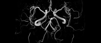
Cerebral angiography is a medical examination method that shows the condition of the vessels in the circulatory system of the brain. Allows you to identify pathologies of the vascular wall and blood flow disorders at an early stage of development. The resulting images are called angiograms and reflect in detail processes such as the venous, arterial, and capillary phases of blood circulation.
Characteristic
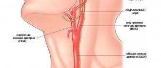
The angiographic method of study today is the best method for diagnosing vascular pathologies. It allows you to obtain a functional and anatomical picture of the current movement. It is carried out under the influence of X-rays or a magnetic field. It is most informative with the introduction of drugs to create contrast with other tissues. The drug is administered through a catheter installed in the vertebral, carotid or femoral arteries. In this case, they talk about the catheterization method. If the drug is administered through a puncture of the vessel being studied, the method is called puncture.
Types of angiography that are used in medicine today:
- Phlebography. The study is aimed at studying the veins of the legs.
- Angiography of internal organs and kidneys.
- Coronography studies the vascular structure of the heart.
- Angiography of cerebral vessels.
The study area is divided into general, selective and superselective. During a general examination, visualization of all cerebral vessels is performed. Selective angiography is aimed at studying the area around a single vessel. Superselective allows you to examine one of the small branches of the main arteries. In this case, simultaneous microsurgical intervention is possible.
Angiography uses various methods: radiography, magnetic resonance and computed tomography. Accordingly, classic, CT angiography and MRI studies are distinguished.
Indications
Frequent headaches, speech and gait disturbances, the appearance of spots before the eyes, and tinnitus are indications for contacting a therapist or neurologist. The specialist will clarify the symptoms, medical history, prescribe additional examination methods, and if there are indications and it is impossible to find out the cause of the disorders, he will issue a referral for angiography of the vessels of the neck and head. Pain in the head, fainting, and impaired gait alone do not serve as a basis for this examination.
An examination is prescribed to check the installed clips and if diseases are suspected:
- aneurysm;
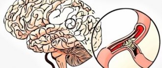
- stenosis;
- atherosclerosis;
- thrombosis;
- hemorrhage;
- occlusion;
- neoplasms;
- vascular malformation;
- epilepsy.
Angiography of cerebral vessels is performed only as prescribed by a doctor.
The essence of the examination
Angiography refers to x-ray research methods. Liquids do not absorb X-rays well, so blood vessels are difficult to see in the image. To clearly visualize the circulatory system, a contrast agent is injected into the patient’s blood. Angiography of the brain is carried out using iodine preparations (Omnipaque, Urografin, Verografin, Ultravist, Gipaque). The medicine is injected into an artery (carotid or vertebral).
After administration of the drug, several x-rays are taken in different projections. The contrast agent moves through the vessels of the brain along with the blood, even penetrating into the capillaries. Therefore, the doctor can evaluate all phases of cerebral circulation (arterial, capillary, venous) and see pathological changes in any part of the circulatory system.
When is angiography prescribed?
Indications for diagnostic testing are:
- chronic dizziness;
- repeated loss of consciousness;
- low blood pressure observed for a long time;
- ringing or noise in the ears that bothers a person for a long time;
- nausea and vomiting accompanied by dizziness or headache;
- frequent headaches;
- epileptic seizures.
X-ray of the vessels of the head and neck is prescribed if the patient has signs of a brain tumor, aneurysm, stenosis (narrowing) or occlusion (blockage) of blood vessels, intracranial hematoma, hemorrhage, brain failure, thrombosis or embolism of a cerebral artery.
The procedure is prescribed after a stroke, traumatic brain injury, before brain surgery and to control the position of clips applied to intracranial vessels.
Who sends for examination?
Diagnostic tests may be ordered by:
- neurosurgeon;
- neurologist;
- angiosurgeon;
- phlebologist;
- oncologist.
Advantages of the method
Angiography of intracranial vessels provides a detailed image of the circulatory system of the brain. Using the procedure, you can identify minimal pathological and anatomical changes in the vessels. Diagnostic testing helps determine certain characteristics of blood flow. It allows you to create an image of the intracranial vascular system, characterizing the dynamics of blood circulation.
A diagnostic procedure helps prevent unnecessary surgery. Since the study does not last long and requires small portions of radiation, it is prescribed even to children and patients in serious condition. After angiography, you can immediately begin treatment of identified diseases.
Preparation
Before angiography of cerebral vessels is carried out, preparation must be carried out, which includes a medical examination of the body. It includes electrocardiography, fluorography. Among instrumental methods, much attention is paid to ultrasound of the kidneys. If there are any abnormalities, the doctor will suggest replacing the examination with the introduction of a contrast agent with MRA or other diagnostic methods.
A urine and blood test is required, including a study of coagulation, biochemistry, blood group, and blood rhesus. Analyzes should not be older than 5 days.
If the procedure is accompanied by the administration of an iodine-containing substance, the drug must be specified. This drug causes severe allergies, so 1-2 days before the examination you need to check your sensitivity to it. To do this, up to 2 ml of the substance is injected intravenously. If swelling, itching, hyperemia, cough, headache, irritation appear, the patient is offered to undergo MRA of the brain vessels or another substance is used for contrast.
It is not recommended to drink alcohol 2 weeks before the examination. You must stop taking medications that affect blood clotting within 7 days. As recommended by your doctor, take an anti-allergy drug and a sedative in advance. Sometimes it is advised to cleanse the intestines in advance.
To avoid vomiting and nausea, eat the last time 8-10 hours before the procedure, drink 4 hours before. Before the examination, it is advisable to wash and, if necessary, remove hair from the site where the catheter is expected to be inserted. Remove all jewelry before the procedure.
Cerebral angiography prescribed for people with chronic diseases requires additional preparation:
- If you have hypertension, you need to normalize your blood pressure.
- To relieve arrhythmia, take potassium supplements, for example, Panangin.
- For heart pain or coronary artery disease, Nitroglycerin and Sustak are prescribed.
- If the patient is diagnosed with chronic respiratory diseases, preliminary antibiotic therapy is carried out.
- If kidney function is impaired, the body is saturated with water - hydration is carried out.
Contraindications
There are no absolute contraindications to the MRI procedure with vascular angiography. Contrast agents are not used during pregnancy. Relative contraindications:
- Being in a serious condition (toxic, septic shock).
- Heart, liver and kidney failure, occurring in severe form.
- Mental disorders in the acute stage.
- Revealed individual intolerance to the contrast agent.
- Wearing pacemakers, inner ear implants, and dentures made of metal.
Due to the use of X-ray radiation, an absolute contraindication for CT angiography of the arteries, veins and vessels that make up the cerebral blood flow system is pregnancy. Other reasons for prohibiting X-ray examination:
- Severe diabetes mellitus.
- Thyroid diseases.
- Malignant blood diseases (myeloma).
- Disturbances in the hemostasis system.
- Inflammatory and infectious diseases in the acute phase.
If the patient is suspected of individual intolerance to contrast agents, preliminary therapy with hormonal drugs (Prednisolone, Medrol) is prescribed. Iodine-containing contrast agents can provoke an allergic reaction. Taking glucocorticoids significantly reduces the risk of developing allergies. Iodine-containing solutions have high viscosity. When using contrast agents, the risk of developing disorders in the urinary system increases.
In patients with impaired renal function, a hydration procedure is recommended before diagnostic studies of cerebral blood flow using contrast agents. In order to prevent nephropathy, an additional liquid is injected into the body, which dilutes the contrast agent and promotes its rapid removal from the body. The day before the examination, the patient drinks at least 2 liters of still water.
Classic angiography
X-ray angiography is a method of obtaining information about vascular disorders using X-rays and a contrast agent. The rays pass through different tissues and are reflected differently. As a result, the silhouette of the organ is displayed on film.
Standard x-rays cannot see soft tissue. To isolate them, an iodine-containing substance capable of reflecting radiation is used.
Technique
Classic angiography of cerebral vessels is performed in a hospital. The subject is placed on a regular couch. If necessary, intramuscular sedatives and antihistamines are given. Lidocaine is injected subcutaneously to numb the puncture site. To prevent spasms and reduce irritation, Novocain is administered.
The puncture site is disinfected. The drug itself is injected into the vessel being tested or through a catheter inserted into the vertebral, carotid or femoral artery. After its introduction, the study begins.
It is permissible to administer a contrast agent 2-3 times. Upon completion of the procedure, the catheter is removed, the injection site is clamped, and a sterile bandage is tightened. It can be removed only when the bleeding stops completely.
What does it show
Contrast vascular angiography is not a routine method, unlike plain radiography or a general blood test. It is prescribed for specific indications, for example, ischemic stroke or vascular malformation.
What does angiography show:
- Dilation or narrowing of the walls of blood vessels.
- Bleeding in the brain.
- Inflammation of the walls of arteries and veins.
- Atherosclerosis: the degree of obstruction and blockage of the lumen.
- Vascular thrombosis is the presence of blood clots in the lumen of a vessel.
- Areas of hematoma.
- Tumors of vascular origin.
- Blood supply to neoplasms.
- Congenital anomalies of the vascular network of the brain.
The angiography method itself is safe for the patient, but associated complications may occur:
- Allergic reaction to the administration of a contrast agent.
- Pain at the site of puncture and injection of the drug.
- Swelling in soft tissues and legs.
Rare complications: cardiac arrhythmia, drowsiness, loss of consciousness.
CT angiography
As a result of a computed tomography scan of the cerebral vessels, a three-dimensional image of them is obtained. It shows detailed structure, tissue condition and blood movement. To perform the procedure, a contrast agent is injected.
A variation of the method is multislice computed tomography. MSCT makes it possible to obtain 160-320 images at a time and track the slightest changes.
Differences from classical radiography:
- Less radiation exposure.
- It is carried out on an outpatient basis.
- The contrast agent is administered intravenously, which is less traumatic compared to arterial administration.
- The result is sharper images.
- The patient can quickly return to normal life.
Technique
In order for the image to be clear, the scanner is adjusted to the heart rate of the subject. The pulse should be smooth, no higher than 65 beats per minute, so before the examination the patient is given medications aimed at reducing tachycardia. If necessary, vasodilator drugs are administered intravenously.
The patient lies down on a movable table, his chest and limbs are secured with straps, and an iodine-containing substance is injected intravenously. Next, the couch is moved into the tomograph. There he and the patient move smoothly, stopping at the moment of filming. At these moments, the diagnostician may ask you to hold your breath. The device reproduces the reconstruction of the received information into three-dimensional images.
You cannot move during the examination. Two-way intercom allows you to inform the diagnostician about discomfort. Time: 30-60 minutes.
Digital (digital) subtraction angiography
Digital subtraction angiography is a method of imaging the vascular system through intra-arterial or intravenous injection of a contrast agent. This type of examination allows a more detailed examination of blood vessels, thanks to computer processing and subtraction (exclusion) of objects that do not have diagnostic value.
The high expansion of the resulting images makes it possible to use less contrast agent and administer it in places more distant from the area of study. Angiography of this type has an undeniable advantage - in the resulting images, differences in contrast are more pronounced, which contributes to a more accurate diagnosis of the disease.
There are three main groups of DSA modifications, depending on which physical variable is used in the image subtraction process:
- time;
- energy;
- depth.
In the case of using one variable to obtain the final image, the exception will be listed as “first-order subtraction”, but if two or more – “second-order subtraction” (hybrid).
The principle of temporary subtraction, consisting of several divisions, deserves special attention:
- traditional subtraction angiography;
- integral method,
- appropriate filtering;
- return filtration.
The main purpose of temporary exclusion is to take a series of images from the moment the contrast agent is visualized on the monitor until it completely disappears. The final image is formed from the difference between images with the highest visualization density and the so-called “mask” - an image without a contrast agent. This principle is the basis of traditional DSA; all other divisions belong to its modified versions, differing in the number of images required for diagnosis.
The integral method is based on the difference between the “mask” images and the image with the introduction of a contrast agent. Images with vascular vibrations that interfere with diagnosis are removed using appropriate filtering.
Recursive filtering involves comparing a series of fragments consisting of two images following each other.
The essence of hybrid subtraction is to combat the display of movements of soft tissues and organs located close to the vessels being studied (heartbeat, swallowing reflex). DSA images can be generated in 3D reconstruction with color coding of the vessels.
This type of angiography does not determine the need for surgical intervention.
Magnetic resonance angiography
This method provides detailed information about the characteristics of blood circulation in the neck and brain. The difference between MRI and classical and CT angiography of the brain is that instead of X-ray radiation, a magnetic field and a radiofrequency pulse are used. The device senses and records the speed of blood flow, providing two-dimensional or three-dimensional images.
There are 3 types of MRA:
- Cerebral venography is aimed at checking the veins and visually assessing the speed of blood flow. Lasts about 60 minutes.
- Time-of-flight studies the arteries of the neck and brain, scanning them perpendicular to the direction of blood movement.
- 4D angiography of the brain is used to study any vessels in dynamics.
Features of the event
No special preparation is needed before magnetic resonance angiography. An exception is the use of contrast agents.
The patient lies down on a movable table. His arms and legs are secured to the couch with straps. It is important to remain calm during the examination, therefore, if control over body movements is impaired, the examinee is given sedatives. If the patient feels discomfort from the hum of the device, he may be offered earplugs. A cushion is placed under the head, and wires are laid along the neck to transmit data to the monitor. The couch is pushed into a wide pipe. The camera is equipped with a built-in air conditioner, so there are no breathing difficulties. The microphone will help you contact a doctor or nurse if necessary.
The essence of the method
People first learned about angiography in 1931, when the young doctor Forsman performed the first such procedure in history. But it found widespread use only 40 years later.
Angiography is an X-ray examination of blood vessels after filling them with contrast agents. This procedure allows you to evaluate the functionality of the vessels, their location, as well as the speed of blood flow. During the diagnostic process, it is possible to detect affected areas, developmental defects, collateral blood flow pathways, as well as the network of neoplasm vessels.
A radiopaque substance containing iodine is injected into the problematic vessel and this is done in one of the following ways:
- Puncture. Contrast is injected into the superficial vessels using a syringe.
- Catheterization. This method is relevant for examining deep arteries or veins. After anesthesia and an incision, a special thin plastic tube is inserted, through which a catheter is then advanced and a certain vessel is filled with a contrast agent.
After the contrast enters the vessels, it begins to be distributed through the bloodstream. From arteries to small capillaries and from small blood vessels to veins. During this entire cycle, many x-rays are taken, which allow one to evaluate the lumen of the vessels and the speed of blood movement through them.
To reduce radiation exposure, we try to take x-rays as quickly as possible
Complications
Cerebral angiography, which uses an iodine-containing substance, leads to complications in approximately 5% of cases. The accidental leakage of medication into tissue from a vessel is called extravasation. It occurs when a vascular wall ruptures or an unsuccessful puncture. If a large amount of medication leaks out, tissue inflammation or death may occur.
In renal pathologies, iodine-containing substances lead to exacerbation of chronic diseases and disruption of the blood supply to the kidneys.
Due to an allergy to iodine, hyperemia and swelling of the skin occurs. Gradually, these symptoms may be replaced by a feeling of lack of air, weakness, drop in blood pressure and loss of consciousness.
In rare cases, spasm of the blood vessels in the head occurs, circulatory disorders occur, and a stroke may develop. The examination may cause vomiting, convulsions, and bleeding.
On the day of the examination using a contrast agent, it is recommended to stay at home, move as little as possible, do not exercise, do not shower or bathe, do not smoke, do not drink alcohol, do not drive a car, and do not have sex.
Decoding
In classical angiography, X-ray images are studied. The radiation passes unevenly, so the density of individual tissues appears differently. Bone tissue is presented in white, the brain in gray, cerebrospinal fluid and blood vessels in black. CT and MRI angiography examine images in three dimensions.
In the photographs, the outlines and shape of each vessel are checked separately. They should be smooth with a uniform change in lumen. Detected narrowing, non-standard bends, branches, and the appearance of abnormal vessels allow us to determine diagnosis and therapy.
The angiogram is interpreted only by a specialist.