All types of circulatory disorders in the human body also affect the substance of the brain, which ultimately affects its integrity and the ability to function normally.
And “starvation” of cells, which is provoked by a violation or complete cessation of blood supply (in medicine, this process is called ischemia), causes a change in the brain substance of a dystrophic nature. That is, degeneration, and sometimes, although very rarely, even the disappearance of tissues and a significant deterioration in their function.
We will talk more about this pathological condition in the article.
Types of changes
In medicine, dystrophic manifestations in the brain substance are divided into two types:
- Diffuse.
- Focal.
In the first case, pathological changes spread evenly to the entire brain, and not to its individual areas. They are caused by both general disturbances in the functioning of the blood supply system and concussion or infections (meningitis, encephalitis, etc.).
Diffuse changes are manifested mainly by a decrease in a person’s performance, a dull headache, difficulty switching to another type of activity, a narrowing of the patient’s range of interests, apathy and sleep disorders.
And what a focal change in the brain substance of a dystrophic nature is can be understood by the fact that it can be caused by various minor pathologies:
- cysts (small cavities that form in the brain),
- small foci of necrosis (tissue death in certain areas caused by lack of nutrients);
- gliomesodermal (intracerebral) scars that occur after injuries and concussions;
- minor changes in the structure of the brain matter.
That is, these are pathologies that cause disturbances in the blood supply in a small area. True, they can be either single or multiple.
What are foci in the white matter of the human brain?
20.03.2019
Lesions in the white matter of the brain are areas of damage to brain tissue, accompanied by a violation of the mental and neurological functions of higher nervous activity.
Focal areas are caused by infection, atrophy, loss of blood supply and trauma. Most often, affected areas are caused by inflammatory diseases. However, areas of change may also be of a dystrophic nature.
This is observed mainly as a person ages.
Focal changes in the white matter of the brain can be local, single-focal, or diffuse, that is, the entire white matter is moderately affected. The clinical picture is determined by the localization of organic changes and their degree. A single lesion in the white matter may not affect the dysfunction, but massive damage to neurons causes a disruption in the functioning of nerve centers.
Symptoms
The set of symptoms depends on the location of the lesions and the depth of damage to the brain tissue. Symptoms:
- Pain syndrome. Characterized by chronic headaches. Unpleasant sensations intensify as the pathological process deepens.
- Rapid fatigue and exhaustion of mental processes. Concentration deteriorates, the volume of operative and long-term memory decreases. It is difficult to master new material.
- Flattening of emotions. Feelings lose their sharpness. Patients are indifferent to the world and lose interest in it. Previous sources of pleasure no longer bring joy and desire to engage in them.
- Sleep disturbance.
- In the frontal lobes, foci of gliosis disrupt the control of the patient’s own behavior. With deep violations, the concept of social norms may be lost. Behavior becomes provocative, unusual and strange.
- Epileptic manifestations. More often these are small convulsive seizures. Individual muscle groups contract involuntarily without threat to life.
White matter gliosis can manifest itself in children as a congenital pathology. The lesions cause dysfunction of the central nervous system: reflex activity is disrupted, vision and hearing deteriorate. Children develop slowly: they get on their feet late and begin to speak.
Causes
Damaged areas in the white matter are caused by the following diseases and conditions:
- Group of vascular diseases: atherosclerosis, amyloid angiopathy, diabetic microangiopathy, hyperhomocysteinemia.
- Inflammatory diseases: meningitis, encephalitis, multiple sclerosis, systemic lupus erythematosus, Sjogren's disease.
- Infections: Lyme disease, AIDS and HIV, multifocal leukoencephalopathy.
- Poisoning by substances and heavy metals: carbon monoxide, lead, mercury.
- Vitamin deficiency, especially B vitamins.
- Traumatic brain injuries: bruise, concussion.
- Acute and chronic radiation sickness.
- Congenital pathologies of the central nervous system.
- Acute cerebrovascular accident: ischemic and hemorrhagic stroke, cerebral infarction.
At-risk groups
Risk groups include people exposed to the following factors:
- Arterial hypertension. They are at increased risk of developing vascular lesions in the white matter.
- Poor nutrition. People who overeat, excessively consume extra carbohydrates. Their metabolism is disrupted, as a result of which fatty plaques are deposited on the inner walls of blood vessels.
- Foci of demyelination in the white matter appear in older people.
- Smoking and alcohol.
- Diabetes.
- Sedentary lifestyle.
- Genetic predisposition to vascular diseases and tumors.
- Constant hard physical labor.
- Lack of intellectual work.
- Living in conditions of air pollution.
Treatment and diagnosis
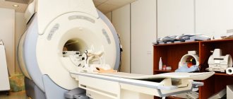
The main way to find multiple lesions is to visualize the medulla on magnetic resonance imaging. On layer by layer
Spots and pinpoint changes in tissue are observed in the images. MRI shows not only lesions. This method also reveals the cause of the lesion:
- A single lesion in the right frontal lobe. The change indicates chronic hypertension or a previous hypertensive crisis.
- Diffuse foci throughout the cortex appear when the blood supply is disrupted due to atherosclerosis of the cerebral vessels or.
- Foci of demyelination of the parietal lobes. It speaks of a disruption in the flow of blood through the vertebral arteries.
- Massive focal changes in the white matter of the cerebral hemispheres. This picture appears due to atrophy of the cortex, which forms in old age, from Alzheimer's disease or Pick's disease.
- Hyperintense lesions in the white matter of the brain appear due to acute disruption of the blood supply.
- Small foci of gliosis are observed in epilepsy.
- In the white matter of the frontal lobes, single subcortical foci are predominantly formed after a heart attack and softening of the brain tissue.
- A single focus of gliosis in the right frontal lobe most often appears as a sign of brain aging in older people.
Magnetic resonance imaging is also performed on the spinal cord, in particular on the cervical and thoracic regions.
Related research methods:
Visual and auditory evoked potentials. The ability of the occipital and temporal regions to generate electrical signals is tested.
Lumbar puncture. Changes in the cerebrospinal fluid are examined. Deviation from the norm indicates organic changes or inflammatory processes in the cerebrospinal fluid ducts.
A consultation with a neurologist and psychiatrist is indicated. The first studies the functioning of tendon reflexes, coordination, eye movements, muscle strength and synchrony of extensor and flexor muscles. The psychiatrist examines the patient’s mental sphere: perception, cognitive abilities.
Foci in the white matter are treated with several branches: etiotropic, pathogenetic and symptomatic therapy.
Etiotropic therapy is aimed at eliminating the cause of the disease. For example, if vasogenic lesions of the white matter of the brain are caused by arterial hypertension, the patient is prescribed antihypertensive therapy: a set of drugs aimed at lowering blood pressure. For example, diuretics, calcium channel blockers, beta blockers.
Pathogenetic therapy is aimed at restoring normal processes in the brain and eliminating pathological phenomena. Drugs are prescribed that improve blood supply to the brain, improve the rheological properties of blood, and reduce the need for oxygen in brain tissue. Vitamins are used. To restore the functioning of the nervous system, it is necessary to take B vitamins.
Symptomatic treatment eliminates symptoms. For example, for seizures, antiepileptic drugs are prescribed to eliminate foci of excitation. In case of low mood and lack of motivation, the patient is given antidepressants.
If lesions in the white matter are accompanied by an anxiety disorder, the patient is prescribed anxiolytics and sedatives.
If cognitive abilities deteriorate, a course of nootropic drugs is indicated - substances that improve the metabolism of neurons.
Didn't find a suitable answer? Find a doctor and ask him a question!
Source: https://sortmozg.com/zabolevaniya/ochagi-v-belom-veshhestve-golovnogo-mozga
Causes of dystrophy
The full picture of the appearance of dystrophic changes is not yet clear to researchers. But numerous observations have led to the conclusion that most cases of this pathology have a genetic predisposition. The action of provoking factors only accelerates the development of the process or enhances its manifestation.
Therefore, the reasons that cause focal changes in the brain substance of a dystrophic nature can be safely divided into genetic abnormalities and acquired ones. Although it should be noted that acquired causes are still a very conditional definition in this case, since they begin their destructive effect only if the patient is predisposed to the specified pathology.
Diagnosis and treatment
Dystrophic changes in the brain are diagnosed using the criterion according to Levin (2007):
- Neurological and psychophysiological disorders are detected using psychological tests. The mental and emotional-volitional sphere of personality is explored. A neurologist checks walking and precision of movements.
- Using a biochemical blood test, metabolic disorders are detected: excessive amounts of fat, sugar.
- Instrumental methods make it possible to visualize foci of dystrophy using computed tomography and magnetic resonance imaging.
- The cause of the disease is eliminated. If it is arterial hypertension, medications that lower blood pressure are prescribed: Obzidan, Metoprolol.
- Pathogenetic treatment aimed at improving blood supply, for example, nootropics: phenotropil, piracetam, glycine.
- Stroke is treated symptomatically: drugs are prescribed to improve the rheological properties of the blood.
Source: sortmozg.com
Focal changes in the brain substance of a dystrophic nature: symptoms of disease development
Symptoms of changes in the substance of the brain of a dystrophic nature most often appear quite clearly, but, unfortunately, this happens when the disease has already progressed significantly. Therefore, it is important to pay attention to the appearance of even small deviations in health.
- Initially, the described focal changes are manifested by headaches that occur during both physical and emotional stress.
- This disease is also characterized by periodic manifestations of paresthesia - numbness or slight tingling in the extremities.
- The patient complains of dizziness and insomnia, and has impaired coordination of movements (ataxia).
- As the disease progresses, the listed symptoms worsen, hyperkinesis (involuntary movements of the limbs) joins them, and paresis and paralysis develop.
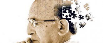
- Further development of the disease leads to memory deterioration, a noticeable decrease in intelligence, and agraphia (loss of the ability to write).
Risk group
Any disease has a risk group, people in it should be extremely careful. If a person has similar diseases, then he is in the primary risk group, if only predisposition, then in the secondary:
- Suffering from diseases of the cardiovascular system: hypotension, hypertension, hypertension, dystonia.
- Patients with diathesis, diabetes mellitus or stomach ulcers.
- Those who are overweight or have a habit of eating poorly.
- Those living in a state of chronic depression (stress) or leading a sedentary lifestyle.
- People over 55-60 years old, regardless of gender.
- Suffering from rheumatism.
For people in the main risk group, first of all, it is necessary to cure the underlying disease, followed by recovery of the brain. Patients with hypertension and all its forms of manifestation should be especially careful.
Is there an age limit for the disease?
It should be noted that single focal changes in the brain substance of a dystrophic nature occur not only in older people, but also in people under fifty years of age.
Stress, injuries, stressful situations, hypertension and other provoking factors can trigger the development of focal changes. The constant overstrain that many able-bodied citizens experience also plays its unseemly role.
Increased brain function against the background of existing vascular spasm in youth, as well as ischemia in old age, can equally lead to the emergence of foci of dystrophic changes with all the ensuing consequences. And it follows from this that timely and properly organized rest is a very important part of the prevention of the described pathology.
Symptoms of focal lesions
Focal brain lesions are caused by damage to blood vessels, which lose elasticity with age. For some, this manifests itself minimally, while for others, the disorders develop into a pathological form. Can appear:
- High blood pressure caused by a lack of oxygen due to the degenerative state of the cerebral vessels.
- Epileptic seizures, during which a person should not put metal objects in his mouth, pour water on him, hit him on the cheeks, etc.
- Mental disorders, memory impairment, distorted perception of reality, atypical behavior.
- Stroke or pre-stroke condition, which can be detected on CT or MRI.
- Increasing throbbing headache in the back of the head, eye sockets, superciliary areas, radiating across the surface of the entire skull.
- Uncontrolled muscle contractions, tremors of the limbs, chin, eyes, neck.
- Ear noise, ringing, congestion leading to nervousness.
- Regular attacks of dizziness leading to nausea and vomiting.
- Photophobia, decreased hearing acuity, blurred vision, double vision, noticeable blurred vision.
- Constant fatigue, apathy.
- Slurred speech.
- Sleep disturbances.
- Muscle paresis, pathological reflex reaction of the limbs.
Many people ask what diseases are caused by focal brain damage, what it is, and why it occurs. It is known that the causes of this disorder may lie in:
- Vascular disorders associated with natural aging, cholesterol accumulations in the walls of blood vessels.
- Osteochondrosis of the neck.
- Oxygen starvation.
- Neoplasms.
- Injuries, open and closed head injuries (age is not important here).
What diseases are accompanied by dystrophic changes in the brain?
Focal changes in the substance of the brain of a dystrophic nature, as a rule, are provoked by very common disorders of the functioning of blood vessels. These include:
- vasomotor dystonia,
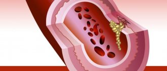
- atherosclerosis,
- arterial hypertension,
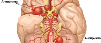
- aneurysm of blood vessels in the brain and spinal cord,
- cardiocerebral syndrome.
Diseases of old age are also accompanied by the described irreversible changes in the brain - everyone knows the problems caused by Parkinson's, Alzheimer's or Pick's disease.
Methods of combating pathology
Gradually affecting the human brain tissue, the disease can cause irreversible consequences. To prevent vascular changes in the white matter of the brain, it will be necessary to stop the symptoms that arise and improve blood flow with the help of medications and physical therapy. Treatment must be comprehensive, which means you will have to change your lifestyle. To do this you will have to follow these rules:
- Active lifestyle. The patient should move more and play sports. After eating, it is advisable to go for a walk, and it doesn’t hurt to do the same before bed. Water procedures, skiing and running have a good effect. Treatment with an active lifestyle improves general condition and also strengthens the cardiovascular system;
- Properly formulated diet. For successful treatment, you will have to give up alcoholic beverages and reduce your consumption of sweets, preserves, as well as smoked and fried foods. You can replace them with boiled or steamed food. Instead of store-bought sweets, you can make homemade pie or eat fruit;
- Avoiding stress. Constant mental stress is one of the causes of many diseases, so it is advisable to relax more and not overwork;
- Healthy sleep. A person should sleep at least 6-8 hours a day. In the presence of pathology, it is advisable to increase sleep time by 1-2 hours;
- Annual examination. If a change in the white matter of the brain is diagnosed, the patient should undergo an MRI twice a year. It is imperative to follow all the doctor’s recommendations and take the necessary tests on time.
Treatment of focal changes usually involves changing lifestyle and eliminating the cause of their development. It is advisable to detect the problem immediately in order to be able to slow down its progression. To do this, you should undergo a full examination annually.
How is the diagnosis made?
The diagnosis of “focal changes in the brain substance of a dystrophic nature” is quite difficult to establish. This requires identifying the signs of the pathologies listed above and excluding other somatic diseases and possible neuroses. By the way, people with diabetes and rheumatism are also at risk.
The doctor must assess the patient’s condition, his neurological status, and also conduct the necessary examinations. The most accurate indications are provided by an MRI study, where lesions can be identified, as well as their size and location. Tomography makes it possible to determine changes in the density of brain tissue even in the initial stage of the disease. Correctly reading the MRI results is an important step in starting treatment for the problem described.
Causes and signs of pathology
Nervous tissue is provided with intensive blood supply; a feature of perfusion in the brain is the bridges between the vessels. In case of emergency oxygen starvation, blood circulation is restored by another vessel.
Neurons are vulnerable even to a short-term lack of nutrition; cell death leads to irreversible processes in the brain - motor abilities and intelligence are impaired.
With age, blood vessels become less elastic and fragile. A common cause of damage to the cerebral cortex is concomitant diseases of the vascular system.
Reasons leading to defeat:
- Arterial hypertension;
- Vascular atherosclerosis;
- Hyperhomocysteinemia (high levels of homocysteine in the blood);
- Metabolic disorders;
- Inflammatory diseases of the meninges (leptomeningitis, pachymeningitis, arachnoiditis);
- Amloid angiopathy (amloid is deposited in the arteries);
- Multiple sclerosis;
- Damage to the cerebral cortex (trauma);
- HIV infection;
- Circulatory disorders (stroke, heart attack, dystrophic changes);
- Dyscirculatory changes (impaired blood circulation in the vessels of the skull, as well as in the spinal cord);
- Consequences of ischemia.
Brain tissue receives nutrition from the carotid and vertebrobasilar basins, interconnected in the velisian circle. In case of abnormalities or anatomically undeveloped vessels, there is no way to compensate for hypoxia by restoring blood flow through an anastomosis (connection with another vessel), which leads to focal damage to the brain of a vascular nature.
Symptoms of lesions of brain cells in the initial stage do not have a clear clinical picture, which leads to further destruction of axon bundles (white matter).
Clinical signs:
- Muscle spasms;
- Increased blood pressure;
- Dizziness;
- Mental disorders;
- Epileptic seizures;
- Migraine;
- Speech impairment;
- Decreased memory;
- Paralysis.
Focal changes in the brain substance of a dystrophic nature: treatment
As mentioned earlier, the exact cause of the appearance of this pathology, unfortunately, has not yet been established. And the diseases diagnosed along with it are more likely to be factors that only provoke the onset of its development or intensify processes that have already begun, and not the main cause of the disease.
Therefore, its treatment consists mainly of normalizing the patient’s daily routine and a proper diet, including foods that contain organic acids (baked and fresh apples, cherries, sauerkraut), as well as seafood and walnuts. The consumption of hard cheeses, cottage cheese and milk will have to be limited, since excess calcium causes difficulty in oxygen metabolism in the blood, and this supports ischemia and isolated focal changes in the brain substance of a dystrophic nature.
In addition, the patient cannot do without symptomatic therapy, which involves prescribing drugs that affect cerebral circulation and reduce blood viscosity, taking analgesics, sedatives and B vitamins. However, this is a separate and rather extensive topic.