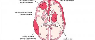
No one will argue that the human brain is quite complex. He is entrusted with many tasks that he must consistently perform throughout his life. For the brain to function properly, it must be provided with adequate nutrition through proper blood supply.
Pathologies associated with brain function are always serious. One of the common problems is expansion of the subarachnoid space. According to the ICD (International Classification of Diseases), expansion of the subarachnoid space in adults is rare; more often, such pathology is found in newborns. We will talk about its causes, diagnosis, treatment and methods of prevention.
Structural features of the brain
To understand the essence of this pathology, it is important to know what membranes cover the brain. There are three of them:
- arachnoid;
- hard;
- soft.
The subarachnoid space is located between the arachnoid and soft membranes. The first covers the entire surface of the brain, which in turn is enveloped by the endometrium. To communicate with other tissues, plexuses under the arachnoid membrane are used - membranes. The system of ventricles of the spinal cord and brain consists of the subarachnoid vascular plexuses. It consists of 4 reservoirs in which cerebrospinal fluid constantly circulates.
Subarachnoid spaces are small cavities in the brain filled with a special fluid (CSF). Their job is to nourish and protect the brain. The cerebrospinal fluid contains nutrients that are used to maintain the vital activity of nerve cells and the ventricles of the brain. Tissue waste products are also removed through the cerebrospinal fluid. If the subarachnoid space is expanded, it begins to compress adjacent tissues and blood vessels. Brain cells that do not receive proper nutrition suffer.
Liquor continuously circulates in the cavities of the brain. This is ensured by heart contractions, breathing, and body position. Normally, the volume of liquid filling the cerebrospinal fluid spaces should not exceed 140 ml.
What is the subarachnoid space?
The subarachnoid space is the distance between the pia mater and the arachnoid membrane of the brain and spinal cord. The second seems to “cover” its surface. From above it is protected by an additional endometrial ball, through which it communicates with other tissues using subarachnoid membranes. They are shaped like vessels and consist of several reservoirs where cerebrospinal fluid circulates.
The human brain is surrounded by three protective membranes - hard, arachnoid and soft. The latter is directly adjacent to the medulla and provides its nutrition. The arachnoid membrane is connected to other membranes of the brain using connective tissue membranes. In areas where there are no membranes, there are cisterns.
The normal amount of cerebrospinal fluid in children is 80-120 ml, in adults 120-160 ml. It is updated 3-5 times per day. The rate of circulation of the cerebrospinal fluid is affected by the position of the body, the intensity of the heartbeat and breathing.
The expansion of the subarachnoid space occurs due to difficulty in the outflow of cerebrospinal fluid. Fluid accumulates in the subarachnoid spaces, dilating the ventricles of the brain. The mechanisms of development of the disorder are different: the tumor mechanically blocks the paths for the flow of cerebrospinal fluid, inflammation provokes increased production of cerebrospinal fluid. Intracranial pressure may increase.
What does this diagnosis mean?
Most often, the diagnosis “Expansion of the subarachnoid space” is made to infants. This pathology can be caused by birth trauma or a deviation in brain development. If an enlarged subarachnoid convexital space is suspected, an ultrasound scan of the brain is performed. This is the main diagnostic method.
If there is an expansion of the cerebrospinal fluid spaces of the brain, the cerebrospinal fluid is unevenly distributed and spills out of the subarachnoid space. The result is hydrocephalus (dropsy), increased intracranial pressure, and dilation of the ventricles of the brain. In this case, the cerebrospinal fluid system does not work correctly, which is why brain tissue and internal organs suffer.
The expansion of external liquor spaces leads to various pathologies (asymmetry of the cranium, impaired vision, speech, coordination, some brain functions, mental development, etc.). The degree of development of such pathologies directly depends on how much the subarachnoid space is expanded. Weak and moderate expansion of external liquor spaces is amenable to complex treatment if it is started in a timely manner. If the ventricles are not dilated, then there is a chance that by the age of two the baby’s brain condition will normalize and hydrocephalus will go away.
It is important that parents do not expect everything to go away on its own. You could waste precious time. The bones of the skull will become stronger, but the dropsy may remain. It is imperative to conduct a full diagnosis and, if necessary, undergo a course of treatment.
Sometimes expansion of the subarachnoid spaces can be observed with a tumor, cystic formation or inflammatory process. This is extremely dangerous, as it often leads to death. If medical care is provided on time, the prognosis is quite favorable.
During an inflammatory process, such as meningitis, more cerebrospinal fluid is produced than necessary. A large amount of fluid leads to expansion of the space (dilatation). If the problem is a tumor, then it interferes with the proper circulation of fluids inside the brain, creating a physical barrier to it. Other reasons may be an abscess, a hematoma, due to which cerebral edema began.
Reasons for expansion
Expansion of the subarachnoid space in an infant or an adult occurs with pathologies leading to an increase in the amount of cerebrospinal fluid, as well as disruption of its outflow due to deformation. Main reasons:
- : meningitis, encephalitis, tuberculosis process.
- Neoplasms that have grown into the space between the meninges.
- Chronic metabolic disorder.
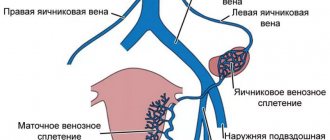
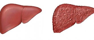
- Kidney and liver diseases.
- Edema syndrome during fasting, ARVI, heart failure.
- Traumatic brain and birth injuries.
- Birth defects.
- Extensive hemorrhagic or ischemic stroke in an adult.
Infections of the central nervous system, such as syphilis, tuberculosis, meningitis, cause a natural process - inflammation. The immune system responds to pathogens by increasing the permeability of blood vessels in the brain, as well as the arachnoid membrane. Histamine and enzymes are released that promote the formation of microholes in the capillaries (fenestra). Blood plasma leaks through them, as well as leukocytes. Edema occurs first interstitial, i.e. intercellular. The amount of cerebrospinal fluid in the subarachnoid spaces increases, resulting in their local or general expansion.
Neoplasms of the central nervous system can disrupt the flow of cerebral fluid, causing its stagnation, as well as dropsy. promotes uneven moderate or sometimes strong expansion of the subarachnoid spaces of the convexital surface of the brain (under the roof of the skull) or the basal, interhemispheric. This depends on the location of the pathological formation, as well as its size.
Edema syndrome caused by heart failure can also lead to dropsy of the brain, and, as a consequence, expansion of the interhemispheric fissure and the entire subarachnoid space.
Excess fluid in kidney diseases - pyelonephritis, glomerulonephritis - leads to intracranial hypertension. In this case, moisture is released through the capillaries of the arachnoid membrane, leading to liquorodynamic disturbances. Brain edema provokes compression of the white, gray matter, i.e., neurons and their processes by which they are connected to each other.
Liver diseases lead to an increase in the hormone aldosterone in the blood and a decrease in the content of albumin in the plasma. This creates conditions for the formation of dropsy of the brain. During fasting, protein does not enter the body, so the synthesis of albumin from it, which prevents edema syndrome, stops. This contributes to intracranial hypertension or hydrocephalus.
Craniocerebral and birth injuries deform the cerebrospinal fluid pathways, leading to stagnation of intracerebral fluid.
Diagnostics
Nowadays, brain pathologies are quite easily diagnosed. For this purpose, hardware methods are used (ultrasound, MRI), and, if necessary, lumbar puncture. The latter allows not only to detect a tumor, but also to examine all its layers and structure. This method allows you to select the treatment regimen for cystic and other formations as accurately as possible.
Basic diagnostic methods:
- Neurosonography. Duration of the procedure is ~ 15 minutes. It is carried out when it comes to a newborn, and consists of attaching a special ultrasound sensor to the patient’s head. Through the open fontanel, it allows you to collect information about the state of the brain. The advantage of this method is that it can be performed frequently, without any consequences for the baby. Now neurosonography is done in the maternity hospital to exclude pathologies of brain development. The result is deciphered by a pediatrician or neurologist.
- CT, MRI. These methods, although effective, are expensive. They are mainly used to diagnose children over 3 years of age and adults. Now considered the most accurate. To diagnose infants, using CT or MRI is very problematic, since the patient must lie absolutely still during the procedure. If such a diagnosis is indicated for a small patient, it is performed under general anesthesia.
- Cisternography. The purpose of the procedure is to determine how correctly the flow of cerebrospinal fluid is directed. It allows you to accurately determine the type of hydrocephalus in a particular patient.
- Angiography. With this diagnostic method, a special contrast agent is injected into the artery. The goal is to identify deviations in vascular patency.
- Neuropsychological examination. The patient is examined and the doctor interviews him. This examination is carried out in children over 3 years of age and adults. The doctor collects the data from all tests and the results of a visual examination. The goal is to identify disorders in the functioning of the brain.
The results of an ultrasound or MRI should only be deciphered by an experienced doctor. Self-diagnosis is unacceptable and extremely dangerous here. It is very important to accurately establish the cause of the pathology and immediately begin to eliminate it. This directly affects the course of recovery and further functionality of the brain.
A blood test is also carried out, the patient’s behavior, the presence of symptoms, and their severity are assessed.
Alarming symptoms
When the convexital spaces expand, the following symptoms are observed:
- constant headache (it appears immediately after waking up);
- nausea;
- vomit;
- dizziness;
- memory impairment (in adults);
- irritability;
- drowsiness;
- fatigue;
- in children, the size of the skull increases;
- high sensitivity to light and sound.
At first, the disease occurs without visible symptoms. Then they make themselves felt, but the intensity may vary. It depends on the degree of brain damage and the amount of cerebrospinal fluid released. If the lesion is local and minor, symptoms may be minimal. This condition responds well to treatment, but it is important to start it at the first sign of pathology, before irreversible structural changes occur. The larger the accumulation of fluid, the more significant these changes are. Over time, the cavities may become larger. External changes may occur in infants - the cranium enlarges (especially its frontal or posterior hemisphere), and basal brain functions suffer.
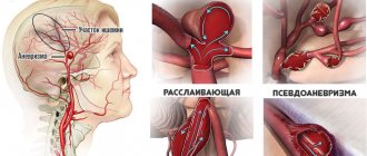
The causes of pathology at different ages differ. In infants, this is most often birth trauma, meningitis, arachnoiditis, or developmental pathologies (genetic code is disrupted). In adults – mechanical trauma, pseudocyst or tumor.
There are different degrees of uniform expansion of the subarachnoid space:
- light (1-2 mm);
- average (3-4 mm);
- severe (4 mm or more).
Localization also varies (interhemispheric, posterior, anterior, etc.). Different amounts of fluid may accumulate, and the external manifestations will also be different. Sometimes the skull becomes enlarged and there is pronounced asymmetry.
Features of the clinical picture
The following symptoms will help to suspect abnormalities in the functioning of the brain and expansion of the subarachnoid space in a newborn baby:
- irritability to moderate or low-pitched sounds and noises;
- increased sensitivity to light;
- excessive regurgitation;
- sleep is disturbed;
- pupils of different sizes or squint;
- increase in head size;
- anxiety about changing weather;
- the fontanel slowly overgrows and there is swelling;
- trembling of the limbs and chin.
The fact that the subarachnoid spaces are expanded in an adult is characterized by the following symptoms:
- headache after waking up in the morning;
- nausea and vomiting, as a result of severe headache that goes away after vomiting;
- dizziness;
- drowsiness, a dangerous symptom of intracranial pressure, indicating the progression of the disease;
- visual impairment;
- dementia, observed after a head injury, sleep is disturbed, a person confuses day with night, memory loss occurs;
- apraxia of walking, the patient in a supine position shows how to walk, but when getting up he sways, shuffles, and walks with his legs wide apart.
The presence of at least one of the symptoms in a child or adult is a reason to urgently contact a neurologist.
Treatment
In order for treatment of the enlarged arachnoid or subarachnoid space to be as effective as possible and tissue damage to be minimal, you should seek the help of a neurologist as early as possible. His consultation is mandatory if there has been an injury, including a birth injury, there is a suspicion of an inflammatory process, or if the listed symptoms are bothering you.
Please note that this pathology can be asymptomatic for a long time.
For successful treatment, it is important to establish the exact cause and eliminate it. Liquor dynamics must be taken into account. It can express the degree of expansion, show how much the surrounding tissues, vessels, and nerves suffer. Often, sinusitis, intracranial pressure, otitis media, and infectious diseases can provoke expansion in a child. With this development, antibacterial drugs and B vitamins are prescribed. Treatment can be quite long. It is prescribed purely individually, taking into account the nature of the pathology and the age of the patient. The patient must be constantly under the supervision of doctors; in the first stages of treatment, he may be placed in the neurology department.
It is important to limit the spread of cerebrospinal fluid, protect the hemispheres and sulci of the brain from compression, and clear the path for fluid to drain. To do this, it is important to determine exactly which area is affected, which lobe of the brain suffers from compression. This could be the hypothalamus, the cerebellum, several departments at once, etc.
This deviation in children is treated with a complex of drugs:
- means for removing excess cerebrospinal fluid (Asparkam, Veroshpiron, Diakarb);
- agents that improve brain trophism (Pantogam, Cavinton).
For the treatment of children over 3 years of age and adults, slightly different tactics are chosen. They are shown:
- barbiturates;
- diuretics;
- saluretics;
- glucocorticosteroids;
- plasma substitutes (solutions);
- painkillers;
- vasoactive agents.
Not all of the listed drugs are included in the treatment regimen. Their selection directly depends on the identified cause. If the problem is hydrocephalus, diuretics are prescribed; when the cause is an infection, antibiotics are prescribed.
It is advisable to supplement treatment with medications with physioneurological procedures. They reduce symptoms, restore the metabolism of cells and brain tissue. The main goal of treatment is to restore blood supply to the brain and normal drainage of cerebrospinal fluid. This will stabilize intracranial pressure and restore the metabolism of cells and tissues.
The prognosis for the outcome of treatment, despite the complexity of therapy, is quite favorable. The main thing is to start it in a timely manner and continue until the brain condition returns to normal.
Sometimes it happens that drug therapy does not bring the desired effect. In such cases, surgical intervention may be required.
This disease absolutely cannot be treated on its own. The help of a competent doctor, complete diagnosis, and long-term complex treatment are required.
Please note that in an advanced state, such a pathology leads to dementia, lack of coordination, speech defects, mental retardation, urinary incontinence and a number of other undesirable manifestations.
One of the most dangerous complications is hydrocephalus. It can cause a number of irreversible vicarious changes, for example, blindness, speech disorders. It can also cause the child to lag behind in development.
Why is this dangerous?
Advanced expansion of subarachnoid convexital spaces and untimely treatment in infants can lead to more serious complications:
- manifestation of chronic diseases;
- tumor;
- arachnoiditis;
- hydrocephalus;
- meningitis;
- delayed psycho-emotional and physical development of the infant.
Timely diagnosis and treatment will reduce the risk or eliminate complications of the disease, promote a favorable course and outcome of the disease, so that it will not affect the functioning, vital activity and physical development of the child and, as a rule, disappears by the age of two years of the child’s life.