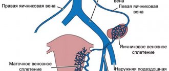
Varicose veins of the small pelvis are provoked by a phenomenon such as the reverse flow of blood through the ovarian vein, which occurs against the background of compression of blood vessels.
The trigger mechanism for the development of the disease is considered to be pregnancy, namely the hormonal changes observed during this period and the growing pressure of the uterus on the pelvic vessels. This type of varicose veins is manifested by prolonged and intense pain in the lower abdomen, usually appearing after constant heavy physical exertion or before menstruation.
Meanwhile, the quality of the patient’s future life, his ability to enjoy the intimate side of relationships with a loved one and the opportunity to continue his family largely depend on the timely detection of pelvic vein varicose veins.
Reference
Pelvic varicose veins in women (PVVV) is a complex medical problem. Many cases of successful treatment of this chronic disease have been described. However, URVMT is often not diagnosed. With this disease, there is a high probability of diagnostic errors, especially at its early stage.
HRVMT is caused by two main reasons:
- Obstruction of the veins of the pelvic organs (ovaries, tubes, uterus), causing an increase in pressure in the underlying areas and their expansion.
- Blockage of large venous trunks, in which an extensive network of “bypass” (collateral) venous outflow pathways develops with their expansion.
ARVMT occurs more and more often with age. So, it can be detected in 20% of girls aged 17 years. In perimenopausal women (45 – 50 years old), the incidence of ARVMT is already 80%.
Causes of the disease
Pathology occurs when the functioning of large main veins that collect blood from the genital organs is disrupted. Also, the disease often occurs from a uniform weakening of the entire venous network of the small pelvis. Most often, the causes of the disease are hereditary predisposition and the presence of connective tissue dysplasia. The latter causes underdevelopment of the vascular wall and a gradual decrease in its density.
The following reasons can also cause varicose veins:
- severe hormonal disruptions, hormone-dependent ovarian tumors and other dishormonal diseases;
- pregnancy and childbirth (due to increased pressure in the pelvis);
- gynecological diseases - endometriosis, chronic endometritis, previous surgeries;
- physical labor, high sports loads;
- obesity;
- sedentary work;
- lack of orgasms, especially for many years;
- abnormalities in the position of the uterus - backward bending, prolapse.
Classification of pathologies
Congestive syndrome (dilated pelvic venous system) is divided according to several characteristics:
- According to the form of the course: Primary veins are dilated - an increase in pelvic blood vessels caused by genetic pathology or valvular insufficiency that has developed as a result of changes in the body.
- The secondary form is dysfunction of the gynecological system.
- dilation of the veins surrounding the vulva (in advanced cases, the flow affects the vessels on the thighs and perineum);
Classification and stages of development
Varicose veins of the small pelvis can manifest themselves in two forms: varicose veins of the vulva and perineum and venous congestion syndrome. In more than half of the cases, both of these forms determine and support each other. Isolated vulvar and perineal varicose veins often result from reflux of blood through the saphenofemoral anastomosis involving the external pudendal vein and the tributary of the great saphenous vein.
It occurs in 30% of pregnant women and persists after childbirth in 2-10% of women. The main provoking factor for varicose veins of the perineum and vulva is the pressure of the growing uterus on the iliac and inferior vena cava. The pathomorphological prerequisite for varicose veins of the small pelvis is the reflux of blood through the ovarian vein.
There are 3 degrees of severity of varicose veins of the small pelvis, taking into account the diameter and localization of venous ectasia:
- 1st degree - dilated vessels have a diameter of up to 0.5 cm and a tortuous course; the lesion may affect any of the venous plexuses of the pelvis;
- 2nd degree - dilated vessels have a diameter of 0.6-1 cm; the lesion may be total in nature or affect the ovarian plexus, or parametric veins, or arcuate veins of the myometrium;
- 3rd degree - dilated vessels have a diameter of more than 1 cm with varicose veins of the total type or main type (parametric localization).
Symptoms
Typical symptoms of varicose veins and pelvic vessels that require visiting a doctor for treatment include:
- Chronic pain . Pain sensations are most often localized in the lower abdomen, sometimes “radiating” to the groin and lower back. They intensify in the second half of the menstrual cycle in women, after sexual intercourse or prolonged standing.
- Discharge from the genital tract . This is a typically “female” symptom. In this case, the discharge is normal in appearance and has no foreign odor. The patient is only alarmed by their unusually large number.
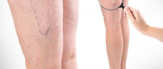
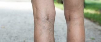
- External signs of the disease - spider veins or increased venous pattern on the thighs, in the perineum - occur in approximately half of the patients. Men may experience slight dilation of the veins on the penis.
- Menstrual irregularities in women and urinary disorders in patients of both sexes are rare and indicate an advanced form of the disease.
Symptoms of varicose veins of the pelvis
In women, there are two variants of the course of this disease - venous congestion of the pelvic organs and varicose veins of the perineum. The symptoms of these conditions are approximately the same, as are the causes that cause them. Most women experience the following symptoms of varicose veins of the small pelvis:
- pain in the lower abdomen;
- copious vaginal discharge;
- severe premenstrual syndrome;
- menstrual disorders;
- urinary disorders.
These symptoms may manifest themselves in different combinations in different patients. Some patients experience almost all of them, while others experience only one or two. The most characteristic sign of the disease is the presence of debilitating pain in the lower abdomen, not explained by any visible changes. Another symptom that predominates in most patients is copious mucous discharge.
The most pronounced symptoms characteristic of this vein pathology make themselves felt after physical activity, as well as at the end of the menstrual cycle. The presence of varicose veins in the legs may suggest the presence of this particular disease. After all, the causes of the development of both diseases are the same factors.
During examination, changes in the superficial veins are usually detected in the buttocks, in the perineum and on the posterior outer surface of the thigh.
Threat to normal pregnancy
Varicose veins of the lower extremities are very often combined with vaginal varicose veins, which indicates the presence of congestion in the pelvis and the involvement of other organs of the female reproductive system in general and the uterus in particular in the pathological process.
Damage to the vessels of the uterus can have negative consequences and pose a threat to the fetus and normal gestation, therefore varicose veins during pregnancy acquire a special status, where the venous vessels of the lower extremities play a leading role. After all, it is with them that everything begins, and the small pelvis is already involved in the process.
It seems incredible to say that varicose veins can interfere with pregnancy and cause infertility, but, nevertheless, it is true. In addition, dilation of the veins before or during pregnancy often creates an obstacle to normal implantation of the embryo and disrupts the correct formation of the placenta.
Description of the disease, its prevalence, statistical data
The phenomenon of chronic pelvic pain has been known to doctors for a long time. But only relatively recently has its most probable cause become known - varicose veins of the small pelvis. This disease was first described in 1975 and is still not well understood.
It is most likely that this pathology develops according to the following algorithm:
- The venous plexus in the pelvis is a complex formation that includes both large vascular trunks and smaller veins extending from them. At the same time, the venous system of the pelvis in men and women differs in its structure, which determines the gender specification of the disease - ARVMT is much more common in the fairer sex .
- Due to compression of blood vessels, complete or partial blockage of the venous bed, changes in the tone of the vascular walls, the outflow of blood from the deep pelvic veins is disrupted.
- As a result, the veins cease to perform their functions in full: the insufficiency of the venous valves progresses and the normal outflow of blood is disrupted.
- In the pelvic veins, stagnation begins, associated with the reverse flow of blood through the vessels - this provokes dilation of the veins and the development of varicose veins.
Symptoms of varicose veins (varicose veins) of the internal organs of the pelvis are much more common in women of reproductive age than in men.
In this case, the ovarian (“ovarian”) veins are most often affected - in 85% of cases. The leading symptom is pain, which is recorded in more than 90% of patients . But the prevalence of this disease among the population has not yet been clarified: according to various studies, it ranges from 6 to 80%. Such a large discrepancy in “indications” is explained only by the insufficient qualifications of diagnosticians when making a diagnosis.
Diagnostics
According to American researchers, in the early 2000s, only 2% of patients with ARVMT were initially given the correct diagnosis. Sometimes the consequence of a diagnostic error was the removal of the reproductive organs in women, although this could have been avoided if the most accurate methods for diagnosing pelvic varicose veins had been used:
- Ultrasound and Doppler examination of veins - makes it possible to suspect varicose veins;
- Phlebography is an invasive study that allows you to accurately determine the presence and extent of the disease;
- Laparoscopy is indispensable in the differential diagnosis of URVMT from gynecological diseases with similar symptoms (endometriosis, fibroids, colpitis).
- Selective ovariography - studying the condition of the veins using the injection of a contrast agent, is considered the most objective diagnostic method.
- Computer or magnetic resonance imaging makes it possible to clarify the details of the course of the disease and differentiate it from other non-gynecological pathologies with similar symptoms (articular diseases, Crohn's disease, etc.).
How does this pathology affect pregnancy and childbirth?
Pregnancy planning with a diagnosis of pelvic varicose veins must be approached responsibly. Before becoming pregnant, you need to consult an obstetrician-gynecologist and complete gynecological examination. Pregnant women with URVT should appear for routine medical examinations more often. At each visit, they undergo an ultrasound examination of the pelvic organs and veins of the lower extremities, and, if indicated, a blood clotting test (coagulogram) is performed. In 35% of patients with pelvic varicose veins, a serious complication of the disease occurs - infertility. In this case, they resort to in vitro fertilization (IVF) methods.
In 6-8% of women, the disease can lead to miscarriages or recurrent pregnancy loss. In these situations, there is a high probability that varicose veins are not the only cause of miscarriage, and the patient has severe concomitant pathology. In this case, an extended diagnostic search is carried out to determine additional causes. It is possible that a woman cannot become pregnant or bear a fetus due to antiphospholipid syndrome, thrombophilia, incompatibility of spouses’ antigens, or a number of other diseases.
In the first and second stages of pelvic varicose veins, childbirth is carried out through the natural birth canal, but only if the woman does not have blood clots in the lumen of the vessels or other concomitant pathology (cardiovascular, endocrine, ophthalmological, etc.). In severe cases of the disease, especially complicated by thrombophlebitis, the doctor completely eliminates the period of pushing and the method of delivery becomes a planned cesarean section.
Complications
If you observe at least one of the above symptoms, consult a doctor for advice; do not ignore the condition. Pelvic varicose veins, left to chance, have several unpleasant consequences:
- dysfunction of the uterus;
- contraindication to natural childbirth;
- the appearance of concomitant diseases such as varicose veins and hemorrhoids;
- mental disorders (increased anxiety, irritability, sleep disturbances, impaired attention, fear of sexual intercourse due to painful sensations).
What is the danger and what are the consequences?
This type of vascular pathology cannot be called a deadly disease .
If detected in time, it lends itself well to medical correction. But the problem is that it is not so easy to detect. Insufficient knowledge of the disease and low awareness of most diagnosticians play a role in this. So it turns out that patients suffer from this disease for years without even knowing it. Meanwhile, a number of irreversible changes occur in their body:
- Varicose veins progress, adjacent areas are included in the pathological process - dilation of the veins of the reproductive organs (for example, varicose veins of the penis), perineum and lower extremities appears.
- Persistent dysfunction of the internal genital organs appears, which can lead to infertility or the inability to carry a pregnancy to term in women.
- Against the background of pain, various psycho-emotional disorders such as neurasthenia develop.
- Due to chronic pain that worsens during intimacy, a person may refuse sex altogether.
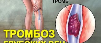
- The rarest and at the same time the most severe complications of pelvic varicose veins are vein thrombosis and pulmonary embolism. They occur in approximately 5% of cases, but are always deadly .
How to treat varicose veins of the pelvis?
For the treatment of pelvic varicose veins in women, there are many methods, each of which is effective in its own case, but the greatest effect is achieved by a combination of various methods as part of complex therapy.
The most important goals of therapy for varicose veins of the pelvis are:
- Restoring vascular tone.
- Elimination of stagnant processes.
- Preventing blood clots.
- Improving tissue trophism.
The disease is treated using an integrated approach. For this, medications are prescribed. The patient should normalize her lifestyle, eat right and use special compression garments. It should be remembered that if the disease is diagnosed, it is strictly forbidden to use traditional medicine methods. They can only be used after consultation with a doctor, as additional therapy.
Medicines
When diagnosing varicose veins, drug therapy is also used. Several groups of drugs are most often prescribed:
- Phleboprotectors. They have an anti-inflammatory effect and improve blood circulation.
- Phlebotonics. Tablets are indicated to improve the elasticity of the walls of blood vessels.
- Enterosorbents. Indicated for binding and removing toxins in the intestines.
- Non-hormonal anti-inflammatory drugs. Help relieve pain and inflammation.
Vitamin complexes are also prescribed to help support the immune system. To prevent the formation of blood clots, antiplatelet agents are prescribed. Medicines are taken only as prescribed by a doctor in the indicated dosages.
Normalization of lifestyle
The reasons for the inability to conceive with varicose veins of the pelvic vessels and the appearance of other symptoms of the disease are an incorrect lifestyle. Experts recommend:
- To refuse from bad habits.
- Avoid standing or sitting for more than three hours.
- Avoid heavy physical activity.
- Do gymnastics daily.
- Do not take a hot bath. The procedure can be replaced with a contrast shower. Changing the water temperature will help keep the vessels in good shape.
- Get rid of excess weight. To do this, you can follow a diet and exercise.
It is not easy to cure the disease. To do this, you must follow all the doctor's recommendations. The doctor also prescribes a set of special exercises that will help restore blood circulation.
Nutrition
In the treatment of the disease, a balanced diet with the exclusion of fatty foods and alcoholic beverages is of great importance.
The menu should include dishes high in fiber and vegetable fats. The main products in the diet should be vegetables, fruits, natural juices, and green tea. Sweet baked goods, dishes with a lot of spices, and fast food are not recommended. Meals should be frequent, but in small portions.
Foods high in vitamin C (citrus fruits, black currants), seafood (oysters, shrimp, seaweed) are beneficial. Dishes should be prepared with a minimum amount of salt.
Gymnastics
In addition, patients with ARVMT need to do gymnastics daily, doing:
- exercise “birch tree”;
- exercise "bicycle";
- scissors exercise;
- exercise in a supine position - stretch your legs, then bend your knees and pull them towards your chest;
- exercise in the position on the stomach - alternately lifting the top of one leg and then the other;
- walking around the room: on your toes, on your heels, and then with your knees raised high (at least 15 minutes);
- while lying on your back, raise your straightened legs, bend your knees and hip joints and straighten them upward again.
Folk remedies
Traditional medicine is effective in overcoming the symptoms of the disease if used in the early stages. Teas and decoctions of chaga and dandelion root, and tinctures based on horse chestnut are very effective.
The most accessible way to use chaga is medicinal tea based on it. To do this, pour 3 tablespoons of dried chaga into 0.5 liters of boiled water and leave in a warm place for several hours. Take half an hour before meals, no more than 2-3 times a day.
A dessert spoon of crushed dandelion root is poured with a glass of boiling water, left for several hours and drunk three times a day 10-15 minutes before meals. This tea is contraindicated for people suffering from gastritis or biliary tract disease.
To prepare horse chestnut tincture, the fruits are peeled, finely chopped, placed in a dark glass bottle and filled with vodka in a ratio of 1:10. Duration of infusion – 14 days. 25-30 drops of the resulting tincture are mixed with a small amount of water and taken three times a day. The duration of treatment should not exceed 30 days.
Examination of patients with ARVMT
Before starting to prescribe tests, ultrasound, x-rays and other research methods, the doctor must conduct a thorough survey and examination of a woman with suspected varicose veins of the small pelvis. The following parameters are taken into account:
- patient's age;
- weight, body mass index;
- presence of other chronic diseases;
- heredity (presence of varicose veins in close relatives);
- profession, whether there is a sedentary lifestyle;
- bad habits;
- number of pregnancies, births, abortions, miscarriages;
- features of the menstrual cycle, its regularity;
- history of surgical interventions, their nature;
- the presence of hemorrhoids, varicose veins of the legs.
After talking with the patient, the doctor begins to examine her. It offers visual and bimanual diagnostics on the gynecological chair. This may reveal changes such as:
- dilation of the vessels of the perineum, their tortuosity, hemorrhoids extending beyond the rectum;
- cyanosis of the vaginal walls;
- prolapse of the uterus and vagina;
- reaction to palpation of the abdomen above the womb, in the area of the projection of the uterus, ovaries, vagina;
- smoothness, swelling of the vaginal vaults.
All these signs indicate that, most likely, the patient has varicose veins of the small pelvis. To confirm the pathology, they resort to laboratory and instrumental examination methods.
Test indications
All women, regardless of complaints, are prescribed general clinical blood and urine tests. They are necessary to assess the general condition of the body.
If there is a suspicion of hormonal imbalance, the doctor may recommend a steroid profile, which includes testosterone, progesterone, estradiol, LH, FSH, prolactin, cortisol, as well as a number of thyroid hormones. When VURVD occurs while taking contraceptives or during menopause, this list is mandatory.
To exclude pathology from the blood coagulation system, a coagulogram is taken. It shows how the most important processes occur. Using a coagulogram, one can assume the presence of blood clots in dilated vessels in order to correct the situation in a timely manner.
Instrumental examination
The following methods can be used to diagnose varicose veins of the small pelvis:
- Ultrasound Doppler. Allows you to quickly and non-invasively determine the presence of pathology using an ultrasound machine. Gives a general picture of blood flow and the state of blood vessels in a given area.
- CT method. Necessary for differential diagnosis with pathologies that have a similar clinical picture. Allows you to very clearly see the anatomical features of the structure of organs.
- Selective ovariography. The vein of interest is contrasted with a dye, after which photographs are taken to evaluate the course of the vessels, their walls, as well as the degree of blood flow disturbance.
- Laparoscopic surgery. Extreme method of examination. It is used when all other methods have not provided diagnostically significant information. Laparoscopy allows real-time assessment of the ovarian vessels.
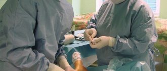
Surgery
Surgical treatment of varicose veins of the pelvis is carried out according to strict indications:
- ineffectiveness of conservative therapy;
- 3 degree of disease;
- inability to relieve pain.
Various surgical techniques are used depending on the location of reflux and venous conglomerates:
- ligation of the ovarian vein (through retroperitoneal access or laparoscopy);
- scleroobliteration of ovarian veins under the control of hagiography (a sclerosing substance is injected into the lumen of the vein, the operation is performed under local anesthesia);
- resection (partial removal) of ovarian veins through retroperitoneal or laparoscopic access;
- clipping of ovarian veins laparoscopically;
- phlebectomy (removal of veins) for vulvar and perineal varicose veins;
- laser and radiofrequency coagulation;
- crossectomy - ligation of the great saphenous vein and its tributaries - with a combination of varicose veins of the perineum and lower extremities.
Signs
Symptoms of varicose veins of the pelvis are varied and often resemble manifestations of lesions of nearby organs. The disease usually occurs with the dominance of signs of a certain form.
Vulvar varicose veins
The main symptoms of this form of the disease include the following:
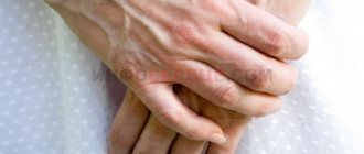
- Venous expansion. The main manifestation of this form is a noticeable expansion of the veins in the vulva or perineum. The patient herself can see them. This disease is accompanied by a feeling of itching of the vulva, heaviness and bursting pain in this area. The doctor can detect swelling of the labia and enlarged veins in this area. The process is often accompanied by hemorrhoids.
- Bleeding. This form of the disease may be accompanied by post-traumatic or sudden bleeding. Since the venous wall becomes very thin, stopping this process is very problematic even with surgical methods.
- Thrombophlebitis. A complication of such varicose veins is acute thrombophlebitis of the perineal veins. This process is accompanied by severe pain, redness and swelling of the skin. The veins in the affected area become dense and painful. Their inflammation may also develop, which provokes an increase in temperature and symptoms of intoxication.
Prevention
- Normalization of working conditions with the exception of long periods of sitting or standing. Industrial gymnastics, regular breaks from work. Avoid heavy physical activity and heavy lifting.
- Normalization of stool, relief from constipation. The diet should contain more vegetable fiber and vegetable oil. Quitting alcohol and smoking. Exclusion from the diet of spicy and salty foods.
- Daily ascending contrast shower on the perineal area.
- Exercises in a lying position with legs raised (“bicycle”, “birch tree”, “scissors”).
- Breathing exercises: slow breathing using the abdominal muscles.
- Wearing special therapeutic tights of compression class II.
- Prophylactic administration of venotonic drugs in courses 3–4 times a year.
Treatment is considered effective in cases where the symptoms of the disease cease, venous outflow improves according to instrumental studies, and the patient’s quality of life improves.
Thrombophlebitis of the lower extremities Varicocele Hemorrhoids Hepatitis C: first signs and treatment regimen Endometriosis Hemorrhoids in men
Stages of the disease
Varicose veins of the pelvic veins in women have three stages:
- The diameter of the veins is approximately 5-7 mm, and the processes cover at this stage only the upper edge of the left ovary.
- At the second stage, the diameter of the veins can reach 10 mm. The mechanism of development of the disease affects the entire left ovary. At this stage, varicose veins may already affect part of the uterus and the right ovary.
- At the third stage, the diameter of the veins can reach 13 mm. The problem already completely affects both ovaries and the uterus.