0
Author of the article: Marina Dmitrievna
2017.07.26
22 028
Diagnostics
Most people undergo an ECG procedure without much preparation. Often doctors don’t even talk about special measures, which is a big mistake. When the patient is tuned in to taking a cardiogram, calm and prepared, the results are truly truthful. Nobody cares when a person gets nervous when he sees a doctor. Many people are afraid of hospitals and procedures, and this affects their psychological, emotional and physical well-being. How to get an ECG for a child without problems?
What can influence the results?
Often, when a patient sees a doctor, after taking an ECG, he observes approximately the following picture: the doctor thoughtfully looks at a graph paper with incomprehensible teeth and shakes his head, and in the end gives out a phrase, the essence of which boils down to the fact that the ECG is not normal, bad or other epithets with similar meaning.
First of all, the following factors can affect the reliability of the results of an electrocardiographic study of the heart:
- incorrect (unsuccessful) attachment of electrodes to various parts of the body, which may be associated with the formation of a film on the skin due to the application of creams, lotions, and other skin care products;
- stress suffered on the eve of the procedure;
- taking antidepressants, nootropics, tranquilizers, sedatives on the eve of the procedure.
Important! Understand that before carrying out the procedure in question, you should definitely inform the doctor about any unusual events that have occurred in the patient’s life.
This is due, first of all, to the fact that any non-standard situations (stress, hormonal imbalance, poisoning, overeating, detection of pregnancy, etc.) can slightly change the electrocardiogram indicators, both bringing them closer to the norm and moving them away from generally accepted norms!
Having studied Elena Malysheva’s methods in the treatment of HEART DISEASE, as well as restoration and cleansing of VESSELS, we decided to offer it to your attention.
The technique of taking an ECG and interpreting the results obtained is an overwhelming task for patients; they cannot interpret it on their own, and therefore people often feel fear, not understanding what awaits them. The doctor himself rarely spends precious time explaining to the patient what is wrong with his analysis. This attitude is found at the medical examination, where patients are examined in a stream.
Many are also concerned about the question of how to improve the cardiogram, since obtaining a position or a new job often depends on the results provided by the medical board. Before we talk about how to influence the results of an ECG, it is worth talking about the reasons why it worsens.
What to do to improve ECG results?
Often, when a patient sees a doctor, after taking an ECG, he observes approximately the following picture: the doctor thoughtfully looks at a graph paper with incomprehensible teeth and shakes his head, and in the end gives out a phrase, the essence of which boils down to the fact that the ECG is not normal, bad or other epithets with similar meaning.
The ECG procedure is carried out only in agreement with the doctor and if necessary
The technique of taking an ECG and interpreting the results obtained is an overwhelming task for patients; they cannot interpret it on their own, and therefore people often feel fear, not understanding what awaits them. The doctor himself rarely spends precious time explaining to the patient what is wrong with his analysis. This attitude is found at the medical examination, where patients are examined in a stream.
Many are also concerned about the question of how to improve the cardiogram, since obtaining a position or a new job often depends on the results provided by the medical board. Before we talk about how to influence the results of an ECG, it is worth talking about the reasons why it worsens.
Reasons why the cardiogram worsens
There are a number of reasons that contribute to poor results, which can manifest as increased heart rate or even pain. The reasons can be divided into two large groups.
The first group includes:
- consumption of alcoholic beverages.
- active smoking.
- active physical activity immediately before the procedure.
- stressful state of the body.
some time before the ECG, it is necessary to lead a measured lifestyle and not be exposed to heavy stress
the patient can influence this group of reasons without the help of a medical professional.
If you take measures, you can completely eliminate the influence of these factors. the only question is what measures are needed.
second group of reasons:
- myocardial infarction in the acute phase.
- blockades with different locations.
- cicatricial changes in the myocardium are a sign of a previous heart attack.
- arrhythmias.
- hypertrophy of the heart muscle.
these reasons require the intervention of a medical professional and a thorough examination, since some of the conditions are a direct indication for hospitalization, and some are life-threatening.
If a child has pathological changes in the heart, a consultation with a pediatrician is necessary. The pediatrician will decide whether it is necessary to undergo additional tests and look for reasons. In case of dangerous changes in the ECG, it is necessary to take measures to stabilize the situation.
how to correct a bad ecg
It is worth understanding that a poor ECG is associated with changes in the heart. That is, it is necessary to influence the reasons why the heart does not work well. This can only be done based on the clinical diagnosis.
The results of the ECG procedure will improve only if the body’s condition stabilizes
After examining an adult or child, the doctor prescribes medications aimed at improving heart function.
The effect of these drugs is limited to a positive effect on metabolic processes in the heart muscle. A similar step is taken to calm nervous patients.
At the medical examination, medications are not prescribed at all, and the employee is sent to the clinic.
Drugs aimed at improving metabolic processes in the heart are not prescribed in countries of the developed world and are not included in treatment regimens there, since their effectiveness is not considered proven.
How can you improve your ECG?
Treatment methods apply to patients with the second group of reasons affecting the ECG condition.
If no heart disease is detected, but the ECG remains unsatisfactory, you must carefully prepare to get the desired results.
An unsatisfactory ECG may be due to functional disorders that do not require treatment. What can you do to increase the likelihood of good results?
Improving ECG results is possible with the correct regimen and the necessary preparation
Many doctors who are only indirectly associated with cardiology believe that there is no need to prepare the patient for the procedure of taking a cardiogram. And this is a wrong opinion. To obtain reliable results, you will have to take measures.
Advice - calm down! Taking a cardiogram is a painless and quick procedure, but stress and fatigue can adversely affect the results.
- Full sleep is necessary before the procedure. It is better to sleep at least 8 hours.
- If you have a habit of doing exercises in the morning, then you need to take a break from exercise for one day. Physical activity affects your heart rate.
- If the ECG is scheduled for the morning, it is better to have breakfast after the procedure or limit yourself to a light snack. If the procedure is during the day, then the last meal should be no earlier than two hours before it.
- It is worth reducing the amount of liquid you drink the day before the procedure.
- Avoid energy drinks, be it tea or coffee, as they speed up and increase the heart rate.
- It is recommended to quit smoking and alcohol at least one day before the procedure.
- You can take a shower before the procedure, but do not apply any creams or other cosmetics to the skin. This is done to ensure good contact between the electrode and the skin. The lack of such contact also has a bad effect on the results.
- It's important to pull yourself together. Breathe at your usual rhythm.
We recommend! You can arrive 15-20 minutes before the start of the procedure so that you have time to mentally prepare.
- It is also important to calm your heart rate after walking.
- The procedure requires undressing, so it is better to choose comfortable and simple clothes. Women are advised to avoid tights so that there is direct contact with the skin. The trouser legs should ride up easily so that you can install the electrodes in the shin area without interference.
- Men are recommended to shave their chests to ensure reliable results.
For children
Taking a cardiogram, like any medical procedure, is particularly difficult when a child is brought in for an appointment.
The child must be warned about the study in advance, explaining that the procedure is painless. If possible, you can be allowed to be present when an ECG is taken in a calmer child.
The main thing to get good results is to calm the child down and explain that they won’t do anything bad to him.
If the child is familiar with the procedure in advance, he will not have fear and the test readings will be accurate. A warm room and a relaxed atmosphere will help improve the results. Often, if a child sees that adults are calm, then he himself is calm and easily tolerates this painless procedure.
It is not so difficult to influence the results of an ECG, moving them in a positive direction, if a person does not have heart disease, and the problems are caused only by a feeling of excitement. To do this, you need to follow simple instructions.
If the results of the cardiogram remain unsatisfactory, then the doctor is asked to repeat the procedure after 10-15 minutes in order to avoid the “white coat syndrome”, characterized by a negative reaction of the patient to the doctor.
Source: https://diagnostinfo.ru/ekg/uluchshenie-rezultatov-ekg.html
How to correct a bad ECG
It is worth understanding that a poor ECG is associated with changes in the heart. That is, it is necessary to influence the reasons why the heart does not work well. This can only be done based on the clinical diagnosis.
After examining an adult or child, the doctor prescribes medications aimed at improving heart function. The effect of these drugs is limited to a positive effect on metabolic processes in the heart muscle. A similar step is taken to calm nervous patients. At the medical examination, medications are not prescribed at all, and the employee is sent to the clinic.
Drugs aimed at improving metabolic processes in the heart are not prescribed in countries of the developed world and are not included in treatment regimens there, since their effectiveness is not considered proven.
The results of the ECG procedure will improve only if the body’s condition stabilizes
How to calm down before an ECG (cardiogram)
Every person has undergone this examination at least once in his life, but the curve on the tape is a mystery to everyone. An ECG is the most popular examination that is prescribed if heart problems are suspected. While a person far from medicine can understand and discern at least something on X-rays or an ultrasound monitor, the curve, which is usually printed on a long strip after an ECG, always remains a mystery to the patient. What can be seen on such a curve and why it is better not to go to the doctor with a hangover, we will look into this on Heart Day, which is celebrated on September 29. Electrocardiogram ECG is one of the simplest and safest heart examinations, which has been actively used in medicine for many years.
How to prepare a child for an ECG procedure?
Taking a cardiogram, like any medical procedure, is particularly difficult when a child is brought in for an appointment.
The child must be warned about the study in advance, explaining that the procedure is painless. If possible, you can be allowed to be present when an ECG is taken in a calmer child.
The main thing to get good results is to calm the child down and explain that they won’t do anything bad to him.
If the child is familiar with the procedure in advance, he will not have fear and the test readings will be accurate
A warm room and a relaxed atmosphere will help improve results. Often, if a child sees that adults are calm, then he himself is calm and easily tolerates this painless procedure.
It is not so difficult to influence the results of an ECG, moving them in a positive direction, if a person does not have heart disease, and the problems are caused only by a feeling of excitement. To do this, you need to follow simple instructions.
If the results of the cardiogram remain unsatisfactory, then the doctor is asked to repeat the procedure after 10-15 minutes in order to avoid the “white coat syndrome”, characterized by a negative reaction of the patient to the doctor.
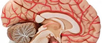
When a person has heart complaints, going to an ECG procedure becomes exciting. We all fear for our health, especially if the doctor diagnoses pathologies. Not all doctors are able to talk about possible problems and ways to solve them in a simple and understandable manner. The frightened patient begins to worry and becomes lost. To avoid such a situation, it is necessary to understand the structural features of the heart, possible changes in its functioning and pathology.
If the results of the cardiogram remain unsatisfactory, then the doctor is asked to repeat the procedure a minute later in order to avoid the “white coat syndrome”, characterized by a negative reaction of the patient to the doctor.
If adults cannot always calm themselves down in time, what can we say about the child? Parents cannot always explain to their child that the procedure will not cause him the slightest feeling of pain. When a child sees that parents or loved ones are worried, he begins to be afraid.
To make the ECG process calm, the child needs to be explained what will happen. It is better for parents to enter the office with their young patients to reassure them.
The doctor must have a positive attitude towards the child so that he does not feel fear. If you fail to pass the ECG the first time, take it a minute later. During this time, the children calm down, as they understand that nothing painful happened.
It is better to take an ECG in the morning, when the child has not yet loaded himself with active loads.
How to improve an ECG for a medical examination
When a person has heart complaints, going to an ECG procedure becomes exciting. We all fear for our health, especially if the doctor diagnoses pathologies. To avoid such a situation, it is necessary to understand the structural features of the heart, possible changes in its functioning and pathology. Quite often the patient is told that sinus rhythm is diagnosed on the cardiogram.
How scary and harmful is this to the heart? A bad ECG most often indicates that the heart is experiencing a blockage. Bad heart cardiogram - what to do to improve the result? Advice! Before the ECG procedure, do not take heart medications so that the result is as reliable and informative as possible.
https://www..com/watch?v=FkTDHlefYT4
It is worth understanding that a poor ECG is associated with changes in the heart. That is, it is necessary to influence the reasons why the heart does not work well. After examining an adult or child, the doctor prescribes medications aimed at improving heart function.
Hello. There was a need to do a cardiogram (and the result needed to be good).
Or will they distort the result (if they distort, then in what direction?) Are there any recommendations for behavior, routine, food intake, etc.
in order to somehow improve the result of the cardiogram? Maybe taking an infusion of VALERIAN or MOONOR on the day before and before the ECG will relieve this anxiety? But this will not affect the ECG itself in any way.
But you should not abuse other drinks, such as water or juice, because overloading the body with fluid may not have the best effect on the cardiogram.
You should not go for an ECG earlier than two hours after undergoing any physiotherapeutic procedures. If at rest your pulse is kept within normal limits, then there is no reason to worry.
Most experts are inclined to believe that we can talk about bradycardia when the heart rate is less than fifty beats per minute.
Arrhythmia, RGC syndrome, tachycardia, hypertension are most often attempted by imaginary patients in ECG diagnostics. Various techniques are used to obtain the necessary false diagnosis. But how effective are their methods?
The most harmless way to simulate arrhythmia is uneven breathing (at first the patient breathes slowly and calmly, then quickly). But there is one drawback: the doctor conducting the diagnosis will notice this and immediately stop the ECG procedure. Therefore, the option is not the most effective for achieving results.
A more dangerous method for deceiving the electrocardiograph is to take large doses of caffeine (medicines, coffee), which has a direct effect on the heart and disrupts the normal rhythm. The danger is that this substance creates additional stress on the heart muscle.
To simulate tachycardia and hypertension, patients drink energy drinks, coffee and Coca-Cola, and perform excessive physical activity before an ECG test. With such manipulations, you can achieve a rapid heartbeat and, as a result, provoke a strong load on the heart.
Medications from the group of alpha-beta adrenergic agonists in overdose can provoke RGC syndrome. Only these drugs do not imitate, but on the contrary lead to pathological abnormalities in the functioning of the heart.
If a healthy person deliberately takes medications from this group to simulate RGC, then he can actually develop this disease.
Then you will have to not deceive the electrocardiograph, but actually undergo a thorough examination to prescribe the correct treatment.
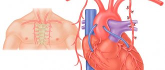
What is an ECG or the essence of the study
An ECG is a recording using an electrocardiograph of electrical impulses generated by the heart. Electrical impulses create currents that spread throughout the body. Their intensity is sufficient for the ECG device to be able to record them on any part of the body. Electric currents are captured by electrodes installed at certain points on the body.
An ECG is a test that provides valuable information about the condition of the heart.
An ECG examination can detect various heart diseases, but not all. For example, heart defects, arrhythmia, tachycardia, bradycardia, RGC syndrome, heart attack, heart failure, abnormalities of the cardiovascular system after lung diseases. RGC can only be diagnosed with an electrocardiogram.
Indications for this procedure:
- frequent fainting, shortness of breath, dizziness,
- causeless repeated occurrence of sudden weakness,
- rapid heartbeat without any emotional shocks or physical exertion,
- the presence of pain in the heart area.
Worse ECG readings
Some conscripts who have received a summons from the military registration and enlistment office want to distort the ECG result in a bad way. They do not want to serve in the army, so they try in any way to deceive doctors and medical diagnostic devices. Most often, conscripts try to spoil the electrocardiogram readings in order to receive a white ticket.
It is possible to deceive the ECG machine, but methods that stimulate arrhythmia can have a bad effect on health
Absolutely healthy guys take medications to increase or decrease blood pressure, medications that contain caffeine, for a short time before diagnosis. They give themselves sleepless nights, regular jogging, excessive physical activity, or a good nervous shock to the body to completely deplete their reserves of strength.
Of course, these manipulations can make the electrocardiogram result distorted, and the results obtained will be bad. But there are also some nuances here.
After all, not every person experiences a significant increase in heart rate from drinking coffee or energy.
Also, if you spend sleepless nights, constantly perform heavy physical activity and come to the procedure absolutely tired, without strength, the doctor conducting the diagnosis will immediately notice this and refuse to carry out heart monitoring.
To protect your own health, you should not try to deceive ECG results using special medications.
Atrial flutter
With obstructive pulmonary disease, it practically does not manifest itself. 3. Atrioventricular block is a delayed excitation of the atrioventricular node for more than 0.09 seconds. There are 3 degrees of blockade of this type. With the highest degree of disease, the ventricles contract more often.
A specialist can identify this pathology immediately, since this is a change in rhythm, the nature of which is foci behind the sinus rhythm. They give extra contractions of the heart muscle.
After this process, a pause doubled in time appears, the name of which is compensatory. Patients believe that such a change in heartbeat occurs due to nervous stress.
Many types of extrasystoles do not inhibit blood circulation and do not reduce the performance of the heart.
Paroxysm is a manifestation of an attack. Doctors distinguish supraventricular and ventricular types of tachycardia. One of the types of supraventricular tachycardia is Wolff-Parkinson-White syndrome. As a result, the heart contracts one extra time.
If the intervals are more than 0.12 seconds, this indicates an arrhythmia. The most striking sign of atrial flutter is high-frequency impulses (250 - 370 beats per minute). They are so strong that they overlap the frequency of sinus impulses.
The letter P represents the activity of the atria. The letter T indicates the restorative capabilities inherent in the myocardium. A bad cardiogram is characterized by deviations from normal values. Heartbeat. It can be determined by the intervals between the P waves. Excessive displacement in any direction means the presence of cardiac problems.
Sinus arrhythmia is a common diagnosis in childhood and adolescence. Up to 40% of sinus arrhythmias should be controlled by a cardiologist.
If bradycardia consists of pauses between contractions of up to 3 seconds during the day and up to 5 seconds at night, there may be a disruption in the supply of oxygen to the tissues, which usually leads to fainting.
It is divided into tachycardia of a physiological and pathological nature.
This is explained by the fact that the rhythm is set not by the sinus node, but by other cells of the atria. The patient's condition also worsens: he becomes weak, sweaty, and dizzy. Shortness of breath and an agitated state may occur. Sometimes loss of consciousness occurs. But the patient feels a strong desire to urinate, during which a fairly large amount of fluid comes out.
Its activity occurs at the moment when the right atrium is already fully engulfed in excitement. In other words, the P peak is sinus excitation that travels along the pathways from the right to the left atria.
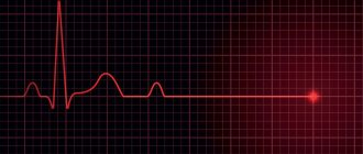
The heart rate is determined by measuring the highest R waves. The P-QRS-T interval can be used to judge how long it takes for an impulse to pass through all parts of the heart. The height of the Q wave indicates excitation in the left heart septum. What a normal ECG looks like is shown in the figure.
The most disappointing diagnosis: heart attack
Most people undergo an ECG procedure without much preparation. Often doctors don’t even talk about special measures, which is a big mistake. When the patient is tuned in to taking a cardiogram, calm and prepared, the results are truly truthful. Nobody cares when a person gets nervous when he sees a doctor. Many people are afraid of hospitals and procedures, and this affects their psychological, emotional and physical well-being. How to take an ECG without problems?
private forms of heart attack on ECG
The most serious diagnosis on the ECG, of course, is myocardial infarction, in the recognition of which the cardiogram plays the main role, because it is it (the first!) that finds areas of necrosis, determines the location and depth of the lesion, and can distinguish an acute infarction from aneurysms and scars of the past.
The classic signs of myocardial infarction on the ECG are the registration of a deep Q wave (OS), an elevation of the ST segment, which deforms the R, smoothing it, and the subsequent appearance of a negative pointed isosceles wave T. This elevation of the ST segment visually resembles a cat's back (“cat”). However, a distinction is made between myocardial infarction with and without the Q wave.
How to make a good cardiogram with a bad heart
0
Marina Dmitrievna
2017.07.26
19 384
Diagnostics
A rare but serious phenomenon - thrombophlebitis of the veins of the face and neck
Most people undergo an ECG procedure without much preparation. Often doctors don’t even talk about special measures, which is a big mistake.
When the patient is tuned in to taking a cardiogram, calm and prepared, the results are truly truthful. Nobody cares when a person gets nervous when he sees a doctor.
Many people are afraid of hospitals and procedures, and this affects their psychological, emotional and physical well-being. How to get an ECG for a child without problems?
If there is a bad cardiogram of the heart, what to do?
When a person has heart complaints, going to an ECG procedure becomes exciting. We all fear for our health, especially if the doctor diagnoses pathologies. To avoid such a situation, it is necessary to understand the structural features of the heart, possible changes in its functioning and pathology.
Quite often the patient is told that sinus rhythm is diagnosed on the cardiogram. How scary and harmful is this to the heart? A bad ECG most often indicates that the heart is experiencing a blockage.
Bad heart cardiogram - what to do to improve the result? Advice! Before the ECG procedure, do not take heart medications so that the result is as reliable and informative as possible. It is worth understanding that a poor ECG is associated with changes in the heart.
That is, it is necessary to influence the reasons why the heart does not work well. After examining an adult or child, the doctor prescribes medications aimed at improving heart function.
Hello. There was a need to do a cardiogram (and the result needed to be good).
Or will they distort the result (if they distort, then in what direction?) Are there any recommendations for behavior, routine, food intake, etc.
in order to somehow improve the result of the cardiogram? Maybe taking an infusion of VALERIAN or MOONOR on the day before and before the ECG will relieve this anxiety? But this will not affect the ECG itself in any way.
But you should not abuse other drinks, such as water or juice, because overloading the body with fluid may not have the best effect on the cardiogram.
You should not go for an ECG earlier than two hours after undergoing any physiotherapeutic procedures. If at rest your pulse is kept within normal limits, then there is no reason to worry.
Most experts are inclined to believe that we can talk about bradycardia when the heart rate is less than fifty beats per minute.
Additional diagnostic methods
The difficulty in using an ECG is due to the fact that many diseases have similar signs that appear on a cardiogram. Because of this, it is quite difficult to draw conclusions based on this method alone.
Usually, if any cardiac pathology is suspected, other diagnostic methods are used.
The main ones:
Holter monitoring.
This method differs from a conventional cardiogram in that it records data throughout the entire day.
The patient can perform normal daily activities. This diagnostic method allows for a more accurate analysis of the condition.
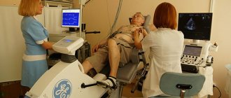
ECG with stress.
In this way, it is possible to detect angina, ischemia and arrhythmia. This procedure is performed using special simulators on which the patient does exercises. Many cardiac abnormalities manifest themselves during exercise, so this diagnostic method is very important.
Ultrasound.
During an ultrasound, doctors find out the features of the organ itself.
Thanks to this method, you can find out the dimensions of the heart chambers, partitions, wall thickness, etc.
Echocardiography.
Through echocardiography, it is possible to determine the characteristics of the valves, wall contractions, and the size of the cavities. Also, using this method, the characteristics of blood flow in the heart are studied.
Scintigraphy.
This diagnostic method allows you to assess the condition of the blood vessels.
A radiopharmaceutical is placed into the blood and its progress is monitored.
Scintigraphy makes it possible to find out the characteristics of the blood supply.
MRI.
Using MRI, doctors identify vascular diseases, detect murmurs and areas of pathological changes. Performing such a procedure is expensive, and in some cases it cannot be avoided.
In addition, the diagnosis uses blood tests (general and biochemistry), urine tests, hormone levels, etc. Detection of heart disease requires attention and knowledge from doctors, so they must take everything into account.
Do you still think that it is impossible to get rid of HEART DISEASES!?
Do you often experience discomfort in the heart area (pain, tingling, squeezing)? You may suddenly feel weak and tired... You constantly feel high blood pressure... There is nothing to say about shortness of breath after the slightest physical exertion... And you have been taking a bunch of medications for a long time, going on a diet and watching your weight...
Source: https://MedLazaret.ru/kardio/kak-uluchshit-kardiogrammu.html
Electrocardiography (ECG): basic theory, recording, analysis, detection of pathologies
If a person knows in advance when the procedure is, then it is better to prepare for it 1-2 days in advance. Many will be surprised, because no one talks about this. The secret is simple - during this time the patient must calm his heart as much as possible.
We suggest you read: Ultrasound of the heart for atrial fibrillation
Advice! Get an ECG from a qualified doctor who can read the information.
Often, due to a medical error, people are treated for diseases of the cardiovascular system that are completely different from those that they have. If necessary, to obtain detailed information, the patient undergoes echocardiography, Holter monitoring, vascular ultrasound, etc. It is impossible to draw a conclusion based only on ECG data.
The first stage of preparation should be the choice of a medical institution or doctor who performs the procedure.
To take a quality photo, you need to know how to improve the result. To do this, the patient must follow the following recommendations:
- Avoid alcohol 1-2 days before the procedure.
- The day before the procedure, do not drink caffeinated drinks.
- If possible, avoid heavy physical activity on the eve of the ECG.
- Minimize the stress factor.
- In the morning before the procedure, take a warm shower (clean skin has better conductivity).
- If men have heavily developed hair on their chest, then shave it so that the contact of the electrodes is better.
- Women should avoid applying greasy creams or balms to their bodies; their skin should be clean.
- Before the procedure, do not take medications to ensure accurate results. Before the ECG, the doctor will clarify which medications to stop for a while.
- While waiting for the procedure, calm down. To do this, inhale deeply and exhale slowly for 10 minutes.
- If necessary, take a bedding towel with you.
These are preparatory moments that help the device obtain accurate information. With such preparation, the data obtained is as true as possible, which is what a cardiologist needs.
Over the past few decades, to effectively study the functioning of the human heart muscle, a relatively inexpensive, reliable and fairly informative diagnostic method has been used - the electrocardiography procedure, familiar to many of us (abbreviated as ECG).
I recently read an article that talks about Monastic tea for treating heart disease. With this tea you can FOREVER cure arrhythmia, heart failure, atherosclerosis, coronary heart disease, myocardial infarction and many other diseases of the heart and blood vessels at home.
I’m not used to trusting any information, but I decided to check and ordered a bag. I noticed changes within a week: the constant pain and tingling in my heart that had tormented me before receded, and after 2 weeks disappeared completely. Try it too, and if anyone is interested, below is the link to the article.
Thanks to this simple and reliable study, thanks to the qualifications of doctors and the correct interpretation of records obtained from cardiograms, doctors have the opportunity to learn a lot about the nature and causes of abnormalities in the functioning of the heart.
Recent reader feedback has made us think that not many people clearly understand what an ECG procedure is.
That is why today we will talk about the specifics of ECG examination of the heart, we will figure out whether any specific preparation for the procedure is necessary.
And also, we will try to clearly explain to the reader what exactly can be seen on a cardiogram - a graphical representation of the electric fields that arise during the work of the heart.
It's no secret that the electrocardiography procedure can be carried out somewhat differently, according to different scientific methods - in other words, an ECG can be of different types:
- most often, doctors use classical cardiography;
- according to indications, intra-esophageal electrocardiography may be prescribed, with the introduction of an active electrode directly into the esophagus;
- Holter electrocardiography, when readings are taken over a long period of time (from a day to a week);
- electrocardiography with additional load or so-called bicycle ergometry;
- high-resolution electrocardiography and others.
Next, I would like to note that there is no long-term specific process of preparation for any type of ECG today.
However, some preparatory conditions must be met by the patient before conducting the heart study in question.
- good rest;
- avoid stressful situations;
- refuse to eat large amounts of food, and the intraesophageal electrocardiography procedure, in general, should be performed on an empty stomach;
- temporarily reduce the amount of fluid consumed (three to five hours before the test);
- immediately before the procedure itself, you should relax, restore your breathing, and possibly meditate.
It is after such simple preparation for the procedure that one can expect that the cardiograph will give the most accurate and objective indicators of the work of the heart muscle during any of the ECG studies.
How is an ECG performed if the appointed time has come?
You will be asked to remove your outer clothing so that nothing obstructs access to the chest, and free your lower legs. The places where the electrodes will be attached will be treated with alcohol, and a special gel will be applied to them.
The next stage comes down to attaching the cuffs and suction cups. They are fixed on the arms, ankles and chest. Ten electrodes will track the heart rhythm and give an encrypted result.
Interpretation of the results will be effective if the patient has followed all instructions for preparing for the ECG
The heart plays the role of an electrical generator. Body tissues have a high degree of electrical conductivity, which makes it possible to detect the electrical impulses of the heart by applying electrodes to areas of the body. The readings of biopotentials are processed by an electrocardiograph and provides data in the form of a summary picture - the distribution of excitation signals throughout the muscle in a graphic image. More specifically, the difference in electrical voltage.
The spread of the impulse throughout the parts of the heart is facilitated by the depolarization of myocardial cells, during which some of the cells acquire a positive charge, the other part - a negative one. This is how a potential difference arises. In the case of complete depolarization (contraction) or repolarization (relaxation) of the cell, no voltage difference is noted. The device records EMF—electromotive force of the heart.
After an ECG is performed, the doctor gets an idea of the functioning of the organ and any abnormalities.
Despite its high diagnostic value, a regular ECG cannot detect changes in heart function outside the time of its taking. Therefore, along with the traditional technique, the patient may be prescribed additional Holter monitoring and exercise testing during the day.
Using this method, it is impossible to recognize heart murmurs, therefore, if structural defects of the valves or septa are suspected, echocardiography, phonocardiography or ultrasound of the heart should be performed.
If it is planned to install a stent or shunt for myocardial ischemia, then coronary angiography is required to determine the localization of the narrowing of the coronary arteries. Tumor processes are diagnosed by X-ray or MRI examination.
Used for practical purposes in the 70s of the 19th century by the Englishman A. Waller, the device that records the electrical activity of the heart continues to faithfully serve humanity to this day. Of course, over almost 150 years it has undergone numerous changes and improvements, but the principle of its operation, based on recording electrical impulses propagating in the heart muscle, has remained the same.
We invite you to read: What is contraindicated for heart disease: diagnosis, signs of the disease, products, recommendations
Now almost every ambulance team is equipped with a portable, lightweight and mobile electrocardiograph, which allows you to quickly take an ECG, not waste precious minutes, diagnose acute cardiac pathology and quickly transport the patient to the hospital. For large-focal myocardial infarction, pulmonary embolism and other diseases that require emergency measures, minutes count, so an urgently taken electrocardiogram saves more than one life every day.
Deciphering an ECG for a cardiology team doctor is a common thing, and if it indicates the presence of acute cardiovascular pathology, then the team immediately turns on the siren and goes to the hospital, where, bypassing the emergency room, they will deliver the patient to the intensive care unit for emergency care. The diagnosis has already been made using an ECG and no time has been lost.
HM ECG - what kind of abbreviation is this so incomprehensible? This is the name for long-term and continuous recording of an electrocardiogram using a portable portable tape recorder, which records the ECG on magnetic tape (Holter method). Such electrocardiography is used to detect and register various disorders that occur periodically, so a regular ECG is not always able to recognize them.
In addition, abnormalities may occur at certain times or under certain conditions, so in order to compare these parameters with the ECG recording, the patient keeps a very detailed diary. In it, he describes his feelings, records the time of rest, sleep, wakefulness, any active activity, notes the symptoms and manifestations of the disease.
The duration of such monitoring depends on the purpose for which the study was prescribed, however, since the most common is recording an ECG during the day, it is called daily, although modern equipment allows monitoring for up to 3 days. And a device implanted under the skin takes even longer.
Daily Holter monitoring is prescribed for rhythm and conduction disorders, painless forms of coronary heart disease, Prinzmetal's angina and other pathological conditions. Also, indications for the use of Holter are the presence of an artificial pacemaker in the patient (control over its functioning) and the use of antiarrhythmic drugs and drugs for the treatment of ischemia.
Preparing for Holter monitoring is also simple, but men should shave the electrode sites, as hair will distort the recording. Although it is believed that daily monitoring does not require special preparation, the patient, as a rule, is informed what he can and cannot do.
Of course, you cannot immerse yourself in a bath; the device does not like water treatments. There are those who don’t even accept showers, all you have to do is endure, unfortunately. The device is sensitive to magnets, microwaves, metal detectors and high-voltage lines, so it is better not to test its strength; it will still record incorrectly. He doesn’t like synthetics and all kinds of metal jewelry, so he should switch to cotton clothes for a while and forget about jewelry.
Sinus rhythm of the heart on an ECG - what does it mean and what can it tell you?
What does it mean and what are the rules?
Have you been struggling with HYPERTENSION for many years without success?
Head of the Institute: “You will be amazed at how easy it is to cure hypertension by taking it every day...
Read more "
Sinus rhythm of the heart on an ECG - what does it mean and how to determine it? There are cells in the heart that create impulse due to a certain number of beats per minute. They are located in the sinus and atrioventricular nodes, as well as in the Purkinje fibers, which make up the tissue of the cardiac ventricles.
Sinus rhythm on an electrocardiogram means that this impulse is generated precisely by the sinus node (the norm is 50). If the numbers are different, then the pulse is generated by another node, which produces a different value for the number of beats.
Normally, a healthy sinus rhythm of the heart is regular with varying heart rates depending on age.
Normal indicators in the cardiogram
What to pay attention to when performing electrocardiography:
- The P wave on the electrocardiogram necessarily precedes the QRS complex.
- The PQ distance corresponds to 0.12 seconds - 0.2 seconds.
- The shape of the P wave is constant in each lead.
- In an adult, the rhythm frequency corresponds to 60 – 80.
- The P–P distance is similar to the R–R distance.
- The P wave in a normal state should be positive in the second standard lead, negative in lead aVR. In all other leads (these are I, III, aVL, aVF), its shape may vary depending on the direction of its electrical axis. Typically, P waves are positive in both lead I and aVF.
- In leads V1 and V2, the P wave will be 2-phase, sometimes it can be predominantly positive or predominantly negative. In leads V3 to V6, the wave is predominantly positive, although there may be exceptions depending on its electrical axis.
- Normally, each P wave must be followed by a QRS complex and a T wave. The PQ interval in adults has a value of 0.12 seconds - 0.2 seconds.
Sinus rhythm together with the vertical position of the electrical axis of the heart (EOS) shows that these parameters are within normal limits. The vertical axis shows the projection of the position of the organ in the chest. Also, the position of the organ can be in semi-vertical, horizontal, semi-horizontal planes.
When the ECG registers sinus rhythm, it means that the patient does not yet have problems with the heart. It is very important not to worry or be nervous when undergoing the examination, so as not to receive false data.
You should not do the examination immediately after physical activity or after the patient has climbed to the third or fifth floor on foot. You should also warn the patient that you should not smoke half an hour before the examination, so as not to get unreliable results.
Violations and criteria for their determination
If the description contains the phrase: sinus rhythm disturbances, then a blockade or arrhythmia has been registered. An arrhythmia is any disruption in the rhythm sequence and its frequency.
Blockades can be caused if the transmission of excitation from the nerve centers to the heart muscle is disrupted. For example, rhythm acceleration shows that during a standard sequence of contractions, the heart rhythms are accelerated.
If the conclusion contains a phrase about an unstable rhythm, this means a manifestation of a low heart rate or the presence of sinus bradycardia. Bradycardia has a detrimental effect on a person’s condition, since the organs do not receive the amount of oxygen required for normal activity.
Why do you feed pharmacies if hypertension is afraid of the usual like fire...
Tabakov has revealed a unique remedy against hypertension! To reduce blood pressure while preserving blood vessels, add to…
If an accelerated sinus rhythm is recorded, then most likely this is a manifestation of tachycardia. This diagnosis is made when the number of heart beats exceeds 110 beats.
Interpretation of results and diagnosis
In order to make a diagnosis of arrhythmia, the obtained indicators should be compared with normal indicators. The heart rate for 1 minute should not be more than 90. To determine this indicator, you need to divide 60 (seconds) by the duration of the RR interval (also in seconds) or multiply the number of QRS complexes in 3 seconds (a section equal to 15 cm in length of the tape) by 20.
In this way, the following deviations can be diagnosed:
- Bradycardia – heart rate/min is less than 60, sometimes an increase in the PP interval of up to 0.21 seconds is recorded.
- Tachycardia - heart rate increases to 90, although other signs of rhythm remain normal. Often there can be an oblique depression of the PQ segment, and an upward depression of the ST segment. It may look like an anchor. If the heart rate rises above 150 beats per minute, stage 2 blockades occur.
- Arrhythmia is an irregular and unstable sinus rhythm of the heart, when the RR intervals differ by more than 0.15 seconds, which is associated with changes in the number of beats per inhalation and exhalation. Often found in children.
- Rigid rhythm - excessive regularity of contractions. RR differs by less than 0.05 sec. This may occur due to a defect in the sinus node or a violation of its neurovegetative regulation.
Reasons for deviations
The most common causes of rhythm disturbances are:
- excessive alcohol abuse;
- any heart defects;
- smoking;
- long-term use of glycosides and antiarrhythmic drugs;
- bulging of the mitral valve;
- pathologies of thyroid function, including thyrotoxicosis;
- heart failure;
- myocardial diseases;
- infectious lesions of the valves and other parts of the heart - the disease infective endocarditis (its symptoms are quite specific);
- overload: emotional, psychological and physical.
Additional Research
If the doctor, when examining the results, sees that the length of the area between the P waves, as well as their height, are unequal, this means that the sinus rhythm is weak.
To determine the cause, the patient may be recommended to undergo additional diagnostics: the pathology of the node itself or problems of the nodal autonomic system may be identified.
Then Holter monitoring is prescribed or a drug test is performed, which makes it possible to find out whether there is a pathology of the node itself or whether the regulation of the autonomic system of the node is disrupted.
For more details about weak node syndrome, watch the video conference:
If it turns out that the arrhythmia was the result of disturbances in the node itself, then corrective measurements of the vegetative status are prescribed. If for other reasons, then other methods are used, for example, implantation of a stimulator.
Holter monitoring is a regular electrocardiogram that is performed throughout the day. Due to the duration of this examination, specialists can study the condition of the heart at different degrees of stress. When conducting a regular ECG, the patient lies on the couch, and when conducting Holter monitoring, it is possible to study the state of the body during physical activity.
Treatment tactics
Sinus arrhythmia does not require special treatment. An incorrect rhythm does not mean that you have any of the listed diseases. Heart rhythm disturbances are a common syndrome characteristic of any age.
A proper diet, daily routine, and lack of stress can greatly help to avoid heart problems. It would be useful to take vitamins to maintain heart function and improve the elasticity of blood vessels. In pharmacies you can find a large number of complex vitamins containing all the necessary components and specialized vitamins to maintain the functioning of the heart muscle.
In addition to them, you can enrich your diet with foods such as oranges, raisins, blueberries, beets, onions, cabbage, and spinach. They contain many antioxidants that regulate the number of free radicals, an excessive amount of which can cause myocardial infarction.
For the smooth functioning of the heart, the body needs vitamin D, which is found in parsley, chicken eggs, salmon, and milk.
If you plan your diet correctly and adhere to a daily routine, you can achieve long and uninterrupted functioning of the heart muscle and not worry about it until you are very old.
Finally, we invite you to watch a video with questions and answers about heart rhythm disturbances:
How to properly prepare for an ECG, what not to do
To undergo an ECG without problems, you need to know which drugs, foods or drinks increase your heart rate.
There are a number of rules that must be followed before the electrocardiography procedure:
- 2-3 hours before the procedure, do not eat fatty, salty or spicy foods.
- It is strictly forbidden to drink energy drinks, coffee and strong tea.
- You can drink plain water, but in small quantities, as a large amount of liquid will cause shortness of breath. Drink no more if the procedure is scheduled for the morning.
- Breakfast on the day of the procedure is light, in the form of a snack.
- 1-2 hours before the procedure, you should not take vasoconstrictor medications, use eye drops or nasal sprays.
- If the patient cannot quit smoking, then stop smoking several hours before the procedure. Nicotine smoke causes vasoconstriction, which will be reflected on the ECG.
- You should not take strong sedatives, as they will significantly reduce the heart rate, which will be a reason to diagnose bradycardia.
The best option would be to stop taking medications that can affect the functioning of the heart. Taking medications for diabetes mellitus cannot be stopped.
For a thorough and in-depth study of the functioning of the heart, doctors have been using one of the most reliable diagnostic methods for many years - electrocardiography (or ECG for short). Thanks to this diagnosis and correct interpretation of the cardiogram, a lot can be said about the nature and cause of abnormalities in the heart.
In a new series of articles, we will tell you about the specifics of the ECG procedure, as well as how to properly prepare for it and be able to independently decipher the results obtained by comparing your own indicators with the norm.
Electrocardiography does not require lengthy preparation, which is one of the advantages of this method. It is removed for emergency reasons in any condition of the patient. But if a planned study is prescribed, then before conducting it it is recommended:
- Do not consume food or caffeine-containing drinks at least 3 hours before the procedure.
- You need to have a good rest before the examination.
- Eliminate physical and emotional stress.
- Take a shower, after which do not use the cream.
Clothing is selected in such a way that it is easy to attach electrodes to the skin of the ankles, wrists and chest.
On the day of the study, drinking alcoholic beverages and smoking is strictly prohibited; you must avoid sports and a hearty breakfast. The best drink to drink is plain drinking water, weak tea or fruit juice.
To take an electrocardiogram, the patient is placed on a couch and a healthcare professional places electrodes on the legs, wrists and chest. If there is difficulty breathing in a horizontal position, the procedure is performed while sitting.
To ensure good contact between the skin and the electrode, the attachment site is degreased with ethyl alcohol and a special conductive gel is applied. After this, readings are taken using an ECG diagnostic device.
The whole procedure takes about minutes.
In order to get a reliable result, you need to be in a calm, relaxed state, and not hold your breath. Muscle tremors from excitement or cold can lead to data distortion.
The generally accepted leads are 3 standard, 3 reinforced and 6 chest. Each lead will record at least 4 cardiac cycles. After this, the device is turned off, the electrodes are removed, and the functional diagnostics doctor is given a signed tape, which he must decipher.
Undoubtedly, there are a number of specific restrictions that describe what is strongly not recommended to be done before conducting an electrocardiographic study of the heart.
Doctors usually do not recommend the following before an electrocardiography procedure:
- drink alcohol or smoke;
- eat too much;
- exercise;
- take medications that the doctor does not know about;
- visit the sauna, take other thermal procedures for recovery.
In addition, it is important to understand that it is better to carry out the procedure in a calm, balanced state, which is why doctors do not recommend their patients to visit any entertainment venues or go to bed late the night before.
We suggest you read: What you can take for heart pain
For the treatment of cardiovascular diseases, Elena Malysheva recommends a new method based on Monastic tea.
It contains 8 useful medicinal plants that are extremely effective in the treatment and prevention of arrhythmia, heart failure, atherosclerosis, ischemic heart disease, myocardial infarction, and many other diseases. Only natural ingredients are used, no chemicals or hormones!
How to prepare for Holter monitoring?
The Holter ECG method is a long-term recording of heart parameters using electrography. The method differs from a conventional ECG in that data is collected throughout the day. Electrodes are installed on the patient's body and connected to a mini-computer. It accumulates the information received within 24 hours.
Important! It is Holter monitoring that allows the doctor to see the slightest changes in the patient’s heart function. It is appropriate to use this method during the medical examination that conscripts undergo.
It is impossible to deceive the device. Often, to obtain a negative cardiogram, conscripts can drink energy drinks or drugs to speed up the heartbeat. Thus, they avoid the army, simulating an attack of arrhythmia, etc.
For monitoring to be successful, you should not give up going to work, because data should be collected taking into account your usual daily routine. The best points can be considered:
- Before the procedure, take a shower, as this cannot be done during the day.
- Clothing should be loose and simple, without metal or synthetic inserts.
- Remove metal jewelry from the body.
- In the morning before the procedure, you should not apply creams or lotions to the skin; it must be clean.
- Quit smoking and alcoholic beverages.
- Do not drink coffee, strong tea or energy drinks.
- You should not overload yourself with excessive physical activity.
- Write down each stage of the day in a diary. The list must include the time of the event. For example: walking, climbing stairs, sleeping, watching a movie, etc.
- Do not take medications that can affect the functioning of the heart (vasoconstrictors, antiarrhythmics, pacemakers).
The attending physician removes the device. It is prohibited to remove the device yourself or rearrange the electrodes. Pay special attention to minimizing the stress factor. During the diagnosis, do not expose yourself to unnecessary experiences or emotions.
Contrary to the opinion of cardiologists, it is generally accepted that an ECG does not require special preparation. Studying the functioning of the heart muscle involves avoiding stress, fatigue, and requires complete rest. On the day of the procedure, you need to get a good night's sleep and ignore morning exercises. If the procedure is scheduled for the morning, then a heavy breakfast should be avoided, or better yet, abandoned altogether. For an upcoming daytime procedure, you should limit yourself to a light snack 2 hours before the session.
Remember to reduce the amount of fluid you drink that affects muscle function. Avoid coffee, tea and other energy drinks. They will stimulate cardiac activity, and the results will be distorted.
It is advisable to take a shower. There is no need to apply care products to the body, because the components of creams and lotions will contribute to the formation of a greasy film on the surface, which will negatively affect the contact of the electrodes with the skin.
Immediately before performing an ECG, try to relax as much as possible. Sit with your eyes closed and restore your breathing - this will ensure an even pulse and objective readings from the device.
What diseases are diagnosed on an ECG?
Using an electrocardiogram, doctors diagnose a lot of diseases, but often an ECG is only a basic method. For a detailed examination, echocardiography is used. An ECG can be used to diagnose:
- angina pectoris
- coronary heart disease,
- blockades,
- arrhythmia,
- bradycardia,
- hypertrophy of the heart muscle,
- scarring in the myocardium,
- myocardial infarction, etc.
If the patient has heart pathology, the doctor prescribes appropriate treatment. When pathological changes are diagnosed in a child, the cardiologist prescribes an additional consultation with a pediatrician. Sometimes consultation with a neurologist and endocrinologist is required.
In addition to the prescribed treatment, the doctor recommends returning the patient to the lifestyle: proper nutrition, moderate physical activity (for obesity), walking, therapeutic and preventive physical exercise.
Hypertrophy of the atria and ventricles. Occurs due to overload of the heart chambers, as blood is transported through the vessels incorrectly. The atria and ventricles increase in size. On the ECG it is manifested by the growth of R waves, deviation of the electrical axis of the organ, and increased excitability vector. Angina pectoris.
When sick, the patient may not feel any deterioration in health for a long time. On the ECG, angina is displayed as: ST segments below the isoline, the T wave changes display. Arrhythmia. Manifests itself in the form of chest pain and rapid heart rate. On the ECG it appears as: the presence of fluctuations in the PQ and QT intervals, the R waves deviate from the norm, the PQ interval decreases. Bradycardia.
In addition to these diseases, ECG diagnoses myocardial infarction, myocarditis, coronary heart disease, and heart failure.
Hypertrophy of the atria and ventricles. Occurs due to overload of the heart chambers, as blood is transported through the vessels incorrectly. The atria and ventricles increase in size. On the ECG it is manifested by the growth of R waves, deviation of the electrical axis of the organ, and increased excitability vector. Angina pectoris.
When sick, the patient may not feel any deterioration in health for a long time. On the ECG, angina is displayed as: ST segments below the isoline, the T wave changes display. Arrhythmia. Manifests itself in the form of chest pain and rapid heart rate. On the ECG it appears as: the presence of fluctuations in the PQ and QT intervals, the R waves deviate from the norm, the PQ interval decreases. Bradycardia.
How to worsen ECG readings?
Some people sometimes have a question: how to deceive an ECG? Moreover, some want to achieve a good conclusion in order to get a job, while others, on the contrary, want to make things worse so as not to serve in the army.
There are other reasons that prompt a person to deliberately deceive not only the doctor, but also the diagnostic apparatus.
Naturally, if you do not follow the doctor’s recommendations for preparing for the procedure, then the results obtained can be distorted, but will this help achieve your goal? And is it possible to simulate angina pectoris, arrhythmia, RGC syndrome and other diseases on an ECG?
Is ECG harmful?
The natural question of whether ECG is harmful can be answered based on the advantages of this diagnostic method:
- reliability of information
- safety and comfort of the session
- efficiency (10 min)
- no health or pregnancy restrictions
As you already understand, it is impossible to harm the health of an ECG, since this method is based on reading heart rhythm indicators and does not produce any radiation or effects on the body. Moreover, people whose work involves constant physical activity undergo electrocardiography almost every day, which once again confirms its absolute harmlessness.
Electrocardiography (ECG) in St. Petersburg
Our specialists will conduct a quick examination and issue an expert opinion. We have everything you need to conduct a qualified, efficient and, most importantly, fast examination. Functional diagnostics doctors at our Center possess modern technologies for diagnosing diseases, which make it possible to accurately identify the cause of the disease and eliminate its impact on the human body. Electrocardiography, cardiogram, ECG is an indispensable measure for surveillance monitoring and control over the functioning of the cardiovascular system. To record an ECG, we use a computer analyzer, which allows us to record a fairly long section of the cardiogram and select the most indicative fragment for printing. In addition, to decipher the ECG, computer registration allows you to examine in detail and, if necessary, significantly increase the indicators of interest.
Good afternoon again. I’ll correct you right away for the future.
Grigory Arutyunov: The insidiousness of this disease is that increased blood pressure accelerates the development of atherosclerosis and increases the risk of cerebrovascular accidents and strokes - approx. In the modern world, a person consumes too much salt, the recommended daily intake is 6 g, the average Muscovite consumes more than 14 g per day, too little - fresh vegetables and fruits less than g per day. Sleep disturbances, obesity, physical inactivity are becoming more and more common; the vast majority of people do not maintain generally accepted standards of adequate physical activity: walking at a speed of kilometers per hour for minutes at least 5 times a week, diabetes mellitus, poorly controlled cholesterol levels. The combination of these risk factors leads to accelerated biological aging and the development of cardiovascular diseases. Grigory Arutyunov: Hypertension is much more common in people over 60 years of age. People with excess body weight, disturbed sleep, and excess salt intake are traditionally more susceptible to this disease. We cannot discount occupational hazards associated with shift work, frequent changes in time zones, etc.
How is the procedure performed?
The process of carrying out the diagnostic procedure in question is familiar to most of us.
To conduct classical cardiography, the patient is asked to undress, freeing the shins, forearms, and chest area from clothing and take a horizontal position of the body.
In this case, doctors treat the places where the electrodes are attached with alcohol and a conductive antiseptic gel for a better fit of the electrodes.
At the same time, the attached electrodes monitor the rhythm of contractions of the heart muscle, producing a graphic image (cardiogram) in the form of an encrypted result describing the work of the organ.
Further, the resulting cardiogram requires a certain interpretation by a cardiologist, who, based on certain deviations, deformations, changes in standard teeth and complexes, will be able to judge the occurrence of a particular cardiac pathology in the patient’s body.
At the next stage, the cuffs and suction cups are attached. Electrodes are fixed on the arms, legs, and chest.
Tags: before, heart, calm
About the author: admin4ik
« Previous entry
What is electrocardiography?
Electrocardiography is a method for studying the physiological state of the heart muscle, as well as its performance.
For the study, a device is used that registers all changes in the physiological processes in the organ and, after processing the information, displays it in a graphical image.
The graph shows:
- Conduction of electrical impulses by the myocardium;
- Heart muscle contraction frequency (HR);
- Hypertrophic pathologies of the cardiac organ;
- Scars on the myocardium;
- Changes in myocardial functionality.
All these changes in the physiology of the organ, and in its functionality, can be recognized on the ECG. Cardiograph electrodes record bioelectric potentials that appear during contraction of the heart muscle.
Electrical impulses are recorded in different parts of the heart organ, so there is a potential difference between the excited areas and the non-excited areas.
It is this data that is captured by the device’s electrodes, which are attached to different parts of the body.