December 8, 2020 Admin Home page » Ultrasound of the chest organs Views: 3818
Hidden heart disease is a dangerous factor that can dramatically change life, once manifesting itself in the form of serious diseases. It is impossible or very difficult to identify them using standard methods. Therefore, stress echocardiography with exercise was developed, which makes it possible to study the performance of the heart under stress conditions. The advantage of the method is that it is non-invasive, easy to use, modern, and gives accurate results.
What is stress echocardiography as a research method? This is the use of two methods simultaneously - bicycle ergometry and ultrasound of the heart to identify hidden deficiencies and diseases. As a rule, stress echocardiography with exercise is used in the following cases:
- silent myocardial ischemia;
- angina pectoris;
- to assess cardiac dysfunction during exercise and rest. For example: heart rate, blood flow speed, blood pressure, etc.;
- assessment of the state of the myocardium after a heart attack, etc.
Characteristics of the method
Most patients, after being prescribed stress echocardiography, are interested in what it is and how the procedure is performed.
Stress echocardiography is a comprehensive diagnostic method that allows you to identify cardiac pathologies at the initial stage through the use of special stress tests. During the study, the heart is simultaneously imaged and its impulses are recorded.
Determination of local disturbances in individual areas of cardiac tissue indicates the development of a pathological process. As a rule, an electrocardiogram is performed at rest, so it is difficult to identify minimal disturbances in the functioning of the organ. Stress echocardiography, on the contrary, is performed while the heart is stressed, causing an increase in its contractions, which helps to accurately detect coronary artery disease at an early stage of development.
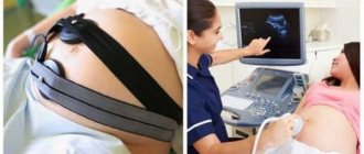
The procedure is performed jointly by a cardiologist and an ultrasound specialist
During the study, the following are used as stress tests:
- pharmacological medicines;
- bicycle ergometry;
- cold stimulation;
- transesophageal electrical stimulation;
- hyperventilation of the bronchopulmonary system.
The method also allows you to determine cardiac endurance, which is why it is used annually among professional athletes.
Indications
With the help of stress echocardiography, it is possible to detect coronary heart disease in the early stages and post-infarction cardiosclerosis - diseases that provoke heart failure and can cause arrhythmias and sudden cardiac death.
It is prescribed in such cases and for the following purpose:
- Before heart surgery to make a prognosis and assess the risk of complications.
- If a regular echocardiogram and ECG showed normal results, but the person is at risk of developing coronary disease (improper lifestyle, heredity, high cholesterol, metabolic disorders, vascular diseases, etc.).
- If the stress ECG turned out to be uninformative.
- To assess the effectiveness of treatment and make a prognosis for cardiac ischemia.
- To test the cardiac endurance of professional athletes and assess human performance.
- To accurately identify functional and disease-affected areas of the myocardium.
Advantages and disadvantages of the test
The use of each type of stress test has both its advantages and disadvantages.
Electrical stimulation of the heart through the esophagus provides clear images without increasing blood pressure
Advantages of the method:
- Allows you to evaluate the activity of the heart in a state of increased physical activity.
- Possibility of performing diagnostics outside the hospital using portable equipment.
- Qualitative level of assessment of the state of the heart muscle.
- High sensitivity of the method.
- No biological effect on the patient and medical staff.
Disadvantages of the procedure:
- The accuracy of the study depends on the skill level of the specialist.
- During exercise, blurred visualization of the left ventricle is possible.
- The development of side effects in the form of dizziness, rapid pulse, discomfort in the chest area.
Despite all the shortcomings, the technique is considered an effective and inexpensive procedure that can diagnose pathological changes in the functioning of the myocardium.
Carrying out the procedure
The research takes place in several stages:
- A regular echography is performed: the patient undresses to the waist and lies on his left side. A gel is applied to the chest and the sensor is moved, the initial parameters of the heart and its structures are studied.
- They give a stress load, its type is chosen taking into account contraindications.
- Immediately after the exercise, echocardiography is repeated.
The duration of physical activity is 10-15 minutes, then an ultrasound is performed. When drugs or electrical stimulation are administered, echography is done immediately. In parallel with the ultrasound, an ECG is recorded for the patient. The total duration of the procedure is 30-60 minutes.
In what cases is it prescribed?
Experts recommend undergoing stress diagnostics for patients who have received normal ECG and Echo kg readings, but at the same time they have symptoms characteristic of heart disease.
Very often, a stress test is prescribed to determine the therapeutic effectiveness after drug treatment.
Indications for performing a stress echo kg are the following conditions:
- Diagnosis of myocardial ischemia.
- Assessment of the degree of damage to the coronary vessels.
- Assessment of the functioning of the heart muscle in patients with organ dysfunction.
- Identification of myocardial areas with a high risk of developing ischemic damage.
- Analysis of the status of chronic ischemic heart disease.
- Preparing the patient for minimally invasive procedures on the chest area.
- Analysis of the effectiveness of angioplasty, stenting and bypass surgery.
- Clarification of the possibility of developing complications after cardiac operations.
- Establishing the timing of surgery in the presence of valve defects.
- Determination of the patient's ability to work.
Which doctor should I contact?
A cardiologist will refer you for an ultrasound of the heart, based on the presence of indications for this examination, so the examination should begin with his consultation. In principle, ordinary echocardiography, which has no contraindications, can be performed by any person if he has a reasonable desire to obtain information about the condition of his heart. To do this, just contact one of the specialized private clinics.
As for stress echocardiography, it is not performed at the patient’s request. The procedure has strict indications and contraindications, which are determined by the doctor, and he also gives directions.
Contraindications to the method
Read more:Technique for performing an ECG with stress
The diagnostic method is contraindicated in the following conditions:
- unstable angina;
- previous myocardial infarction in the acute phase;
- hemodynamic disorders;
- inflammatory process in the myocardium and pericardium;
- expansion of the aorta;
- heart failure in the progressing stage;
- acute febrile condition;
- mental disorders.
Features of the method
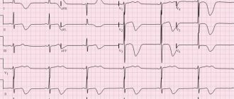
An early sign of weakened blood circulation in the myocardium is a decrease in the number of heart contractions in response to additional load, whereas in a normal physiological state contractions remain unchanged or increase.
The loading method displays myocardial activity in the form of a graph
During diagnostics, changes may be detected in the form of:
- Deterioration in the contractility of the damaged area (visualized by ultrasound).
- Pathological changes during ECG registration (determined by stress test).
- The appearance of pain behind the sternum.
Myocardial movements are preliminarily assessed before testing. After this, the patient is given a medicine that increases the heart rate or is asked to do an exercise test.
In most cases, the administration of medications is more advantageous than performing tests with physical activity. While walking on a treadmill, many patients cannot achieve the required frequency of myocardial contractions, and some experience rhythm interruptions that interfere with the study. All this can negatively affect diagnostic results.
Pharmacological testing is associated with more cardiovascular complications than exercise testing. In the case of a load test, it is recommended to rotate the pedals on a bicycle ergometer in a horizontal position. This makes it possible to quickly move the patient to the couch.
What is the procedure
Echocardiography (ECHOCG) is a non-invasive (skin is not damaged) research method that allows you to view “online” how the heart functions. Exercise stress echocardiography is a combination of ultrasound imaging with bicycle ergometry (on a recumbent or upright exercise bike) or treadmill test (treadmill). This combination allows you to compare the ultrasound image of the heart structure in a stressful situation and at rest.
During the procedure, a sensor similar to a computer mouse generates high-frequency sound waves. When the operator moves it along the chest wall, it sends “ultra” waves through the skin and other tissues to the heart at different angles. Having been reflected from its structures, they return to the sensor in the form of an “echo”. After processing on a computer, the echo signals are converted into an image of the walls of the heart and its valves and displayed on the monitor screen.
During the procedure, one or more echo image acquisition modes may be used. How to conduct stress echocardiography and what imaging methods the doctor will use depends on the diagnostic goals set for the study. The most commonly used ultrasound scanning modes are:
- M-mode is the simplest type of echocardiography . The resulting image can be compared to a sketch of the heart, and not to a “real” picture of it. M-mode is used to determine the size of the atria and ventricles and the thickness of their walls.
- Echo-Dopplercardiography . A method using the Doppler effect allows you to evaluate the movement of blood in the chambers of the heart. In addition, Doppler ultrasound can detect abnormal blood flow, which can be a sign of malfunctioning heart valves.
- Color Doppler mapping is an analogue of the previous technique. The difference is that on the echogram, differences in the direction of blood flow are indicated by a certain color, and the speed is indicated by a change in shades, which makes it easier to interpret the data obtained.
- 2D echocardiography . The visualization method allows you to see the structures of the heart (its various sections), as well as their movement in real time.
- 3D echocardiography . Method for obtaining a three-dimensional image.
Stress echocardiography
First, so-called baseline sonography (echo image of the heart at rest) is performed. The patient is then asked to run on a treadmill or pedal on an exercise bike.
The duration of these “trainings” depends on the target load - reaching a certain heart rate or the appearance of symptoms in the patient that do not allow him to continue “exercising” in full. As soon as these target points are reached, a repeat echographic examination is performed in the same mode, after which the doctor compares the resulting echo images.
Dobutamine stress echocardiography is a type of stress test that uses pharmacology instead of exercise to make the heart beat faster.
Dobutamine is a medicine that can help regulate your heart rate. Typically, it is administered intravenously over 15 to 20 minutes. This option is usually recommended for patients in whom stress echocardiography with treadmill testing cannot be performed due to a musculoskeletal problem. There are other possible reasons.
We recommend reading about tests for ischemic heart disease. You will learn what laboratory tests, electrocardiography and echocardiography, stress tests, and multislice computed tomography (MSCT) will show for coronary heart disease. And here is more information about what an electrocardiogram looks like during cardiac ischemia.
How to prepare for research?
At the preparatory stage of the study, the patient is prescribed medications containing nitrates, which can reduce the number of myocardial contractions and also lower blood pressure. The medication is prescribed to protect the heart muscle from the effects of adrenaline produced during stress, which can cause adverse reactions from various organs and systems.
If you are using transesophageal electrical stimulation, you should tell your cardiologist if you have problems with nasal breathing
To fully prepare the body for the procedure, you must follow these recommendations:
- The day before the procedure, drinks containing caffeine and alcohol should be avoided.
- It is necessary to avoid physical activity for several hours.
- The last meal is no less than 3-4 hours before the procedure.
- Stop smoking immediately before screening.
On the day of diagnosis, patients are allowed to take nitroglycerin to stop a possible attack of angina. However, the use of funds must be agreed with a specialist.
Diagnostic principle
Stress echocardiography is based on the principle that when ischemia forms in the cardiac myocardium, the ability of the left ventricle to contract in the region of the coronary artery deteriorates.
A prerequisite for the development of pathology is disturbances in venous blood flow. Stress echocardiography allows you to record the response of individual zones of the left ventricular myocardium to increased, sudden loads that are placed on the patient’s body. In addition to bringing the cardiovascular system into a state of overstrain, the study simultaneously visualizes the performance of the heart muscle in the form of a graph, which makes the method an effective tool for complex diagnostics.
Load test
When carrying out the method, various stress tests can be used. The type of test used depends on the objectives being pursued and the intended diagnosis. Thus, to detect myocardial ischemia, as well as assess the degree of damage to individual areas after a heart attack, they resort to dynamic loading.
For dynamic loading, use a treadmill
When using a load in the form of a treadmill, the initial readings are taken at rest, and then when regaining strength after stopping the load.
It is preferable to carry out the test with a bicycle ergometer, since the recording of indicators occurs directly during the period of load or at its peak. The best visualization of the organ is achieved when using bicycle ergometry in a horizontal position.
For the category of elderly patients, a treadmill load is used, since it is more physiological and does not cause pain caused by an uncomfortable body position.
Research Modes
To obtain an informative image, the following modes can be used: a simple M-mode displays a sketch view of the heart, visualizes the thickness of the walls, the size of the ventricles and atria, Doppler echocardiography assesses the work of blood vessels and blood circulation speed, on the screen - a black and white image, or Doppler mapping in color, 2D and 3D echocardiography – a two-dimensional or three-dimensional picture of the heart. The choice of mode depends on the cardiologist and the degree to which the hospital is equipped with the necessary equipment and equipment.
Medication tests
In case of intolerance to physical activity, the use of a drug test is recommended, which is quite safe for the body and causes minimal side effects.
Dobutamine test
It is the most widely used test, during which the number of myocardial contractions increases, blood pressure increases, which causes an increase in the organ's need for oxygen. The difference between the heart's need for oxygen and the ability of the coronary vessels to supply it indicates the presence of local pathological processes in the myocardium.
A test with dobutamine allows you to determine the vital activity of the myocardium in the affected area
Test with dipyridamole
The test is done with a gradual increase in the dose of the drug. At each stage, the degree of impairment of myocardial contractility is assessed. In the absence of pathological changes, in order to achieve the required heart rate, an additional 1 mg of atropine is administered. A couple of minutes after the administration of dipyridamole, an intravenous injection with aminophylline, which is an antidote, should be given.
Evaluation of results
The results of the study are displayed in the form of a two-dimensional graph, which makes it possible to fully assess the quality of left ventricular functioning. Interpretation of the results includes an assessment of the degree of thickening and mobility of the heart muscle tissue in individual areas.
A preliminary analysis of the graphs is carried out by a specialist immediately after their registration. After the screening is completed, the cardiologist can view a video recording of diagnostic indicators in slow motion. The obtained data is stored on disks, creating an information base for the patient with further assessment of the dynamics of heart performance indicators.
Thus, stress echocardiography is a modern method for diagnosing coronary artery disease. The study allows us to determine the initial stage of the disease, when other methods reveal low effectiveness. However, before the procedure, possible cardiac complications associated with excessive load on the organ should be taken into account.
Possible complications
In principle, ultrasound examination of the heart is absolutely safe, but in stress echocardiography technology complications are possible that are associated specifically with the action of stress “provocateurs”. And although they do not occur so often, they can be quite serious:
- headache, dizziness;
- shortness of breath, feeling of lack of air;
- heart rhythm disturbances - extrasystole, tachyarrhythmia and even ventricular fibrillation;
- acute coronary syndrome - narrowing of the heart arteries, pre-infarction condition.
Complications are, as they say, “rare, but not unknown,” requiring resuscitation or surgical intervention on the heart vessels. Therefore, before the procedure, as before the operation, the patient must give consent and sign an official document.