Cardiography using an ultrasound sensor is a routine technique based on the effect of high-frequency radiation on the patient’s body and its tissues. Within the framework of the practice of cardiology specialists, we are talking about an indispensable technique. Despite the simplicity of the method, it is extremely informative, convenient and accessible for the patient himself.
ECHO CG of the heart is an ultrasound diagnostic method that allows you to visualize the anatomical features of a muscular organ: the condition of the valves, the myocardium itself and its vessels, therefore the technique mainly reveals defects. Also disorders acquired over the years. For example, with a prolonged increase in blood pressure. There are many options.
In essence, this is a regular ultrasound, only the sensor is used to diagnose cardiac structures.
Since the technique is safe, it is used repeatedly. As often as the situation requires. There are no age or other serious restrictions. Although there are certain contraindications.
What does a patient need to know before echocardiography? How effective is this research?
The essence of the technique and what it shows
As already mentioned, cardiac ECHO is a modification of standard ultrasound of internal organs. However, unlike other methods that are similar in meaning, the cardiography apparatus can operate in several modes.
For example, duplex scanning is available to a diagnostician. Doppler ultrasound is used, among other things, to examine the speed of blood flow. Its quality. This is important when diagnosing, for example, ischemic disease, assessing the condition of an organ after a heart attack.
Like other ultrasounds, echocardiography is completely safe.
What does ECHO show as part of a routine examination:
- Mass, the amount of muscle tissue in the area of the left ventricle of the heart. As a rule, it changes with a prolonged increase in pressure. Untreated hypertension is especially dangerous.
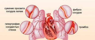
- An echogram of the heart will show the intensity of blood movement through the pulmonary artery. This vessel is one of the largest. Any violation is fraught with immediate complications. Up to and including death. Especially the increase in local pressure.
- Stroke volume. The amount of blood that the heart throws into the aorta and “drives” in a large circle. Many defects change these indicators, so deviations are quite informative.
- Condition of the left atrium.
- Wall thickness.
- Echocardiography of the heart shows even the slightest changes in cardiac structures: the position of the myocardium, heart valves (tricuspid, mitral, etc.) at the moment of contraction and return to a calm state. This indicator is defined as end systolic and diastolic volumes.
- General position of the heart. Its dimensions, anatomical location. Including regarding other organs of the chest.
The technique shows the morphological features of cardiac structures.
What pathologies can be detected
When conducting echocardiography, the following diseases and conditions are detected:
- signs of hypertrophic disease;
- previous myocardial infarction;
- congenital malformations;
- insufficiency of the mitral, aortic, tricuspid and pulmonary valves;
- heart failure;
- cardiac aneurysm;
- dilated and hypertrophic cardiomyopathy;
- pericarditis;
- narrowing of the lumen (stenosis) of the heart valves;
- myocarditis;
- metabolic cardiomyopathy;
- blood clots in the heart cavity;
- aortic aneurysm or dissection;
- infective endocarditis;
- signs of hypertension in the pulmonary artery system (thromboembolism, chronic diseases of the respiratory system);
- hydro- and hemopericardium.
During the examination, an experienced doctor also notices arrhythmias and disturbances in the rhythm of heart contractions. The price of the examination is in the range of 1200-3500 rubles.
What diseases can be detected
Based on the results of echocardiography, specialists make several diagnoses.
Arterial hypertension
This process can be identified indirectly. We are talking about a stable and regular increase in pressure in the vascular bed. If we talk about a full diagnosis, the condition should be called hypertension.
The long course of the pathological process leads to organic changes in the heart - the left ventricle is transformed. The muscle layer at the level of this chamber becomes thicker.
Read more about left ventricular hypertrophy in this article.
This is a kind of compensatory mechanism. This way, the cardiac structures can pump blood with greater force. The intensity of each blow increases. This is not normal, but an understandable phenomenon.
The longer the pathology exists, the worse the situation becomes. Possible cardiomegaly. Excessive growth of a muscular organ. Then they will not be able to perform their functions.
Heart defects
Both congenital and acquired. Mainly those that affect the valves - aortic, mitral, tricuspid, less often the septum between the chambers.
Such conditions are extremely dangerous. Because without treatment early on they lead to generalized dysfunction and circulatory disorders. And this is a direct path to death from heart failure or heart attack. Therefore, immediately after detection, the issue of treatment is decided.
Some congenital anomalies are classified as defects very conditionally. For example, an open oval window. In this case, they usually do nothing. They simply observe the patient from time to time, every year.
Thromboembolism
Dangerous disorder. Its essence is the blockage of large vessels with blood clots. A cardiac echo is a method that allows you to see blood clots in the pulmonary artery and coronary vessels. Thanks to this, you can receive treatment in a timely manner. The patient will remain alive.
Read more about the types of thromboembolism here; blockage of the pulmonary artery, possible risks and treatment methods are described here.
IHD in the form of angina pectoris
Classic situation: disturbance of trophism (nutrition) of the heart and its tissues. Accompanied by severe chest pain, shortness of breath, nausea and other symptoms. It's not a heart attack yet, but it's not that far off. One step left.
Unstable angina is especially dangerous. It proceeds unpredictably, so no one can say in advance how the process will end during the next attack.
An echocardiogram gives a picture in which areas of dystrophy are clearly visible, and areas where blood flow is impaired will be detected by echocardiography with Doppler analysis.
Symptoms of an angina attack and methods for correcting the condition are described in this article.
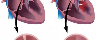
Actually a heart attack
Emergency condition. When it happens to a patient, there is little time for diagnosis. Usually the fact is stated after the initial treatment measures.
The chances of recovery depend on how quickly therapy is started. Through ECHO Kg, a focus of necrosis (death of cardiac structures) is noticeable. The smaller it is, the easier the treatment will be.
Heart sclerosis
Consequence of heart attack, inflammatory processes in cardiac structures. This is a condition in which certain areas of the myocardium become scarred.
Connective tissue of this kind is unable to contract or stretch. Therefore, part of the organ falls out of work.
The condition is dangerous because it provokes further worsening of dystrophy and problems with the nutrition of the heart. Lifelong treatment is required. Echocardiography shows both the focus of cardisclerosis itself and the degree of impairment.
Tumors
Neoplastic processes in a muscle organ, oddly enough, are quite rare. However, they pose a huge danger.
There are two reasons:
- The first is that even benign formations (for example, myxoma), having reached a size of more than 1 cm, compress and squeeze the heart. Hence the violation of the shape of the organ, dysfunction, and insufficient nutrition.
- Secondly, if the tumor is malignant, it grows through the muscle tissue. Which means it destroys them. Compression is also present, the harm is double.
Treatment is urgent, surgical.
Pericarditis
Inflammatory process. Provocateur - pyogenic flora and other agents. As a rule, streptococci or staphylococci are to blame. Rarely seen.
A special case is the accumulation of fluid in the pericardium - hydropericardium. If the pericardial sac fills with effusion and blood, local pressure increases. As soon as the indicator becomes equal to that inside the chambers of the muscular organ, cardiac arrest occurs. Therefore, the condition is classified as urgent.
Myocarditis
Inflammatory disease of the heart itself. Accompanied by severe pain. When scanning, foci of changes are noticeable. If not treated in time, consequences will occur like a heart attack. Perhaps even harder.
Cardiomyopathy
A typical pathological process for those who engage in intense physical labor. For example, for athletes. Alcoholics and heavy smokers are at increased risk.
The essence of the process is a change in the myocardium: the muscle layer grows, becomes excessively large, or stretches.
This is not normal and requires therapy. As a rule, medicinal. Plus lifestyle corrections.
Read more about the types of cardimopathy and treatment methods in this article.
Rhythm disturbance
Various. From atrial fibrillation to paroxysmal tachycardia. Echocardiography alone is unlikely to help here. To identify functional disorders, an ECG will also be required.
Changes in the anatomical position of the heart
For example, mirror (dextrocardia). It can be a defect or a natural and completely normal phenomenon.
Approximately such diagnoses can be made or confirmed based on the results of echography. In addition, other examinations are also needed. ECG, stress tests, bicycle ergometry, monitoring, etc.
Indications for use
Echo CG is performed on all patients with suspected heart disease.
Such people usually come to the cardiologist with complaints of palpitations, chest discomfort or pain, fatigue, swelling, and shortness of breath. These are typical symptoms of chronic heart failure.
Also, a person who has an ECG abnormality may be referred for an ultrasound scan, even if there are no unpleasant symptoms.
It is advisable to undergo echo CG for preventive purposes:
- once every 1–2 years for healthy children and young adults;
- once a year - for athletes;
- once every six months to a year for people over 60 years old;
- once every 6 months for those who suffer from chronic cardiovascular diseases and those who have small congenital heart defects that are asymptomatic;
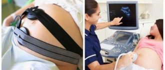
- all pregnant women (ultrasound of the mother’s heart is mandatory and, if indicated, ultrasound of the fetus’s heart).
Indications and contraindications
Since the technique is universal, there are quite a lot of reasons for ECHO CG.
- Chest pain of unknown origin. The patient is not always right in assessing his well-being. Discomfort occurs with stomach diseases, intercostal neuralgia and other conditions. The pain is successfully masked. But it is necessary to check the position of the muscle organ.
Read about how to understand that it is the heart that hurts and distinguish heart pain from others.
- Systematic increase in blood pressure. Hypertension does not happen out of the blue. Secondary forms are caused by kidney disease and hormonal dysfunction. And the primary ones develop in pathologies of cardiac structures. Therefore, you need to check the culprit using an ultrasound technique.
- Heart rhythm disturbances. Using the ultrasound method, organic changes can be detected. Doctors do not always receive information. Therefore, echocardiography is supplemented with an ECG, often also with 24-hour Holter monitoring. When an automatic device reads blood pressure and contraction frequency of a muscle organ throughout the day.
- Visible symptoms of possible heart disease. For example, cyanosis of the nasolabial triangle. Paleness of fingers, etc. Including shortness of breath. That is, those manifestations that usually indicate pathologies of a muscular organ. In this case, the technique is used as a preventive measure.
- Suspicion of a tumor. Indirectly, the same symptoms as above indicate a neoplastic process. Shortness of breath, weakness, blue discoloration of the area around the mouth, pallor, rhythm disturbances. Ultrasound gives a rough idea of neoplasia. The exact same result can be obtained through MRI.
- Exercise intolerance. Decreased tolerance. Accompanies angina pectoris and ischemic disease. An ultrasound is mandatory.
- Treatment being carried out. In this case, echocardiography is used to identify possible complications and side effects of therapy. Also as part of a preventive examination.
- Already established cardiological diagnoses. To identify deterioration (to study the dynamics of the disorder).
- Evaluation of treatment effectiveness. Including surgical.
Why is echocardiography performed?
EchoCG is used to identify changes in the structure of cardiac muscle tissue, dystrophic processes, malformations and diseases of this organ.
A similar study is carried out on pregnant women if there is a suspicion of pathology of fetal development, signs of developmental delay, the presence of epilepsy, diabetes mellitus, or endocrine disorders in the woman.
Indications for echocardiography may include symptoms of heart defects, suspected myocardial infarction, aortic aneurysm, inflammatory diseases, neoplasms of any etiology.
The study should be carried out if there are varicose veins and thrombophlebitis to exclude the danger of thromboembolism.
An ultrasound of the heart must be performed if the following symptoms are observed:
- chest pain;
- weakness during physical activity and regardless of it;
- cardiopalmus:
- interruptions in heart rhythm;
- swelling of the arms and legs;
- complications after influenza, ARVI, sore throat, rheumatism;
- arterial hypertension.
The examination can be done at the direction of a cardiologist or at your own request. There are no contraindications to its implementation . There is no special preparation for a cardiac ultrasound; it is enough to calm down and try to maintain a balanced state.
During the study, the specialist
- the state of the myocardium in the phase of systole and diastole (contraction and relaxation);
- the size of the heart chambers, their structure and wall thickness;
- the condition of the pericardium and the presence of exudate in the cardiac sac;
- functioning and structure of arterial and venous valves;
- the presence of blood clots, neoplasms;
- the presence of consequences of infectious diseases, inflammation, heart murmurs.
The results are most often processed using a computer program.
More details about this research methodology are described in this video:
Who is the study contraindicated for?
Contraindications are minimal, but they still exist.
- Lung diseases. Because patients with respiratory failure find it difficult to lie still for 10-20 minutes.
- Deformation of the sternum. For example, a hump. In this case, there will be problems with visualizing heart tissue.
- Inflammatory processes of the skin of the breast.
- Mental disorders. Excluding adequacy. For example, exacerbation of schizophrenia.
Contraindications are not absolute. Doctors are considering options to perform the procedure.
Carrying out an ultrasound
There are several ways to do an ultrasound of the heart. Transthoracic echocardiography is done through the sternum. The patient undresses to the waist and lies on the couch with his left side. This is the most comfortable position, since the top of the organ is as close as possible to the sternum and you can view all four chambers of the heart.
The doctor lubricates the sensor with special gel for better passage of ultrasonic waves through the body. Presses the device:
- to the jugular fossa, which is located above the sternum;
- to the left of it, where the upper impulse of the heart is located;
- in the V-intercostal space zone;
- to the chest under the xiphoid process.
After processing the data, a picture appears on the monitor in one-, two-, or three-dimensional versions (depending on the equipment and diagnostic method).
Trans-food diagnostics
For examination, a sensor is placed in the esophageal canal in the area of the left atrium. First, the doctor irrigates the person's mouth and throat with lidocaine. The patient is placed in a position similar to transthoracic ultrasound. For better glide and tight contact, the probe is pre-lubricated with gel.
A special mouthpiece is inserted into the patient's mouth. Endoxop is gradually introduced into the esophagus. This is a flexible, long, hollow tube with a sensor at the end. Through it, ultrasound is supplied and data is obtained.
Abdominal diagnostics
During an abdominal examination, a special gel is applied to the woman's abdomen. The sensor moves along the outer wall of the peritoneum. The anatomy of the heart muscle is examined for possible anomalies.
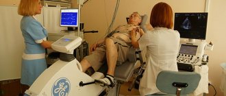
Stress echocardiogram
Before a stress echocardiogram is performed, the patient first undergoes a simple ultrasound of the heart. This is followed by a decoding of the results. After this, special sensors are attached to the patient’s body, which will continuously transmit all the changes that will occur during physical activity. The doctor may ask you to pedal an exercise bike or run on a treadmill.
The load is initially set to the minimum. If the patient copes well with it, then a gradual increase in pace should follow. At the same time, the doctor monitors the increase in blood pressure and the amplitude of heart contractions. If the patient's condition worsens, the study is stopped immediately.
Research quality
The quality of diagnosis depends on many factors. Excess body weight, pulmonary emphysema, severe deformities of the sternum and other anatomical features of the examined patient can distort the picture. This makes it difficult to see the image. In these cases, a transnutritive examination or MRI is recommended.
Using old equipment can let you down. Modern ones are more accurate, multifunctional and the most informative. The results largely depend on the experience of the diagnostician. He must be able to place the patient in the desired position and press the sensor to strictly defined points.
Types of Echo CG and their differences
There are several types of cardiac ultrasound. Basically, methods are divided according to the method of access to the muscle organ.
- Classic or transthoracic form, through the anterior wall of the sternum. This is the most common option. Gold standard for primary diagnosis. The ultrasound sensor is placed on the chest, after which the doctor changes its position. To visualize tissues in different projections and from several angles.
- The second option is an ECHO study with contrast enhancement. In essence, it’s the same transthoracic ultrasound. But this time special substances are injected intravenously. They accumulate in blood vessels, tissues and enhance the reflection of ultrasonic waves. Makes the picture clearer. In general, the method differs little from the previous one. From a technical point of view, everything is the same. But with contrast you can get much more information.
- Finally, transesophageal echocardiography. Invasive research. Due to its high complexity, it is performed only in a hospital setting. Moreover, problems and unforeseen disorders are possible afterwards. It is considered a particularly accurate method compared to others. The technique is used if previous modifications have not produced results.
Another way of classification is by the nature of the study.
- ECHO at rest. Occurs especially often. This is a typical cardiography.
- Assessment of cardiac condition after exercise. Appointed in controversial situations.
What is the essence of echocardiography
The ultrasound sensor has a built-in crystal that changes under the influence of electricity and produces high-frequency sound. Passing through the tissue and reflecting from them, the wave returns back in a distorted form. The sensor records this, and then the information is converted into electricity and displayed on the monitor screen.
There are several methods for performing echocardiography:
- Option M is one-dimensional. It allows you to accurately understand the size of the heart chambers and analyze the activity of the ventricles during contraction. The information is displayed in the form of a graph.
- Option B is two-dimensional. Allows you to identify tumors, aneurysms and blood clots. It shows the thickness of the walls and valves, and reveals the degree of contractility of the ventricles.
- Electrocardiography with Doppler. The test is used to look for heart defects and other dangerous disorders.
A Doppler study is performed to examine blood vessels.
Preparation
No special events needed. Conventionally, we can name the following requirements:
- The day before the test you should not smoke. Otherwise, the vessels will narrow and the doctor will detect false changes. In the coronary and pulmonary arteries.
- The same goes for alcohol. You should give up alcohol a few days in advance. To make the results more accurate.
- On the day of the procedure, you should not engage in intense physical activity. You need to follow a gentle regime. Peace is desirable.
- You must arrive at the appointed time. It is recommended to take a towel or disposable napkins with you. To remove excess gel after echocardiography.
Otherwise, no preparation is needed. You can do everyday, familiar things.
What is echocardiography
Echocardiography (EchoCG) is an ultrasound method for diagnosing morphological and functional changes in the heart, blood vessels, and valve apparatus. Echocardiography is mandatory for children in the first year of life. In the future, echocardiography is prescribed if indicated.
The basics of echocardiography are the reflection and detection of ultrasound waves. The sensor sends a pulse that is reflected from tissues of different densities. The device then captures the signals, converting them into a graphic image that is displayed on the monitor.
EchoCG is performed using several approaches:
- transthoracic;
- transesophageal.
Transthoracic echocardiography is the most comfortable for the patient. The heart is visualized through the chest. The study gives quick, accurate results and does not require special preparation. These facts make this technique preferable for identifying heart pathologies.
Transesophageal echocardiography provides more detailed visualization of cardiac structures. It is carried out to identify vegetation in infective endocarditis, diagnose aortic aneurysm, and also to assess the degree of mitral regurgitation.
Echocardiography can also be performed with or without contrast. Contrast echocardiography is used to diagnose congenital or acquired heart defects and also shows impaired myocardial perfusion.
EchoCG can be performed in various forms:
- in one-dimensional form, echo waves move along a single axis, you can visualize the walls of the heart and large vessels;
- two-dimensional echocardiography allows you to see the heart in two planes, you can evaluate the movements of individual structures;
- hemodynamic parameters are determined in Doppler mode.
At the moment, echocardiography is the leading method for diagnosing congenital heart defects in the compensation stage. Their identification is the basis for successful treatment.
Watch the video of the heart echocardiography process:
Additionally, read the article about ultrasound examination of the heart in a child.
Progress of the study
The patient goes to the office of a functional diagnostics specialist. Next, the procedure is carried out according to the usual scenario for many, like a regular ultrasound.
- You need to lie down on the couch.
- The doctor will lubricate the chest with a special gel. It conducts ultrasonic waves better, so the picture will be more accurate.
- The specialist places a sensor and begins to study the anatomical area.
- During the procedure, the doctor changes the position of the scanner and examines the organ from different angles. Works in several modes. Do not be afraid of the strange sounds that the device makes. This is fine.
- During the scan, the specialist may ask you to hold your breath. Roll over on your side. The patient's job is to follow the doctor's instructions. After the procedure is completed, you can go home.
Other modifications differ. If a contrast study is prescribed, a standard ultrasound is first performed, then contrast is administered and the procedure is repeated. Everything takes about 10-20 minutes. Plus or minus. Transesophageal ultrasound of the heart requires more time.
The echocardiogram is given to the patient after another 10-20 minutes. Because a specialist must give an opinion. Sometimes a person receives only a diagnostic protocol, without the doctor’s explanation.
Preparation for the procedure
If the patient does not additionally undergo an ultrasound of the abdominal cavity and genitourinary system, then he does not need special preparation. However, before the examination, it is advisable to wash the skin of the chest area with soap.
The examination is carried out in the ultrasound diagnostic room by a functional diagnostics doctor. The patient completely removes clothing from the chest and lies down on a special couch. A special gel is applied to the skin, which improves the passage of ultrasonic waves.
Decoding the results
The interpretation is carried out by the treating specialist. Cardiologist. Understanding what's what on your own is extremely difficult. Special medical knowledge is required. To an inexperienced person, the conclusion and protocol will seem like Chinese writing.
Attention:
The results need to be deciphered in a system and not individually. Cardiography alone is not enough to draw far-reaching conclusions. Although there are exceptions.
Normal indicators for an adult are presented in the tables:
Left ventricle and atrium
| Index | Men | Women |
| Myocardial mass | 85-220 g | 65-160 g |
| Volume at rest | 165-195 ml | 60-135 ml |
| Size during diastole | 35-55 mm | |
| Size during systole | 25-35 mm | |
| Left atrium size | 25-35 mm | |
| Ejection fraction | 55-70% | |
| Shortening fraction | 25-40% | |
| Rear wall thickness at rest | 8-11 mm | |
| Interventricular septal thickness at rest | 8-10 mm | |
Right ventricle and atrium
| Index | Meaning |
| Relaxed size | 75-110 mm |
| Pancreas wall thickness | 2-5 mm |
| Right atrium size | 25-45 mm |
| Right ventricle size | 20-30 mm |
| Thickness of the interventricular septum at systole | 10-15 mm |
| Thickness of the interventricular septum at diastole | 6-11 mm |
Blood flow speed
| Name | Index |
| Transmitral | 0.5 – 1.5 ms |
| Transtricuspid | 0.3 – 0.7 ms |
| Transpulmonary | 0.6 – 0.9 ms |
| Transaortic | 1 – 1.7 ms |
Other
| Index | Meaning |
| Normal fluid level in the pericardium | 10-30 ml |
| Aortic root diameter | 20-35 mm |
| Amplitude of opening of the aortic valve leaflets | 15-25 mm |
How is cardiac ultrasound performed?
EchoCG does not pose any difficulty for the patient; you just need to listen to the diagnostician’s instructions.
How is transthoracic echocardiography done:
- The patient needs to expose the upper part of the body, lie on the left side - this will allow you to get a clearer picture.
- The doctor applies a gel to the chest to improve acoustic contact.
- The diagnostician attaches sensors to various positions to examine in detail and record the condition of all departments.
Cardiac echocardiography is a simple and painless procedure
During stress ultrasound, a regular echocardiography is first performed, then sensors are attached to the patient’s body - they record all functional changes and transmit them to the screen. The person is asked to work on a cardio machine, and the loads are gradually increased.
Before performing a transesophageal ultrasound, the doctor treats the oral cavity with a lidocaine solution, after which the patient needs to lie on his left side, a protective ring is inserted into the mouth, and an endoscope is inserted.
Pros and cons of the diagnostic technique
There are many advantages of the study:
- Simplicity. An ECHO device is available in almost any district clinic. Even in the regions, not to mention the capital and large cities.
- Safety. The technique does not create harmful radiation exposure. The study can be carried out as often as the clinical case requires.
- High scanning speed. Everything takes about 10-20 minutes. Plus or minus.
- Information content. Despite the accessibility and simplicity of the technique, it is effective enough to detect most diseases.
- Non-invasive and painless. Not counting the transesophageal method. It is still classified as invasive. But, nevertheless, well tolerated. And the procedure is rarely required.
- Minimum contraindications. They are rather formal.
- Variability. Several scanning modes. For example, echocardiography with Doppler analysis is a way to study not only the heart itself, but also the vessels of the local circulatory network.
There is only one minus - ECHO CG does not provide accurate information in many cases. It is necessary to appoint auxiliary measures. Can this be called a negative trait? Hardly. Because the method copes with its tasks one hundred percent.
ECHO of the heart shows the condition of the myocardium, the entire organ and the local circulatory network. The initial parts of the aorta, pulmonary artery. This is a universal, safe and effective diagnostic technique.
What are the disadvantages
The methods have the same not only advantages, but also disadvantages:
- Limited resolution. This is explained by the longer ultrasonic wavelength compared to x-ray examination.
- Devices that propagate waves are calibrated by the average speed of transmission through tissue, although in reality the indicator is variable, which can slightly distort the picture.
These techniques are not usually used to examine the lungs.
- Tracing the inverse relationship between survey depth and resolution.
- Problems with the ability to study gas-containing organs and structures (lungs, intestines), which is why ultrasound or ECG are very rarely prescribed for them.