Tachycardia is characterized by an increase in heart rate from 95 to 150 beats per minute. The reasons for this phenomenon can be completely different. The simplest division of this phenomenon is:
- tachycardia as a pathology;
- tachycardia resulting from high physical exertion or emotional experiences.
Tachycardia is not a separate disease in itself. This is just a symptom of completely different diseases, such as pathologies of the endocrine system, autonomic nervous system and various forms of arrhythmia.
An attack of supraventricular tachycardia occurs suddenly.
Development mechanism
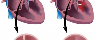
The natural center of heart automatism is the sinus node (first order pacemaker). The generation of an impulse from such an excitation source ensures rhythmic contraction of the atria.
Then the impulse passes to the AV node (pacemaker of the second order), which temporarily delays it and gives time to the atria to push blood into the ventricles; then the impulse passes through the His bundles, Purkinje fibers and enters the ventricles, contracting them and pushing blood into the aorta.
Failure in the functioning of pacemakers occurs due to structural changes in the heart muscle or activation of trigger factors - electrolyte imbalance, ischemic damage, exposure to certain medications. As a result of this, the atria contract faster - supraventricular tachycardia develops.
Supraventricular tachycardia occurs through two main processes:
- The re entry mechanism is the formation of an additional pathological path for conducting an electrical impulse, accompanied by re-entry and circular movement of the excitation wave; The re entry mechanism is most often diagnosed.
- The formation of ectopic (pathological) foci of excitation and the occurrence of trigger activity - such processes provoke the development of paroxysmal or ectopic forms of arrhythmia.
To clarify the mechanisms of development and determine ectopic foci of tachycardia, electrophysiological, often invasive, research methods are used . Some of them have been diagnosed - these are Bachmann's bundle, anterograde and retrograde pathways in the AV node.
Classification of supraventricular arrhythmia
The pathology is quite variable, so it is divided according to the nature of its manifestation into three types: tachycardia, bradycardia, extrasystole.
All extrasystoles differ in frequency, location and number of ectopic foci, as well as the time of occurrence of the attack.
By frequency
The frequency of premature contractions, which are counted per minute, divides all extrasystoles into:
- single - there are no more than five extrasystoles;
- paired - the ECG shows two extrasystoles located one after the other;
- group - premature contractions occur several in a row;
- multiple - more than five extrasystoles are determined.
According to the location of the outbreak
It can be determined directly in the atria or in the atrioventricular node. In any case, they talk about supraventricular arrhythmia.
Atrial arrhythmias are localized in the corresponding part of the heart
Atrioventricular arrhythmias are characterized by the location of an ectopic focus in the septum located between the atria and ventricles.
By number of outbreaks
When determining one ectopic focus using special research methods, they speak of monotopic extrasystole.
The presence of two or more pathological foci indicates polytopic extraordinary cardiac contractions.
By time of occurrence
Using an ECG, the time period during which the extraordinary contraction occurs is determined. There are three types of extrasystoles, distinguished by the time of formation:
- early - occur at a time when the atria contract;
- interpolated - the localization site is at the junction of the contractions of the atria and ventricles;
- late - characteristic of the period of ventricular contraction; they can also form during relaxation of the heart (in diastole).
Causes and risk factors
In some cases, paroxysmal supraventricular tachycardia occurs in clinically healthy people. It is considered physiological under the condition of rare and rapidly passing paroxysms with complete restoration of the sinus node.
But in 70% of cases, supraventricular tachycardia is pathological. Its causes can be both congenital and acquired organic heart lesions; Also among the causes are extracardiac pathologies.
Cardiac causes include the following:
- IHD (post-infarction cardiosclerosis, progressive angina);
- Heart defects;
- Chronic heart failure (CHF);
- Inflammatory lesions of the heart muscle - myocarditis;
- Cardiomyopathy;
- Pulmonary heart;
- Mitral stenosis.
Congenital heart abnormalities include Brugada syndrome and Wolff-Parkinson-White syndrome (WPU syndrome).
Extracardiac causes include:
- Pathologies of the endocrine system - thyrotoxicosis (increased production of thyroid hormones), pheochromocytoma (tumor of the adrenal glands, accompanied by increased production of adrenaline and cortisol);
- Electrolyte disorders – imbalance of minerals in the blood (magnesium, potassium, calcium, sodium);
- Overdose of drugs (antiarrhythmic drugs, antidepressants);
- Dyscirculatory disorders of the brain (against the background of vegetative-vascular dystonia);
- Anemia accompanied by very low hemoglobin levels.
In addition to the main causes, there are provoking factors for tachycardia, the presence of which significantly increases the risk of its development. Among them:
- Frequent stressful situations;
- Depression;
- Excessive physical activity;
- Drinking alcohol and drinks containing caffeine;
- Tobacco smoking.
Non-modifiable risk factors are female gender and family history.
Treatment
Without medical care, it is impossible to predict how the paroxysm will end if the patient has organic heart damage. Without cardiac pathologies, you can eliminate the attack yourself.
Emergency care involves the use of special medications:
- Calcium channel antagonists - normalize heart rate, reduce the conductivity of pacemakers. The main drug is Verapamil.
- Antiarrhythmic drugs - block biological reactions that increase heart rate (Novocainamide, Quinidine).
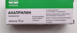
- Beta blockers - slow down the pulse and heart rate, reduce myocardial conductivity (Anaprylin, Egilok).
- Cardiac glycosides - reduce the conductivity of pacemakers, slow the pulse.
Sometimes an attack can be stopped without medication. For this purpose, various tests are carried out:
- Valsalvas - holding the breath and straining are done simultaneously.
- Ansher (eye-heart reflex) - gentle pressure on the eyeballs after three breaths while holding the breath for three, rarely six seconds. The pulse is measured one minute before the test and one minute after. This is also a diagnostic procedure.
- The patient is asked to breathe deeply, immerse his face in cold water, or squat down.
- Massage in the area of the carotid artery (carotid node).
Do not use for glaucoma, severe heart attack or stroke, decompensated heart failure.
The patient is hospitalized if emergency care does not have the desired effect, if the attack is prolonged, the course is recurrent, or a complication has developed.
Treatment of supraventricular tachycardia is aimed at eliminating risk factors, treating the underlying disease, restoring hemodynamics and heart rhythm, and preventing or eliminating complications.
The basis of treatment consists of medications of the same groups that were used to relieve paroxysm. Other drugs may be selected and the dosage adjusted. A dietary diet is prescribed excluding fried and fatty foods, salty marinades, coffee, and strong black tea. Alcohol is completely excluded.
As maintenance therapy, to prevent relapses, lipid-lowering agents are prescribed to correct lipid metabolism. Angiotensin-converting enzyme inhibitors and angiotensin receptor antagonists are used to treat hypertension and heart failure, and statins are used to prevent atherosclerosis.
Drug therapy is carried out under ECG control, which allows timely replacement of the drug or adjustment of the dose.
If therapeutic methods are ineffective, a surgical approach is used. In 98% of cases, severe prolonged paroxysms of SVT are relieved using transesophageal electrical stimulation. Laser therapy is used for the same purpose (destruction of “wrong” pathways).
In 95% of cases, uncomplicated SVT can be completely cured using radiofrequency ablation. The operation is minimally invasive and is done under general anesthesia. It involves passing a catheter with an electrode through the femoral artery to the heart. Using an electrode connected to an electrocardiograph, a pathological focus is detected. The area conducting the tachyarrhythmia is cauterized (40–60 degrees), as a result of which the pathological electrical impulse is blocked and normal conduction is restored. A scar forms at the site of the intervention. After the procedure, the electrode is removed and the wound is sutured. The manipulation lasts 1.5–6 hours. The patient is under observation for at least four days.
If indicated, a pacemaker (cardioverter-defibrillator - ICL or artificial pacemaker - IVL) is installed. The devices are installed in patients with severe forms of tachyarrhythmias, fibrillation, post-infarction cardiosclerosis, sustained bradycardia, atrial fibrillation, and unknown etiology of arrhythmia.
Surgical treatment is indicated if the patient cannot tolerate antiarrhythmic drugs, their effect is insufficient or absent, and there are contraindications to their use. For example, taking Verapamil is prohibited for WPW syndrome.
Classification
First of all, supraventricular tachycardias are classified depending on the location of the ectopic focus:
- Sinoarthrial form - the focus of excitation is located in the area where the sinus node is located.
- Atrial paroxysmal form - the pathological focus of excitation is located in the atria.
- Atrioventricular paroxysmal form - the ectopic focus is localized in the AV node.
Depending on the characteristics of the ventricular complex, tachyarrhythmias are divided into the following:
- Rhythm disturbance with a narrow QRS complex, where anterograde conduction of the impulse through the AV node is observed (with a supraventricular paroxysmal form).
- Rhythm disturbance with a wide ventricular complex, in which anterograde conduction of the electrical impulse is observed through additional pathways (with atrial and ventricular tachycardias).
The nature of the course of atrial tachycardia also varies. Based on this, several forms are distinguished:
- Paroxysmal form - characterized by the sudden onset and cessation of an attack that lasts from several minutes to 2 hours; in the absence of organic pathologies and the provision of adequate care, it has a favorable prognosis.
- Persistent form - characterized by prolonged attacks of up to several days: clinical signs appear more clearly, paroxysm of supraventricular tachycardia is more difficult for patients to tolerate.
- Chronic form - accompanied by an ongoing course, paroxysmal attacks are observed almost constantly; has a less favorable prognosis, as the risk of complications increases significantly.
At the initial stages, the paroxysmal form can be easily corrected by lifestyle; in this case, you can do without taking medications. While the chronic course requires a serious approach to treatment, often with surgical intervention.
In addition, atrial flutter and atrial fibrillation are separately distinguished, which are variants of atrial fibrillation - another form of supraventricular rhythm disorder.
Classification and types
There are many forms and types of NVT, divided according to different criteria. According to the course, they are divided into paroxysmal (occurring in paroxysmal) and non-paroxysmal (continuing continuously). The following forms of supraventricular tachycardia also occur: sinus, reciprocal, flutter and atrial fibrillation.
Paroxysmal reentrant AV nodal tachycardia is the most common form of SVT among young healthy people. You can read more about why it occurs and how it is treated here.
Paroxysmal supraventricular tachycardia
The concept of paroxysmal supraventricular tachycardia includes atrial (with the exception of fibrillation and flutter) and atrioventricular failures that occur in paroxysms. Separate identification of paroxysmal forms is of clinical significance, since these arrhythmias pose a greater threat to human life than continuously flowing ones.
The unification is also due to the fact that all paroxysmal SVT (with the exception of SVC syndrome) have common features:
- sudden appearance and equally sudden cessation of rapid heartbeat;
- higher heart rate compared to constant SVT;
- similar clinical symptoms accompanying the attack;
- identical treatment regimens to stop arrhythmia.
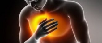
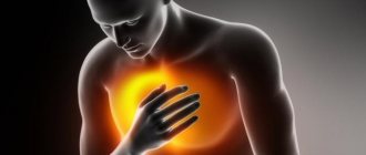
Most of my patients say that during an attack they experience a sharp heartbeat, discomfort, compression in the heart area, and general weakness. In more severe cases, when blood pressure decreases due to an irregular heart rhythm, the patient experiences dizziness, spots before the eyes - the so-called presyncope, and sometimes loses consciousness (fainting).
An attack of tachycardia may also be accompanied by sweating, nausea, and trembling in the body. I often meet patients who urinate more frequently after it.
In approximately 10-15% of patients during paroxysm, the main symptoms are abdominal pain, nausea, vomiting and frequent urge to defecate. Often they are even hospitalized in the surgical department with a diagnosis of “acute abdomen.”
Many of my patients do not feel any manifestations of rhythm disturbances at all, although when a cardiogram is taken, a paroxysm of supraventricular tachycardia is recorded in them.
In older people with cardiac disease, an attack can cause heart pain, worsening symptoms of heart failure (shortness of breath, fatigue, swelling in the legs), hypotension, pulmonary edema and shock. In such patients, it is important to correctly determine the source of the symptoms: they often suffer from a large number of pathologies, each of which may have similar manifestations. How not to get confused? The key is asking the right questions. It is necessary to detail each complaint and delve into the process of its origin. Do not forget about the patient’s external signs, which often help to quickly determine the main cause of the patient’s suffering.
Atrial tachycardia (AT)
This type includes NVT, in which the source of the rhythm disturbance is located in the atria. Depending on the number of these sources, monomorphic and polymorphic atrial tachycardias are distinguished.
About 10-15% of all cases of SVT are represented by PT. Minor PT events can also be observed in healthy people.
According to the clinical course, they are paroxysmal and constant, paroxysmal is more common. With a permanent form, a person may not experience any discomfort.
The predominant number of atrial tachycardias have the same causes as all arrhythmias. Sometimes they develop after atrial surgery. Polymorphic PT in most cases occurs with severe bronchopulmonary pathology.
The polymorphic variant is an unfavorable sign that increases the likelihood of death. However, the severity of the situation is often associated not with the arrhythmia itself, but with the course of the underlying disease that caused the tachycardia.
Consequences of a long-term non-paroxysmal course of PT: expansion of the cavities of the heart and deterioration in the ability of the myocardium to pump blood normally.
Clinical picture
The severity of clinical manifestations of paroxysmal supraventricular tachycardia depends on the heart rate and the presence of underlying diseases. Thus, paroxysms with a pulse of up to 150 beats/min are often asymptomatic and are tolerated quite easily by a person.
If an attack occurs with a pulse of more than 200 beats/min, a clinical picture occurs. The appearance of certain symptoms depends on the form of tachycardia, the body’s reaction and the presence of concomitant pathologies.
So, the occurrence of an attack is characterized by the following signs:
- The feeling of palpitations is the main symptom that occurs with any form of tachycardia;
- Difficulty breathing;
- Dizziness;
- Hand tremors;
- Increased sweating;
- General weakness.
With the development of paroxysm against the background of cardiovascular diseases, the following symptoms are added:
- Pain in the heart area of varying intensity, sometimes radiating to the left arm and shoulder blade (more often in patients suffering from coronary artery disease);
- Shortness of breath with difficulty breathing (observed in people suffering from CHF);
- Severe dizziness, syncope.
An attack of supraventricular tachycardia against the background of congestive cardiomyopathy or myocardial infarction can trigger the development of cardiogenic shock.
Causes of supraventricular arrhythmia
It was noted above that in the development of arrhythmia the primary role is played by a provoking factor, which can have a functional or organic origin.
Functional reasons:
- autonomic dysfunctions;
- excessive consumption of alcoholic and caffeinated drinks;
- irregular work schedule with insufficient rest;
- hormonal disorders (most often in women and adolescents);
- infectious processes accompanied by high fever.
Organic reasons:
- heart defects;
- myocarditis and endocarditis
- postoperative disorders;
- traumatic heart lesions;
- tumor processes located in the heart area;
- pulmonary heart disease, which developed against the background of pulmonary hypertension.
Some metabolic disorders, such as a lack of potassium, magnesium, and renal failure in the blood, can also contribute to the development of NFA.
The use of cardiac glycosides, sympathomimetics, theophylline and antiarrhythmic drugs in incorrect doses can cause supraventricular arrhythmia.
If the patient has a state of hypoxia, which often occurs with anemia, heart failure and bronchopulmonary pathology, the activity of the heart can be disrupted by the type of arrhythmia localized in the atria.
Diagnostics
Supraventricular tachycardia can be suspected by the feeling of palpitations, shortness of breath or dizziness. If such symptoms develop, you should consult a doctor who will conduct a comprehensive examination.
Clinical
Basic (clinical) diagnostics includes the following methods:
- Collection of complaints - information about the features of the clinical picture is clarified;
- Collecting anamnesis of life and disease - determining the nature of nutrition and lifestyle, risk factors
- Diseases, presence of concomitant pathologies; the features of the course of attacks are clarified;
- Physical examination - an assessment of the general condition, skin, and the functioning of the main body systems is carried out;
- Auscultation of the heart - a possible pulse deficiency is diagnosed, the frequency and rhythm of the heartbeat is assessed.
Additional
After collecting basic information, additional studies are prescribed:
- Clinical and biochemical blood tests are carried out to diagnose concomitant diseases.
- Blood test for hormones - diagnosis of hyperthyroidism, pheochromocytoma.
- Ultrasound of the heart and thyroid gland – diagnosis of organic pathologies.
- Holter monitoring is a daily study of the electrical activity of the heart using portable equipment, which is carried out to determine the characteristics of paroxysms.
- An electrophysiological study (EPS) of the heart - stimulation of the myocardium with physiological doses of current through special electrodes with recording of the response to an ECG - is carried out to diagnose rhythm disturbances.
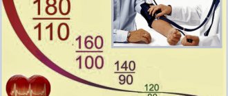
The main instrumental method for diagnosing tachycardia is an electrocardiogram (ECG), which also studies the electrical activity of the heart. All forms are diagnosed with an increase in heart rate (on average, from 150 to 250 beats/min), but other ECG signs will differ:
- Before the paroxysm of sinoarticular tachycardia, an atrial extrasystole is observed on the ECG;
- The atrial form is characterized by deformation of the P wave - it becomes negative, its amplitude decreases; possible prolongation of the PQ interval (sign of 1st degree AV block);
- In case of tachycardia against the background of VPU syndrome, deformation of the QRS complex, a decrease in the PQ interval and the appearance of a delta wave are diagnosed;
- In the case of atrial flutter or fibrillation, the absence of P waves is observed - f waves are formed instead; the ventricular complex remains normal, but the RR distance is different.
Differential diagnosis is carried out with ventricular forms of tachycardia, sick sinus syndrome, and ventricular overexcitation.
Prevention of supraventricular arrhythmia
There are certain principles for the prevention of rhythm disturbances that apply to all forms of arrhythmia, including supraventricular;
- Proper nutrition should consist of lean meats, fish, and a sufficient amount of plant foods should be included in the diet.
- Physical activity must be at an acceptable level for the patient so that the heart is supplied with sufficient oxygen and nutrients.
- Maintaining blood glucose and total cholesterol levels within normal limits. Also, body weight must correspond to age and physiological norms.
Video Therapy of extrasystoles and supraventricular tachyarrhythmias
4.75 avg. rating ( 93 % score) - 4 votes - ratings
First aid
If paroxysm occurs, first aid consists of the following techniques:
- Unfasten the tight collar, remove the scarf from your neck;
- Provide access to fresh air - open a window or vent;
- Help the person lie down and calm down;
- Measure blood pressure and heart rate.
To relieve paroxysm, the use of vagal tests is effective:
- Massage of the carotid sinus, which is located in the area of the carotid arteries;
- Holding your breath while straining at the same time (Valsalva maneuver);
- Pressing on closed eyeballs for several seconds (Aschner test);
- Holding your breath and lowering your face into a basin of cold water;
- Artificially induced cough and gag reflex.
If such tests are ineffective, supraventricular tachycardia is stopped with Anaprilin (20-40 mg sublingually) or Verapamil (40-80 mg orally). The dosage of the drug depends on hemodynamic parameters (heart rate and blood pressure).
Paroxysm and first aid
The attack begins as follows:
- The patient feels pressure in the chest, a sharp push.
- Significantly at one moment the heart rate increases to 150-200 beats per minute.
Then the accompanying symptoms of the syndrome appear. It is better to start measures of primary, pre-hospital influence on your own. But calling an ambulance is necessary; you won’t be able to eliminate the phenomenon completely on your own.
How to act in such a situation:
- Remove tight clothing from the neck, loosen collar or jewelry.
- Open a vent or window to provide fresh air.
- Measure blood pressure and heart rate.
- Take a tablet of Anaprilin and Verapamil, immediately and together. They will allow you to quickly stop the attack and restore the functional activity of the heart. Do not exceed the dosage, it can be dangerous.
- Try using vagal techniques. Deep breathing (5 seconds per movement), pressing on the eyeballs (only if there are no ophthalmological problems).
- Lie down and relax. Do not make sudden movements, cardiac arrest is possible.
Do nothing else until the ambulance arrives; complications are likely. Next, the issue of transporting the patient to the hospital is decided.
Treatment of supraventricular tachycardia
Therapy for supraventricular tachycardia includes taking medications and using physical therapy techniques. Surgery is performed according to indications.
Conservative treatment
The basis of conservative treatment is medications. The choice and dosage of tablets depends on the form of tachycardia, the presence of background pathologies, and the individual characteristics of the body.
Treatment includes a combination of beta-blockers and antiarrhythmic drugs.
From the group of beta blockers, one of the following drugs is prescribed:
- Bisoprolol 2.5-5 mg 1 time per day;
- Atenolol 25-50 mg 1 time per day;
- Metoprolol 100-200 mg 2 times a day;
- Anaprilin 20-40 mg 2-3 times a day.
Among the antiarrhythmic drugs, the drugs of choice are:
- Amiodarone – 200 mg 1 time per day;
- Diltiazem – 60 mg 3 times a day or 90 mg 2 times a day;
- Propafenone – 150 mg 3 times a day.
In case of VPU syndrome, the use of Verapamil and cardiac glycosides is contraindicated.
As an emergency medical aid to relieve paroxysm, medications intended for intravenous administration are used:
- ATP – 5-10 mg intravenously as a bolus for several seconds;
- Verapamil – 5 mg IV bolus, repeat the dose if necessary;
- Novocainamide 10% - 10 ml IV slowly over several minutes or Digoxin 0.025% 0.5 ml IV slowly.
All drugs are administered in dilution with 0.9% sodium chloride or 5% glucose under continuous monitoring of hemodynamic parameters.
Drug therapy is often carried out in conjunction with physiotherapeutic methods: rubbing, dousing, circular shower, hydromassage baths.
Surgery
Another treatment option for tachycardia is surgery. Indications for surgical intervention are:
- Continuously recurrent paroxysms;
- Poor tolerance of attacks by the body;
- Ineffectiveness of drug therapy;
- Contraindications to taking antiarrhythmic medications.
Currently, the main surgical method for treating tachyarrhythmia is radiofrequency ablation (RFA), which is one of the methods of endovascular surgery.
The operation is performed under local anesthesia as follows: a catheter is installed in a peripheral vein and an electrode is inserted through which the ectopic focus is cauterized and an artificial zone of necrosis is created, after which it can no longer create pathological impulses.
Advantages of RFA:
- Minimally invasive surgery, which reduces the risk of postoperative complications;
- Short rehabilitation period due to good tolerance and rapid recovery;
- The procedure is painless - unpleasant sensations arise only at the time of catheterization.
The disadvantages of RFA are the various consequences of surgical intervention. Among them are violation of the integrity of the myocardium, thrombus formation, pulmonary artery stenosis, bleeding from a catheterized artery.
Less commonly, surgery to install a pacemaker (pacemaker) is performed. A special device is implanted into the heart, which plays the role of an artificial pacemaker. The supply of its electrical impulses prevents the occurrence of a wave of excitation from pathological foci, preventing the development of paroxysm.
Treatment methods
The therapy is complex. It is represented by medications, surgery, and lifestyle changes.
Use of drugs. Funds from the following groups are assigned:
- Cardiac glycosides. Lily of the valley tincture, Digoxin. Normalize the frequency of organ contractions.
- Calcium antagonists. Verapamil or Diltiazem. Only one thing.
- Potassium and magnesium complexes. At the discretion of the doctor. Asparkam and others.
- Carvedilol, Anaprilin, Metoprolol, Amiodarone.
These are the first methods of restoring rhythm in pathologies of low intensity (persistent tachycardia and the onset of chronic).
Other medications are added as needed: statins, vitamins, antithrombic drugs, and those that normalize the rheological properties of blood.
Surgical therapy is represented by radiofrequency ablation. The focus of the atypical signal is cauterized. Open operations are required for heart defects or severe abnormalities in the functioning of the organ due to myocarditis and destruction of cardiac structures.
For your information:
Supraventricular tachycardia is NOT treated with folk remedies. This is a waste of energy and time.
Lifestyle changes are required in the early stages and can be the main method of eliminating processes.
- Quit smoking and alcohol, especially psychoactive components.
- Normalization of sleep: 8 hours per night.
- Walking, physical activity or exercise therapy every day.
- Drinking regimen - 2 liters.
- Adequate nutrition. Everything is possible, but with restrictions and in reasonable quantities.
- Salt no more than 7 grams.
It is better to discuss steps to normalize the condition with a cardiologist. Also with other specialists, depending on the origin of the problem.
Forecast
Paroxysmal supraventricular tachycardia has a different prognosis and depends on the following factors:
- Features of the course of tachycardia;
- Causes of tachycardia;
- The number of risk factors that provoke paroxysmal attacks;
- The age of the person;
- Presence of concomitant diseases;
- Individual characteristics of the body.
For rare paroxysms in middle-aged people who do not have organic heart damage, the prognosis is favorable.
Elderly people suffering from coronary artery disease, heart failure or chronic pulmonary diseases have a less favorable prognosis - in such cases the risk of complications and death increases significantly.
Causes
The causes of the development of all types of arrhythmias overlap. Sustained arrhythmic episodes are always the result of an underlying abnormality: most often these are diseases of the cardiovascular and nervous system, or problems with blood supply.
Factors contributing to the development of arrhythmic syndrome:
- smoking tobacco;
- hormonal imbalance;
- consumption of alcoholic beverages;
- abuse of caffeine or other psychostimulants;
- taking certain medications that cause general overexcitation of the nervous system;
- genetic risks (congenital predisposition);
- consequences of injuries in the thoracic area;
- lack of vitamins and microelements;
- electrolyte ailments;
- stress.
Main concomitant diseases:
- hypertension, hypertension;
- autonomic disorders;
- diabetes mellitus type 1 and 2;
- hypoxia in various types of anemia;
- renal failure;
- congenital or acquired heart defects;
- endocrine diseases;
- rheumatic heart disease;
- cardiac ischemia.
Complications
The vast majority of complications of supraventricular tachycardia are associated with increased thrombus formation. Thromboembolism (blockage of a vessel with a blood clot) leads to the following conditions:
- Myocardial infarction – acute ischemia of the heart muscle followed by necrosis; manifests itself as chest pain of varying intensity, a feeling of fear, and shortness of breath.
- Cerebral infarction (ischemic stroke) is an acute disorder of cerebral circulation, the symptoms of which may be impaired speech and swallowing, paresis of the limbs on one side, facial asymmetry, and impaired consciousness.
- Pulmonary embolism (PE) is a sudden occlusion of an artery, accompanied by acute hemodynamic disorders; clinically manifested by chest pain, shortness of breath, dizziness, drop in blood pressure and increased heart rate.
In addition, an attack of tachyaarthymia can lead to the development of arrhythmogenic shock - circulatory failure with a sharp decrease in cardiac output and hemodynamic disturbances.
Its main symptoms are severe pallor of the skin, impaired consciousness, decreased diuresis, clammy sweat, decreased blood pressure and increased pulse.
Symptoms of the disease
The vast majority of patients experience a paroxysmal increase in heart rate. In this case, the attack develops suddenly and ends the same way. Its duration varies from 2 - 5 seconds to 3 days.
At first, a blow to the heart is felt, which gives way to a strong heartbeat. The contraction frequency can exceed 200 beats per minute.
At this time, the following complaints appear:
- headache and heart pain,
- dizziness,
- intense sweating,
- nausea,
- fainting state.
After attacks, a lot of light-colored urine is released. This occurs due to both activation of the sympathetic nervous system and increased production of atrial hormone. It removes sodium from the body and is formed when there is high pressure in the right atrium, which is noted at the time of crisis.
Treatment of supraventricular tachycardia
- For paroxysms of an unstable nature with an unexpressed clinical picture, drug therapy is not carried out.
- To relieve the symptoms of supraventricular tachycardia, calcium channel blockers and beta-blockers are prescribed.
- A decrease in heart rate is achieved by administering digoxin; it is only contraindicated in case of glycoside intoxication.
- In ⅔ of patients, the attack is stopped with the help of antiarrhythmic drugs IA, 1C or III.
Drug therapy is ineffective in the case of refractory supraventricular tachycardia. Then atrial programmable pacing is used.
Today, radiofrequency ablation is considered a radical treatment method for supraventricular tachycardia. Using minimally invasive surgery, ectopic lesions that cause disruption of the normal heart rhythm are removed.
How to restore heartbeat?
The patient can eliminate the paroxysm of supraventricular tachycardia on his own using reflex tests, which involve stimulation of the vagus nerve. If activated, the heart rate will slow down. If paroxysm occurs, it is recommended to perform the following actions:
- hold your breath and strain at the same time;
- apply gentle pressure on the eyes for 5 seconds for 2 minutes;
- inflate a balloon;
- imitate gagging.
Return to contents