Modern medicine has stepped far enough forward and now everyone can use one or another high-tech methods in order to prevent or detect any disease at the initial stages.
New medical equipment allows us not only to look into the very depths of the human body and nature, but also to do it as painlessly as possible, so that the patient has only positive emotions from the procedure.
General information about Doppler ultrasound
to radiography and ultrasound (ultrasound) in studies of the human body for diseases . Both methods are constantly being improved and are capable of producing clear and accurate results. It is difficult to imagine how many human lives are saved by each of these research methods.
The most difficult task for doctors has always been the identification of vascular diseases and the functioning of blood in the body . After all, it is deviations in the functioning of blood vessels that can lead to serious diseases, which in some cases result in death. The venous network is several times more microscopic than the organs of the human body, which means it is much more difficult to study.
But the modern method of Doppler ultrasound is capable of identifying and visualizing deviations in the movement of blood through the vessels and predicting possible diseases.
It is worth learning more about what it is before contacting an ultrasound scan yourself.
Indications for use
You can understand what MAG ultrasound is, what it is and why the procedure is needed in general if you know the situations in which it is prescribed. Doppler ultrasound may be prescribed if:
- frequent headaches;
- cerebrovascular insufficiency of any form;
- the need to plan operations;
- compression of blood vessels due to pathologies of surrounding external tissues or prerequisites for it;
- pathology of the jugular veins or suspicion of it.
Doppler ultrasound of the great vessels may be prescribed if the patient is at risk. This includes people with hypertension, diabetes, and also if the person is a smoker or is obese.
Transient ischemic attacks
This is an acute form of ischemic cerebral blood supply disorder. An episode of such disorders can be triggered by ischemia of a specific area of the spinal cord or brain. These attacks are characterized by the fact that they are not accompanied by a stroke, since otherwise they would be classified differently.
Hypertonic disease
You will have to find out what MAG ultrasound is if a person suffers from high blood pressure. Hypertension is dangerous because the load on the organs increases significantly and hemorrhage can occur. It is important to determine whether hypertension is primary or secondary (caused by certain factors).
Vascular malformations
They are congenital and represent the so-called vascular glomeruli, which consist of abnormal vessels intertwined with each other. They can cause the development of steal syndrome or begin to compress the brain tissue.
Neurological reversible deficit
Otherwise, the condition is called a “minor stroke.” It is characterized by the fact that in the acute form, cerebral circulation is disrupted. Although after a minor stroke, impaired functions are restored within about one month, it is best to prevent the condition and select adequate treatment. USDG MAG
(what it is was said above) will provide sufficient information about the condition of the vessels and the presence of abnormalities.
Previous strokes
People who have had a stroke are at risk. For them, undergoing an ultrasound scan is a necessity, since this procedure allows them to keep their health condition under control and prevent a possible relapse.
Cerebral atherosclerosis
The disease is dangerous because it affects the arteries of the brain and leads to dementia, or vascular dementia. The reason for the development of this condition may be the formation of atherosclerotic plaques, around which connective tissue grows, and calcium salts are deposited on the walls of the vessel. Dopplerography of the great vessels of the head can be prescribed to people after 60 years of age, since the development of this disease most often occurs at this age.
#!UZIseredina!#
What is it used for and what does it show?
Ultrasound Dopplerography of blood vessels is carried out both according to a doctor’s indications and for the purpose of self-monitoring of one’s health. This procedure is based on the Doppler technique , as is the operating principle of all ultrasound machines.
The procedure involves sound waves , but do not be afraid of them, they are absolutely safe for the body (ultrasound is prescribed even to pregnant women). The waves pass through the tissues of the human body, but are reflected from moving objects. In this case, we are interested in the flow of blood in the vessels.
An image is transmitted to the monitor that lines up the vascular network and shows in two colors how and at what speed the fluid moves, whether there are any difficulties or obstructions (blood clots) in its path, and also, you can clearly see the blood flow itself.
Modern Doppler ultrasound devices are capable of giving the doctor performing the research a complete picture of the functioning of your body , which is what you will use as a basis when choosing treatment.
This study is prescribed for both adults and children . The primary task of such a procedure is to identify the presence of disturbances in the body’s blood supply system, or to refute their existence.
Having a particular disease, people want to establish its causes in order to choose the right treatment, and Doppler ultrasound also helps to understand this issue.
The study helps to identify the following deviations from the norm:
- increased or decreased speed of blood flow;
- increased intracranial pressure;
- vascular lesions of an inflammatory or atherosclerotic nature;
- vascular abnormalities;
- obstruction of patency;
- pathological tortuosity.
This method is recognized as the fastest and most painless way to check blood vessels for proper functioning of the blood in them. In terms of effectiveness, it can only be compared with surgical intervention, which many people try to carefully avoid.
Vessels of the cervical spine and their diagnosis
The most important arteries that supply the brain and neck are the carotid arteries. They are separated by two common carotid arteries (internal and external).
The external carotid artery supplies the facial part of the head and neck. This artery also has its own branches (anterior, posterior and medial). It is located in the area of the parotid gland.
The internal carotid artery is responsible for feeding the brain and eye area. The internal artery is divided into cavernous, cerebral, cervical and petrosal.
In addition, the posterior and middle arteries of the brain are also distinguished, which are responsible for blood circulation in the cerebral hemispheres, temporal and occipital lobes.
The appearance of pain in the neck may indicate problems with the vessels of the cervical spine. Ultrasound examination of the vessels of the cervical spine will help diagnose this disease.
Experts recommend regularly undergoing ultrasound of the neck as a preventative measure. Such measures will reduce the risk of ischemic stroke by 80%
With the help of ultrasound, a specialist has the opportunity to assess the general condition of your blood vessels and determine whether there is a possibility of disturbances in blood circulation.
Ultrasound is also used to determine:
- Is there a violation of the outer lining of blood vessels, arteries and veins (tissue condition)
- Compliance of the vessel lumen with standards (lumen diameter)
- Vessel patency
- Vessel geometry
- Condition of the vascular network
- Aneurysms
The scope of application of ultrasound includes research:
- Facial vein
- Subclavian artery
- Internal and external carotid arteries
- Jugular veins
- Basilar artery
- Arteries of the spinal column
Read about hypoplasia of the left vertebral artery - causes, symptoms and treatment here.
This procedure also has another, more precise name, Doppler ultrasound (USDG). Doppler ultrasonography involves a combination of classical ultrasound with Dopplerography (visual identification of blood vessels)
Methods used, which parts of the body are examined
When performing Doppler ultrasound, the capabilities of two devices are used at once - Doppler and an ultrasound sensor. This paired work is determined by achieving the desired effect: the ultrasound sensor shows how the vessels are located, what their outlines, sizes and contours are , and Doppler allows you not only to see, but also to hear the movement of blood through the vessels, and also to understand its consistency.
During the procedure, many
patients hear a characteristic whistle. Some people attribute it to a feature of the equipment, but in reality it is the sound of blood moving through the vessels.
The vessels of the head and neck (brachiocephalic arteries) are mainly examined by ultrasound. It is in these areas of the body that the most important areas of the blood supply are concentrated, by studying which one can identify a predisposition to certain diseases.
Periodically, patients are prescribed ultrasound scanning of the vessels of the lower extremities or arms. The procedure can be performed on any vessels of the body.
Indications and contraindications
This study is absolutely painless and harmless , therefore it is recommended to be carried out both for completely healthy people, for the purpose of prevention, and for a clear diagnosis of those who suffer from any diseases.
Doppler ultrasound has no age restrictions and can be prescribed to both newborns and older people.
There are no contraindications for this study , however, if you cannot remain in a lying position for a long time (small child, sore spine), then it is better to clarify this point with the doctor conducting the study to find the optimal solution to the problem.
If we talk about indications for conducting research, there are a huge number of them.
Today, diseases associated with blood vessels occur in a huge number of people, mainly due to an unhealthy, inactive lifestyle. Also, heredity, poor nutrition and poor ecology play a role.
A visit to Doppler ultrasound will be useful for people suffering from:
- aneurysms;
- thrombosis;
- frequent migraines and headaches;
- vegetative-vascular dystonia;
- dizziness;
- fatigue;
- noises in the head;
- tinnitus;
- motion sickness;
- weather dependence;
- fainting;
- sleep disorders;
- cerebral circulatory disorders;
- after traumatic brain injury;
- frequent nervous breakdowns.
If we talk about children, then in addition to the above problems, we can add to the list:
- restlessness;
- decreased attention;
- poor memory;
- delay in the development of the speech apparatus;
- rapid decrease in vision.
Putting off a visit to the doctor is dangerous for your health and life, because the consequences of the symptoms that appear are critical.
Preparation rules
Despite the fact that the procedure is painless and does not represent anything complicated, it is necessary to pay attention to preparation.
It is better to carry out Doppler ultrasound , but it is not always possible to end up in the hospital in the morning. If your appointment is scheduled for lunch and evening, do not deny yourself food, only limit the consumption of foods rich in glucose.
A week before the proposed study, you must give up alcohol and cigarettes . These negative factors affect the condition of the blood vessels and cannot contribute to giving an objective assessment of what is happening.
The day before the procedure, you should not take vascular medications, as well as sedatives and sleeping pills.
Otherwise, the Doppler ultrasound procedure has no requirements for the patient, except calmness and the desire to get to the bottom of the truth.
How does the procedure work?
How to do an ultrasound scan of blood vessels - step by step:
- You need to sit comfortably on the couch so that your arms are extended along your body and your gaze is directed clearly upward.
The legs should be brought together, the back should not be tense. Do not tilt your head, try to fix it by resting the back of your head on the couch. - The doctor performing Doppler sonography will apply a special gel to the areas being examined (neck, head), which will help in conducting the study. Therefore, grab napkins in advance or after the procedure, ask your doctor for them.
- You will have sensors attached to you that will read information about your heartbeat and breathing.
- Using a sensor, the areas where the gel is applied will be carefully examined, and the image will be displayed on the screen.
- The doctor may ask you to do certain things. For example, hold your breath, turn your head slightly, breathe frequently, raise your arms, strain your neck muscles. Sometimes, the specialist himself can press on certain points on the neck and head to check the patency of the blood flow.
In total, the procedure can last from twenty minutes to one hour . Time is allocated individually, depending on your health condition and the ability to conduct research.
How is Doppler sonography done?
The Doppler ultrasound procedure is done as follows:
- The patient should lie down on the couch and take a comfortable position. The position can be changed at the request of the doctor, it depends on the area being studied.
- The desired area of the body must be left without clothing.
- The doctor applies a special gel to the ultrasound sensor. In paid clinics, the client is offered napkins to wipe it off; in free clinics, most often you are required to bring your own.
- The device is applied to the patient’s skin, and the necessary organs are viewed.
- The ultrasound specialist may require the patient to change the breathing rate and tension of certain muscles. This is necessary for analyzing the blood flow of peripheral vessels and large arteries.
- The readings received through the sensor are recorded by a specialist and deciphered for the patient.
Principles for interpreting results
You can get a transcript of the results within a few minutes after the procedure . When examining your health condition, the location of blood vessels and arteries, their size, patency, homogeneity, as well as the consistency of the blood and the speed of its passage are assessed.
Upon completion of the study, a summary table is compiled, which shows your indicators according to these criteria , and also leaves a transcript of what is within the normal range and what is unacceptable.
The result of Doppler ultrasound is the basis for contacting a doctor with a request to prescribe treatment if the study reveals abnormalities.
By carrying out a similar procedure annually, you can monitor the dynamics of the functioning of the circulatory system, which will allow you to identify the risk of certain diseases in old age.
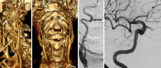
Duplex and triplex
With Doppler ultrasound, an ultrasound scanner performs Dopplerography in duplex or triplex. First, we find the vessel in B-mode, then to obtain information about the blood flow, we turn on the color Doppler mode, then in its lumen we set the area of the required scanning depth (control volume mode for data measurement) and obtain the flow spectrum. Duplex scanning is a combination of any two scanning modes (B + spectral or B + color Doppler), triplex scanning is the simultaneous use of all three modes (B-mode + spectral + color).
Dopplerography picture of cerebral vessels using triplex mode
As we see, ultrasound and Doppler Dopplerography of the main arteries (vessels) of the head are carried out primarily to detect anomalies in the circulatory system, measure its pathogenetics and hemodynamics. This helps in monitoring the effectiveness of treatment, angiography and the involvement of a vascular surgeon when considering surgical treatment.
Establishing diagnosis
Your doctor should pay close attention to the results obtained from Doppler ultrasound. Thanks to this diagnosis, you can identify:
- aneurysms;
- diseases of the arteries of the extremities;
- diseases of the arteries of the head and spine;
- thrombosis;
- diseases that contribute to occlusion of the carotid arteries;
- varicose veins
All of these conditions require treatment, so once diagnosed, you should be prescribed appropriate medications.
It’s great if an ultrasound with Dopplerography of blood vessels is performed at a certain interval . Then, seeing the dynamics of your condition, the doctor can not only make a diagnosis, but also predict your future condition. In this case, you will be given recommendations on how to keep your body healthy.
Learn more about Doppler ultrasound, as well as its differences from duplex and triplex studies, from the video:
Average prices in Russia and abroad
The cost of ultrasound examination of veins and arteries may vary depending on the region of our country . In addition, the price may depend on the equipment used to conduct the study. The newer, more expensive and better quality the device, the higher the cost. What can we say about the professionalism of the doctor - an ultrasound diagnostic specialist.
The average cost of the procedure varies from 2000 to 3400 rubles. But remember that price does not always mean quality.
If you are able to have this procedure done overseas, you can expect to spend between $50 and $80 for the procedure (in Europe) and between $45 and $65 in the US .
Our health is a very fragile thing, and we cannot always see or feel the beginnings of the disease. Today there are thousands of ways to check your condition and Doppler ultrasound is one of the innovative ones .
This type of diagnosis is available to everyone, so do not delay your visit to the hospital. Perhaps today you will see the makings of some kind of illness on a black and white monitor and cope with them in a matter of days. Be healthy and take care of yourself.
Two research modes
Scanning the main arteries of the head is a high-quality way to adequately assess the condition of the cerebral vessels and potential risks and threats to human health. This method can be used if necessary in several modes:
- two-dimensional mode;
- duplex scanning.
Two-dimensional scanning is also called B-mode. It allows you to determine the location of blood vessels, deformations of the blood flow that are significant for hemodynamics, the structure of the walls of the vascular system, and the presence of atherosclerotic plaques or blood clots. Also, at the first stage of the examination, the doctor determines the thickness of the inner and middle linings of the arteries, this is the so-called intima-media complex (IMC). The specialist observes and describes the degree of differentiation of the CMM into layers or its absence. When plaques are detected, the doctor uses ultrasound to determine their structure, surface contour, height and extent, as well as the degree of narrowing of the lumen of the vessel. At the same stage, it is possible to describe the place of attachment of the base of the thrombus, its structure and size, and signs of flotation (mobility). This allows us to draw conclusions about the potential threat of fragmentation (separation) of a blood clot, blockage of the distal branches of blood vessels with subsequent ischemia of the organs supplied by these vessels.
If necessary, three-dimensional duplex light scanning is then used to examine the direction and nature of blood flow in the veins and arteries, determine the speed, peripheral resistance index, pressure gradient and other indicators.
Doppler ultrasound is a modern, accessible, informative method for examining the vascular system of the brain.