Clinic
Most often, at the initial stage, the inflammatory process occurs against the background of a slight increase in body temperature. The patient experiences general weakness and malaise, myalgia.
The symptom complex occurs against the background of chest pain. Pain is most often observed in the left chest area, but can radiate to the trapezius muscle. In some patients, the pain is pleural in nature and intensifies during deep inspiration. For most people, the pain decreases when sitting, especially if the body is tilted slightly forward. Many people hold their breasts and constantly try to put something on them, for example, a pillow or other object.
In addition, the presence of fluid in the heart is accompanied by:
- dry cough;
- pale skin;
- shortness of breath in a horizontal position;
- anxiety.
In severe cases of constrictive pericarditis, many patients experience swelling in the legs. This is due to the fact that the heart muscle cannot cope with the incoming volume of blood. If there is no treatment, there is a huge risk that pulmonary edema will begin.
Preventive measures
After correct and timely treatment of pericarditis, there will be no trace of this pathology. But there are times when the disease is too advanced. For example, with tamponade, the heart can completely lose its pumping function. The fluid around the pericardium compresses the heart muscle so much that it is unable to eject blood. If treatment is started correctly, normal heart function can be restored within a few months.
Sometimes pericarditis is diagnosed in a fetus that is still in the womb. Doctors are able to notice such changes using ultrasound as early as 20 weeks of pregnancy.
Important! A fetus may be diagnosed with pericardial effusion if there is increased coronary blood flow or increased abdominal volume. In this case, appropriate treatment and therapy is prescribed.
Pericarditis can recur, for example, in the case of an incompletely eliminated disease. Do not think that a common cold or flu is not capable of causing great damage to the body. On the contrary, when such viral diseases are not completely cured, the likelihood of the proliferation of pathogenic microorganisms only increases. They remain in the body for a long time. This is especially true for various oral infections. Caries or stomatitis can also cause an inflammatory process, since these diseases are provoked by bacteria.
Causes
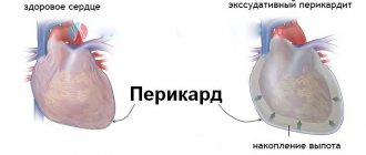
What does fluid in the heart mean, what is the danger? In normal condition, the two-layer shell of an organ should contain no more than 30 milliliters of liquid. If the amount of this fluid increases, swelling appears. Pericarditis can appear not only against the background of an increased amount of fluid, but also due to the appearance of adhesions and other inflammatory processes.
Most often, the pathology appears against the background of a viral disease, more precisely, viral pericarditis, or after the flu. The pathology occurs quite often in HIV-infected individuals. There is a possibility of the disease developing against the background of a parasitic, fungal or bacterial infection.
Fibrinous pericarditis most often occurs against the background of the presence of Staphylococcus aureus in the body. This form of pathology, on the contrary, is characterized by the fact that the fluid leaves, therefore this type is also called dry pericarditis. Fibrin is deposited on the petals, they become dull and may become covered with villi.
Most often, in the absence of proper treatment, fibrinous pericarditis turns into an exudative or constrictive form, and these types of pathology already threaten the patient’s life.
Symptoms of the disease
Signs of pericarditis depend on the form and stage of the process.
Acute inflammation of the pericardium usually produces fibrin secretions, and as the process progresses, inflammatory fluid accumulates.
Considering that inflammation of the cardiac membrane is rarely an isolated disease, its occurrence may go unnoticed.
The severity of the symptoms of the process depends on the amount of fluid in the lining of the heart, on the speed of appearance of this fluid and on the severity of the underlying disease.
Given the inflammatory nature of the process, at the onset of the disease there may be symptoms such as fever, severe general weakness, muscle pain, and headaches.
Rare causes of pathology development
Rarely, but still, a number of diseases can provoke the appearance of fluid in the heart muscle:
- tumors;
- Dressler's syndrome, after a heart attack or surgery;
- Mediterranean fever;
- blood diseases;
- renal failure;
- long-term treatment with certain medications;
- problems with the thyroid gland;
- injuries in the chest area.
And in some patients it is not possible to establish the cause of the pathology at all.
Fluid in the heart appears in children for the same reason as in adults.
Signs of pathology
When the contents of the pericardial cavity increase to 100 ml, there may be no symptoms of hydropericardium. Patients note deterioration of their condition only with rapid or significant fluid intake:
- feeling of heaviness in the chest
- pressing pain in the heart, aggravated by bending forward,
- increasing weakness
- difficulty breathing during exercise and at rest,
- attacks of cardiac asthma (suffocation),
- swelling of the limbs and face,
- swelling of the veins of the neck with visible pulsation,
- difficulty swallowing.
The progression of the disease leads to compression of the heart by accumulated fluid - tamponade occurs. It can be suspected by severe shortness of breath, a drop in blood pressure, rapid heartbeat, the appearance of agitation, and cold sweat in patients.
When listening to the heart, the sounds become muffled and arrhythmia occurs. If resuscitation measures are not started, the outcome of tamponade can be cardiogenic shock and cardiac arrest.
Watch the video about the treatment of pericarditis:
Diagnostic measures
Due to the high risk of death, it is very important to make a timely and correct diagnosis. Especially when it comes to exudative, compressive, purulent form. It is very important to differentiate, to distinguish pericarditis from other diseases.
Initially, the doctor collects anamnesis, examines and listens to the heart rhythm. Laboratory tests are also carried out:
- blood chemistry;
- immunological test.
A blood test will help clarify the nature and causes of the disease.
The ECG is of great importance. With the help of PCG it is possible to check diastolic and systolic murmurs.
X-ray of the lungs allows you to determine the increase in the size and silhouette of the heart muscle. Thickening and calcification can be detected on MRI, chest CT and cardiac MCT. Echocardiography allows you to determine the presence of excess fluid, even if the volume of excess is about 15 milliliters.
Diagnostic methods
To confirm the diagnosis, instrumental diagnostic methods are prescribed. The most informative, allowing for quick implementation, are chest radiography, echocardiography (ultrasound of the heart), and ECG.
To determine the cause of fluid accumulation, general clinical examinations of blood and urine, biochemical and immunological complexes are prescribed. When performing a pericardial puncture, the resulting fluid is analyzed to obtain an idea of the source.
When up to 70 ml of fluid accumulates, the contours of the heart do not change. If there is more of it, then there is an expansion of the boundaries of the heart shadow, straightening of the left contour. The heart looks like a triangle and its pulsation is low.
Signs depend on the amount of effusion in the pericardial cavity:
- a little - free space behind the left ventricle,
- moderate amount - a gap is added on the anterior wall, which is better visible during systolic contraction,
- significant transudate – there are zones of divergence of the pericardial layers in various projections both in systole and diastole.
Low amplitude ventricular complexes, or signs of fluctuations in QRS, P and T voltage due to changes in the position of the heart, its movement in the chest with a large volume of pericardial contents.
Research is being conducted on the following indicators:
- presence of tumor cells (atypical),
- microbiological culture,
- immunological tests.
Pericarditis: symptoms and treatment in adults
Depending on the cause of the pathology, therapeutic measures are prescribed.
In the acute stage of the disease, non-steroidal anti-inflammatory drugs are used, in particular Ibuprofen. Initially, 300 to 800 milligrams of the drug are prescribed every 6-8 hours. Treatment with this drug continues until the pericardial effusion disappears.
The effectiveness of taking NSAIDs can be assessed only after 2 weeks. Conclusions about the need to replace the drug used with a new one can be made only after this period.
At the end of the course of treatment, the drug is not canceled immediately; it should be taken for another 1 week, gradually reducing the dose.
Elimination of symptoms and treatment of pericarditis in adults can be supplemented with Colchicine. It is taken 0.5 mg twice a day. The drug gives a positive effect even in the acute stage, and has much fewer side effects when compared with NSAIDs.
Depending on the cause of the fluid in the heart, treatment may include corticosteroid medications. Drugs of this type are prescribed in cases where the disease appears against the background of connective tissue pathology, uremia, or in the presence of autoimmune processes.
Diuretics are also prescribed to remove excess fluid from the body. They also reduce the load on the heart muscle, which improves the patient’s overall well-being. This could be Furosemide or Veroshpiron. Patients are also advised to limit the amount of salt consumed; it is not recommended to consume more than 2 grams per day. Along with salt, it is better to reduce the amount of water consumed.
Narcotic analgesics can be prescribed for severe pain in the heart area. But such drugs are prescribed quite rarely, as they can cause the development of drug addiction.
If the pathology is of an infectious nature, then treatment is carried out using antibacterial drugs. If the cause of the development of the pathology is tuberculosis, then the treatment of this pathology is carried out first.
Prevention
Prevention is the prevention of the manifestation of diseases that provoke the occurrence of a given disease. Correct and adequate treatment of infectious diseases, as well as protecting the child from injury, is an opportunity to reduce the likelihood of heart pathologies.
Children who have had pericarditis require secondary prevention. The goal of preventive measures is a medical examination of the child by specialists, a systematic check of ECG and echocardiography, improvement of the sources of chronic infection, a healthy lifestyle and light physical activity.
If fluid continues to accumulate
In most cases, if the cause of the fluid in the heart has been identified, conservative treatment is effective. But in some patients, fluid continues to accumulate even after therapy. In this case, pericardiocentesis may be recommended. The procedure involves inserting a needle into the pericardial cavity under local anesthesia and removing excess fluid from there. In most cases, the procedure is performed under echocardiography guidance.
It is possible that the patient's condition will require the creation of a so-called pericardial window. This is a small operation in which the surgeon makes an incision in the pericardium and inserts a shunt there, which is brought out into the abdominal cavity. As a result, the accumulated excess fluid drains into the stomach.
Pericarditis in children: what is the difficulty of diagnosis
Pericarditis in children is an inflammation of the pericardial sac, a membrane of connective tissue surrounding the organ. The prevalence of the disease according to various sources is from 1 to 5-6%.
Pericarditis occurs as a concomitant disorder accompanying a viral or bacterial infection. As it progresses, the heart sac fills with fluid (exudate). This causes compression of the heart, negatively affects its activity and can lead to the death of the child.
Treatment comes down to eliminating the causes of the disease and its main symptoms. In some cases, a puncture of the heart sac or surgery may be required. In most cases, pericarditis ends in recovery.
The most common causes of the disease are viruses: Coxsackie, Epstein-Barr, influenza. The next most common microorganisms in pericarditis are intracellular bacteria. It can also be provoked by protozoa (dysenteric amoeba and others) and helminths.
In rare cases, inflammation can be caused by non-infectious diseases and lesions:
- allergy;
- cancer tumor;
- radiation;
- heart attack;
- injuries;
- medications based on steroid hormones;
- metabolic disease;
- lack of vitamin C as a result of an unbalanced diet.
Such pericarditis is called aseptic.
Pericarditis can manifest itself in different ways. Its course depends on the cause that caused it. The most typical symptoms that parents need to pay attention to include:
- the child complains of heart pain;
- shortness of breath for no apparent reason;
- noises when exhaling, reminiscent of creaking or crackling;
- general signs of fever: fever, malaise, weakness;
- swelling, swelling of the veins in the neck, the appearance of a bulge in the heart area;
- high blood pressure (arterial hypertension).
Diagnosis is made by blood test, general examination, ECG and x-ray. If necessary, fluid is taken from the pericardial sac for additional research. It is necessary to distinguish pericarditis from atrial septal defect. manifesting itself in a similar way in children over 3 years of age. For this purpose, a high-precision study is used - echocardiography.
Depending on the course of pericarditis, the following types are distinguished:
- dry (fibrinous);
- effusion (exudative).
Fibrinous pericarditis is characterized by the formation of fibrin, a thread-like protein, between the layers of the heart sac. Normally, the organ itself is covered with one leaf, and the bursa is formed by the second. There is a fluid in the space that prevents friction of the heart. secures it and serves to absorb shock. Fibrin threads make it difficult for the organ to push inside the bag.
Exudative pericarditis is characterized by increased fluid secretion into the cavity between the leaves. In the first case, it is also present there in excess, but gradually dissolves, leaving behind only fibrin threads. The disease manifests itself as severe pain in the heart as a result of its compression. It is often accompanied by extrasystole, one of the types of arrhythmias.
The disease rarely develops before age 6 years. Infants suffer from pericarditis only in an acute form, accompanied by an extensive purulent process. Its cause in newborns is hospital infections and infection in maternity hospitals.
The primary sources are staphylococci and streptococci. The difficulty of diagnosing the disease in infants is manifested in the absence of specific symptoms. Even an X-ray of an infant's heart does not give a clear picture. Since the disease develops quickly in infants, at the first signs of it, a quick response from a doctor and the prescription of antibiotics are necessary.
In children over 6 years of age and adolescents, inflammation of the heart sac occurs in the same way as in adults. It is provoked in most cases by acute respiratory viral infections and acute respiratory infections, common in children's groups. Rheumatoid pericarditis is sometimes observed in adolescents. It develops in 10-25% of cases, as a consequence of symptoms of rheumatism.
If the primary diagnosis of pericarditis is carried out by a general practitioner, then only a specialized doctor - a cardiologist - can treat it. While in an adult the disease, like any other inflammation, can go away on its own, this rarely happens in children. This is due to the fact that their immunity is not yet fully formed.
Depending on the severity of the disease, a specialist may prescribe the following types of therapeutic procedures:
- taking antibiotics;
- taking non-steroidal drugs;
- taking diuretic medications to drain fluid;
- puncture of the pericardial sac to drain excess lymph;
- surgery to remove fibrin strands.
A diet for pericarditis may be indicated in individual cases. It is prescribed for problems with metabolism and food allergies. The diet for such a diet should be discussed with your doctor (allergist, endocrinologist) and therapist.
Prevention of pericarditis comes down primarily to timely treatment of the concomitant disease. General preventive measures include strengthening the immune system. For this purpose, hardening procedures, daily walks in the fresh air, proper healthy eating, and adherence to a daily routine will be useful.
Etiology of pericarditis. There are infectious, aseptic and idiopathic pericarditis. Pericarditis in newborns in most cases is secondary in nature and most often develops against the background of a generalized septic infection (infectious pericarditis), primarily of a staphylococcal nature. Among the causative agents of viral infection, Coxsackie viruses, cytomegalovirus, and influenza virus prevail.
Aseptic pericarditis includes allergic ones that occur with systemic connective tissue diseases, vasculitis, and blood diseases.
With a bacterial infection, morphologically, pericarditis is purulent in nature, and in the presence of a viral infection, a serous effusion appears. Often, a viral infection is accompanied by the development of small-volume serous pericarditis, which are discovered accidentally during an ultrasound examination of the heart. The pathogenesis of such pericarditis is not clear; it is assumed to be related to the body's hypersensitivity to a viral infection. In most cases it is mild and disappears after a few weeks.
Pathogenesis of pericarditis. The mechanism of occurrence of pericarditis is different. There may be an introduction of an infectious agent into the pericardial cavity through the blood or lymphatic vessels, a sensitizing effect of microbial or protein breakdown products with the development of hyperergic inflammatory reactions, the spread of the inflammatory process from adjacent organs, exposure to toxic substances from the blood on the pericardium, and impaired permeability of the vascular walls.
The most common is effusion pericarditis. Intensive involvement of the pericardial layers in the inflammatory process causes the formation of fluid and a decrease in the possibility of its reabsorption. The effusion, depending on the etiology of the disease, can be serous-fibrinous, hemorrhagic or purulent. If the amount of fluid in the pericardial cavity reaches such a level that the heart's work becomes difficult, cardiac tamponade develops. As fluid accumulates in the pericardial cavity, an obstacle is created to the filling of the ventricles of the heart with blood during diastole, venous pressure in the vessels of the pulmonary and systemic circulation increases, which leads to a decrease in cardiac output. With dry pericarditis, a small effusion is reabsorbed and fibrin is deposited on the pericardial layers.
There is no clearly defined clinical picture for pericarditis. Usually it is hidden behind the symptoms of the underlying disease with worsening clinical symptoms in the form of increasing intoxication, shortness of breath, heart failure and peripheral circulatory disorders. Such a specific symptom as a pericardial friction rub is rarely heard in newborns; muffled heart sounds are considered more characteristic. On physical examination, the severity of clinical symptoms depends on the amount of exudate in the pericardial cavity. Low pulse blood pressure, absence of precordial pulsation, muffled heart sounds and paradoxical pulse indicate a significant amount of fluid.
Diagnostics. Clinical diagnosis is difficult. The most informative methods remain ultrasound examination of the heart and radiography.
The ECG shows a variety of changes. A characteristic electrocardiographic sign is the low voltage of the QRS complex, which is caused by the attenuation of the electrical signal as it passes through the fluid layer in the pericardial cavity. Fluid pressure exerted on the myocardium can cause a slight elevation of the ST segment from the baseline in the precordial leads. Generalized T wave inversion is caused by concomitant myocarditis. With a small pericardial effusion, no changes are detected on the ECG.
Echocardiography visually determines the volume of effusion between the epicardium and pericardium. The effusion accumulated in the posterior part of the cavity is recorded behind the LV epicardium to the junction of the LV and the atrium. The effusion accumulated in the anterior section is located between the chest wall and the anterior wall of the pancreas.
Treatment of pericarditis is aimed at treating the underlying disease. For exudative pericarditis, along with antibacterial treatment, the prescription of anti-inflammatory drugs is required. If exudative pericarditis is suspected, a puncture of the pericardium is performed for therapeutic and diagnostic purposes, followed by evacuation of the exudate.
The prognosis for purulent pericarditis is complex.
Pericarditis in children is an inflammation of the outer protective membrane of the heart (the pericardial sac). It is the result of the consequences of other diseases. Very rarely occurs as an independent pathology.
Surgical intervention
If the causes of fluid in the heart cannot be eliminated, or treatment does not produce adequate results, then such patients are subject to surgery. Today, two types of operations are possible:
- wide excision of the altered leaf;
- limited resection.
Patients with compressive pericarditis are required to undergo surgical intervention. The main goal of the procedure is the most complete release of the heart muscle from the altered pericardium.
What to do if there is fluid in the pericardium
In the human body, everything is anatomically arranged in such a way that the heart is located in a kind of bag - the pericardium. The membrane consists of two sheets, between which there is always a certain volume of transparent serous yellowish fluid, with a small amount of protein and fibrin. Approximately 15-50 ml is necessary to perform the main function - easy gliding during contractions of the heart muscle. Fluid in the pericardium of the heart can significantly impair myocardial contractile function. In this case, shortness of breath is observed, systolic arterial pressure decreases and venous pressure increases, and blood stagnation appears in the organs. In addition, a bacterial infection may occur, which will lead to a more severe condition of the patient and a worse prognosis.
Possible complications
The most serious consequence is constrictive pericarditis. This is the most severe form of the disease, in which the pericardial layers become scarred and calcified. As a result, the heart is compressed and unable to perform its assigned functions. Patients with this form of the disease suffer from edema, they have heart failure, severe shortness of breath and rhythm disturbances in the organ. Fluid can accumulate and stagnate in the lungs and abdominal cavity.
Another serious complication requiring immediate surgical intervention is cardiac tamponade. The condition is characterized by an abundant amount of fluid that compresses the organ; it cannot contract. As a result, if urgent measures are not taken, then everything can end in death.
Treatment
To get rid of pericarditis, you must first determine the cause of its occurrence. By curing the underlying disease, the complication can also be eliminated. For optimal and correct treatment, the patient must be hospitalized for observation.
If the disease is not cured in a timely manner, it becomes chronic, posing a great danger to the patient’s life.
Pericarditis: clinical guidelines
The latest recommendations were issued in 2020 by the European Society of Cardiology. The document analyzes and summarizes all evidence available at the time of drawing up the recommendations. Despite the fairly high incidence rate, there is little epidemiological data. But still, it was possible to clearly establish that men are 2.2 times more likely to suffer from this pathology.
The main classification is based on dividing the types of pathology into two types:
- non-infectious nature of origin;
- infectious.
Basic treatment is based on the use of NSAIDs and Aspirin. Colchicine is still the main drug in anti-inflammatory therapy.
Classification
Pericarditis can be infectious (caused by some pathogen) and aseptic (occurring against the background of an allergic or systemic disease).
Pericarditis is conventionally divided into:
- Dry or fibrous.
- Exudative, which are divided into:
- serous;
- purulent.
- Adhesive (develop during fusion of both layers of the pericardium).
The course of pericarditis can be asymptomatic, acute or chronic.