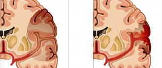
Portal hypertension occurs in situations where some kind of barrier arises during blood circulation in the liver area. The pressure in the portal system is approximately 7 mmHg. If the indicator exceeds 20 mm, blood stagnates in the veins. Since the walls in these vessels are not strengthened by muscles, they expand. A rupture may occur. When cirrhosis develops, in almost 90% of cases the expansion occurs in the area of the esophagus and other parts of the gastrointestinal tract. Sometimes bleeding occurs and there is a high probability of death.
Portal hypertension?
Problems with the movement of blood in the veins can occur below the liver, in the organ itself, or in the area of the vena cava.
The disorder is classified according to the following criteria:
- If blood clots form in the liver vessels, a blockage occurs above the organ. Inflammation of the pericardium provokes high pressure in the blood vessels. Blood leaves the liver slowly.
- Problems arise inside the organ due to cirrhosis, inflammatory processes, neoplasms, in adhesions after damage and surgery.
- Toxins destroy liver cells, increasing resistance to blood flow through the vessels. Hepatocytes are highly viable and can be regenerated.
- Even if a fragment of the liver is destroyed, the remaining part continues to function normally, and over time the functions of the organ are fully restored.
- Problems can arise due to regular exposure to toxins and inflammation. Formed connective tissues are not able to cleanse the body.
Inflammatory processes in the peritoneum can cause blockage of blood flow before reaching the liver. Compression of several branches of the portal vein occurs. The vessels stack abnormally, and complications arise after unsuccessful surgery. People often experience isolated thrombosis, which may be caused by an intra-abdominal infection.
Classification
According to its forms, portal hypertension is divided into:
- Prehepatic portal hypertension is a violation of portal blood flow in the portal vein before it enters the portal of the liver;
- Intrahepatic portal hypertension is a disturbance of blood flow in the portal vein that occurs inside the liver;
- Posthepatic portal hypertension - impaired blood flow through the hepatic veins or the inferior vena cava;
- Mixed portal hypertension – the presence of a combination of the above forms of portal hypertension.
The intrahepatic form of portal hypertension is divided into several types:
- presinusoidal intrahepatic portal hypertension - disturbance of blood flow in the portal vein before it enters the hepatic lobe;
- sinusoidal intrahepatic portal hypertension - disturbance of blood flow in the portal vein at the level of the hepatic lobe;
- Postsinusoidal intrahepatic portal hypertension is a violation of blood flow in the hepatic vein, which exits the hepatic lobule.
According to stages, portal hypertension is divided into:
- The preclinical or initial stage, which is characterized by the absence of complaints, is confirmed only during examination;
- Moderate or compensated stage - characterized by the appearance of symptoms of liver circulatory disorders, enlargement of the liver and spleen;
- Severe or decompensated stage - all symptoms of portal hypertension are pronounced, minor bleeding;
- The terminal stage is massive, prolonged bleeding from the veins of the gastrointestinal tract.
Symptoms
Signs of portal hypertension caused by pathology, causing increased pressure in the vessels. As the pathological process develops, clinical symptoms characteristic of different forms of the syndrome arise:
- The size of the spleen increases, the concentration of platelets and red blood cells decreases, and problems with clotting arise.
- Varicose veins in the stomach and rectum.
- Anemia worsens and venous bleeding occurs.
- Ascites.
Clinical stages:
- Preclinical, in which the patient feels heaviness in the right side under the ribs, malaise occurs, and the abdomen swells.
- Severe symptoms appear, pain, malaise, and the tummy swells.
- All signs of portal hypertension are present, ascites, no bleeding observed.
- Major bleeding occurs.
In the hepatic form, the disorder often occurs in childhood, develops mildly, and has a positive prognosis. The portal vein is pinched, complications become more frequent, bleeding occurs in the vessels in the lower parts of the esophagus, the portal vein is compressed, and coagulation improves.
Portal hypertension is characterized by signs of cirrhosis. These are the main symptoms of the disease. The dynamics of development are determined by the intensity of the occurrence of hypertension. The disease is accompanied by bleeding, ascites is present. The skin turns yellow, this indicates that the liver does not perform its functions normally, resulting in liver failure. Initially, yellowing appears on the palms of the hands and under the tongue. Chiari pathology is mainly the cause of the suprahepatic form of PH. Death causes organ failure and bleeding.
Dangerous consequences
Increased pressure in the hepatic vein is fraught with life-threatening complications. Dysfunction of the spleen leads to massive destruction of blood cells, resulting in:
- anemia;
- insufficient blood clotting;
- immunodeficiency states.
Delayed treatment of hepatic hypertension is dangerous due to dilation of the esophageal veins, erosion of its walls, and bleeding. At stages 3 and 4 there are:
- abdominal dropsy;
- liver failure;
- bacterial peritonitis;
- secondary kidney dysfunction.
Portal hypertension syndrome in children in 7 out of 10 cases is caused by intrauterine development disorders, thrombosis of the peri-umbilical veins. Late detection and treatment leads to death.
Features of the development of the disease
Portal hypertension causes disorder throughout the body. In the first stages, disturbances in peripheral vessels cause difficulties with liver function. Problems with general blood flow appear, inflammation in the portal vein is observed. Destructive processes in the liver begin, and regulatory function deteriorates. Connective tissue appears on the surface of the organ.
There are several types of disease development:
Blood circulation in the liver is aggravated for various reasons:
- Increases resistance to portal vessels.
- There is too much blood draining.
- Ascites appears, which often helps identify diseases.
- Collaterals are created.
- Problems with nervous activity due to liver disorder.
- The size of the spleen increases, congestion occurs.
Symptoms and development of the problem
The initial signs of portal hypertension are closely related to the cause that led to the pathological changes. As the disease progresses, concomitant symptomatic manifestations begin to appear:
- In patients, the spleen increases significantly in size.
- Blood clots poorly, which is especially dangerous with regular bleeding.
- Varicose veins expand in the area of the stomach and rectum.
- The consequence of frequent bleeding is anemia.
- Fluid begins to accumulate in the abdomen, causing it to significantly increase in size.
Diagnostics
To detect portal hypertension, several tests need to be performed. Specialists will have to collect a detailed medical history and determine possible liver disorders in relatives. Only after this the patient is sent for diagnostics. First, a blood test is done to identify viral infections and immunoglobulin. Then the patient is sent for an x-ray. To determine ascites, an ultrasound examination of the peritoneum is performed. Using Doppler ultrasound of problem organs, it is possible to determine the quality of blood circulation.
Stages of the disease
Subcompensated
Portal hypertension includes three stages:
- Compensated is the initial stage, which is characterized by a slight increase in pressure in the portal vein system; at this stage, the liver, with the help of its reserve capabilities, copes with this increase; an enlargement of the spleen and liver is observed (the liver may not enlarge).
- Subcompensated - the manifestations of this stage include the following: high pressure in the portal vein, enlarged spleen, varicose veins of the esophagus, as well as the stomach, which clinically may be accompanied by bleeding from these vessels, but it may not exist, the liver no longer has enough reserves for this to cope with the pressure.
- Decompensated is the most unfavorable form of the disease in terms of clinical and prognosis, manifested by a significant enlargement of the spleen, liver, varicose veins of the esophagus, stomach, which clinically may be accompanied by bleeding from these vessels, but it may not exist, ascites appears, to significant, serious circulatory disorders in the portal vein system are associated with disturbances in central circulation (i.e., heart function).
Therapy
The chances of successful recovery increase if portal hypertension is treated promptly. Only a specialist should be trusted to carry out therapy. If you self-medicate, you can seriously harm your body. Therapy for portal hypertension involves the following procedures:
- Use Propranolol 2 times a day. In this case, ligation or sclerotherapy of vessels susceptible to varicose veins is performed.
- When bleeding occurs, the patient must be injected with 1 mg of Terlipressin. This therapy is performed every 4 hours throughout the day. This treatment method is stable and does not cause side effects.
To prevent bleeding from occurring so often, you need to inject about 250 mg of somatostatin; after a few hours, you can put in a drip with this substance. Injections can be given over 3-4 days. Such treatment methods disrupt the ratio of water and salt in the body, so you need to carefully monitor what foods you eat during the treatment process. If ascites develops, this technique is not used.
If portal hypertension is too advanced, medications cannot change anything, and you have to use invasive therapy or undergo surgery.
We list the most effective procedures:
- Endoscopic sclerotherapy is considered the most popular treatment method for portal hypertension. The point of surgery is to perform tamponade, for this purpose Somatostatin is used. The patient is chipped with a sclerosant that promotes the gluing of blood vessels. Blockage begins, and death is soon observed. The procedure is 80% effective.
- Esophageal tamponade. This technique is performed using a Blackmore probe. Special devices are placed in the stomach through which air enters the organ. As a result, part of the esophagus compresses the stomach vein. The device pumps air throughout the day.
- Endoscopic ligation is performed on the stomach and vessels prone to varicose veins. The procedure is complex and effective. It will be possible to contain the bleeding and prevent the subsequent development of the disease.
- Elective surgical procedures are performed to prevent possible bleeding, while fragments of the liver or blood vessels are cut out.
- Liver transplantation is considered the most radical method of solving the problem of portal hypertension. The operation is performed in a situation where the pathology is complicated by constant bleeding or cirrhosis develops.
Treatment
As a treatment, it is especially important to initially establish the underlying cause, which led to such pathological manifestations.
Treatment for portal hypertension is as follows:
- Propranolol 20-180 twice a day, accompanied by ligation of varicose vessels.
- For bleeding, the following are prescribed: terlipressin 1 mg intravenously, 1 mg every 4 hours during the day.
- The “gold standard” in the treatment of PG is endoscopic sclerotherapy. Tamponade is performed and somatostatin is administered. This is one of the most effective methods.
- Esophageal tamponade using a Sengstaken-Blakemore probe. The probe is inserted into the stomach cavity, after which air is released into it, thereby pressing the veins of the stomach against its walls. The balloon is kept in the cavity for no more than 24 hours.
- Endoscopic ligation of varicose veins from the inside with special elastic rings. It is also one of the most effective methods of treating portal hypertension, but not possible in all cases, especially with rapid blood loss. Bandaging subsequently prevents relapses with bleeding.
- Surgical operations for portal hypertension. Prevents recurrent bleeding.
- Patients with liver cirrhosis and regular bleeding are indicated for hospital surgery and organ transplantation. In this case, patients are given a blood transfusion. This treatment method is used abroad in Israel and Germany.
Favorable treatment for portal hypertension will directly depend on the main cause that led to such consequences. PG is a rather serious disease and if you do not consult a doctor in a timely manner, if you do not follow all his recommendations and if there is regular bleeding, it often leads to death. It is impossible to answer the question with accuracy how long people with PG live, since each individual case has its own characteristics and causes of the disease. Therefore, it is so important to identify the disease in time and cure the disease in a timely manner.
Complications
If left untreated for a long time, additional health problems may arise. Most often, the liver and other internal organs become clogged. We list the possible problems:
- bleeding in different parts of the gastrointestinal tract;
- ascites;
- hypersplenism, due to which the functioning of the immune system deteriorates;
- liver failure, in which part or all of the organ cannot do its job;
- during hepatic coma, blood purification is not performed;
- problems with the organ cause problems with mental activity;
- stomach ulcers and tissue integrity problems;
- nonspecific colitis;
- cystitis;
- death;
Prevalence and prognosis of portal hypertension
Portal hypertension, complicated by esophageal varices, is associated with a high risk of death for the patient.
The incidence of portal hypertension is unknown. Its main cause, liver cirrhosis, ranks 10-12 among other causes of death.
The higher the Child-Pugh class of the disease, the worse the prognosis. Thus, in children with class A congenital pathology, the frequency of esophageal varicose veins was 40%, and in class C – already 85%. With compensated cirrhosis, these figures are 30%, with decompensated cirrhosis – 70%.
The rate of varicose veins is about 8% during the first 2 years after the onset of the disease and by the 6th year it is already 30%. The risk of bleeding in the first year is 30%.
Regarding gender distribution, 60% of patients with portal hypertension are men, and the main reason for this is alcoholism and viral hepatitis. In women, veno-occlusive disease and primary biliary cirrhosis come to the fore. In children, the main causes of pathology are portal vein thrombosis and secondary biliary cirrhosis of the liver.
Variceal bleeding is the most common complication associated with portal hypertension. Almost 90% of patients with cirrhosis develop varicose veins, and about 30% have varicose veins that bleed. The estimated mortality rate for a first episode of variceal bleeding is 30-50%.
After the first episode of bleeding, it recurs within a year in 60-80% of patients. About 30% of them are fatal.
Contribute to increased mortality and complications of portal hypertension:
- hepatic encephalopathy;
- aspiration pneumonia;
- renal failure;
- systemic infections and sepsis;
- bacterial peritonitis;
- ascites (accumulation of fluid in the abdominal cavity);
- hepatorenal syndrome;
- cardiomyopathy, hypotension, heart rhythm disturbances.
Surgical treatment of ascites
The development of cirrhosis complicates the treatment of ascites. At some point, the pathology does not respond to the action of medications. Fluid in the peritoneal area limits the diaphragm's ability to move, causing problems with oxygen absorption. The patient breathes too quickly, does not have enough air, and has problems sleeping because it is difficult to remain in a horizontal position. This condition requires immediate removal of accumulated fluid by laparocentesis. After this procedure, approximately 80 liters of transudate are removed from the body.
Breathing problems go away and the patient feels better. After two to three weeks, the fluid again collects in the abdomen in the same volume. The function is repeated. Sometimes in such situations, a drain is made in the abdomen so that the transudate is constantly removed without the need for multiple surgical interventions. If fluid is removed too often, protein will be excreted from the body, resulting in increased hypothermia and general health deteriorating. Therefore, the secondary occurrence of tense ascites is prevented by conservative methods.
Carrying out the operation
Surgical treatment in patients with cirrhosis involves complications. Often such patients suffer from many illnesses and vital organs do not function normally. There are serious risks involved in the operation. To reduce the likelihood of death or complications, peritoneovenous shunting should be performed. Since ascites fluid is similar in characteristics to plasma, filtration mechanisms and injection back into the vascular bed are used. In this case, lymph stagnation can be eliminated; fluid will no longer be directed into the abdominal cavity.
Signs and clinical manifestations
Most often, patients encounter the following signs of pathology:
- splenomegaly (in some cases with PG, the first symptom is considered to be an increase in the size of the spleen);
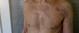
- varicose veins of the venous capillaries of the esophagus, stomach, abdomen (head of the jellyfish) and anus (hemorrhoids);
- ascites - a pathological accumulation of fluid in the abdominal cavity, which can be combined with hydrothorax;
- pain in the abdomen and right hypochondrium;
- lack of appetite, bloating;
- dyspepsia;
- symptoms of intoxication;
- general weakness;
- weight loss;
- swelling of the ankles;
- erosions on the mucous membrane of the digestive system.
If the cause of portal hypertension is blood stagnation due to blockage of the hepatic ducts, the patient's skin turns yellow. Sometimes hypersplenism is diagnosed - a decrease in the number of red blood cells, leukocytes, and blood platelets due to increased spleen function.
Signs of PG in children deserve special attention. They are only extrahepatic, manifested by an enlarged spleen, varicose veins of the venous capillaries of the esophagus, and hemorrhages. Bleeding occurs extremely rarely.
Endoscopic ligation of vessels prone to varicose veins
For different types of bleeding, hemostasis techniques are used. It is necessary to give preference to adequate methods, since there is no other way to control the bleeding. To identify sites of hemorrhage, a FEGDS procedure is performed. Using this method, varicose veins of the esophageal vessels are diagnosed. The area of damage and the nature of bleeding are determined. In the simplest cases, an endoscopic procedure can be performed quickly. Ligation is considered the most effective way to stop bleeding of vessels susceptible to varicose veins. Effective technologies reach 90%, and the number of relapses is minimal.
Parabasal sclerosis
For preventive purposes, operations are performed to reduce pressure in vessels with several ductal anastomoses. The meaning of this procedure is to create additional pathways for the outflow of blood from the location of the portal vein by creating:
- Direct anastomosis.
- Mesenteric forging.
- Splenoreal.
The formation of direct anastomoses allows you to quickly reduce portal pressure due to blood transfer to the inferior vena cava area. At the same time, it is necessary to prevent deterioration of liver function. The appropriate size of the anastomosis must be determined. Recently, portacaval devices with a width of 6, 8 and 10 mm have become popular. Anastomoses allow partial drainage of portal blood into the vena cava area. In this way, complications can be prevented.
Splenorenal anastomosis requires removal of the spleen, which may be difficult in certain settings. Sometimes during surgery the patient loses a lot of blood.
Possibilities of ultrasound examinations for portal hypertension in children
Ultrasound scanner HS70
Accurate and confident diagnosis.
Multifunctional ultrasound system for conducting studies with expert diagnostic accuracy.
Portal hypertension is one of the main causes of severe esophageal and gastric bleeding in children, often leading to mortality. In addition, the outcome of portal hypertension in children with liver disease may be liver or kidney failure. In this regard, it is extremely important to timely recognize the syndrome itself and establish the causes of its development.
The term “portal hypertension” was first introduced in 1928 by the English surgeon A. McJandoe. Its development is based on an obstruction to the flow of blood through the portal vein system. Usually a blockade of a mechanical nature is assumed, but there are cases of a functional, spastic origin of a blockade of any part of the portal system. The following types of portal hypertension are distinguished: intrahepatic (liver cirrhosis, congenital liver fibrosis, severe fatty infiltration, intrahepatic tumors); extrahepatic (thrombosis of the portal and splenic veins, isolated thrombosis of the splenic vein, malformation of the portal vein, compression of the veins by a tumor or lymph nodes); suprahepatic (Budd-Chiari syndrome, compression of the hepatic veins by a tumor); mixed (when the malformation of the portal vein is complicated by chronic viral hepatitis, and then cirrhosis of the liver, or the abnormal development of the portal vein is combined with fibrosis of the liver).
The pathogenesis of portal hypertension with different types of blockade of the portal system is different. The most common cause of the hepatic form of hypertension is cirrhosis of the liver, when, against the background of inflammation, sclerosis occurs with subsequent fibrosis of the portal tracts, the development of regeneration nodes, disruption of the structure of the hepatic lobules, compression of vascular structures, including intrahepatic branches of the portal vein. Violation of the organization of the lobules leads to changes in intralobular blood circulation and is the cause of the development of necrosis in the center of the lobule, fibrosis and pronounced cellular infiltration with proliferation of Kupffer cells. The latter can protrude into the lumen of the sinusoids, narrowing them, leading to an increase in pressure in the portal system. Obstruction of blood flow through the portal vein system and increased portal pressure lead to enlargement and hyperplasia of the spleen. Further, portal hypertension causes the development of collaterals, both natural and new portocaval connections: through the coronary vein of the stomach to the venous plexus of the esophagus and further through the azygos and semi-gypsy veins with the superior vena cava system. The veins of the esophagus dilate. Vv.epigastricae participate in the development of portocaval anastomoses, forming the “head of the medusa”, more often observed in adult patients. Cruveillier-Baumgarten syndrome is most characteristic of childhood with congenital liver fibrosis. With the development of portacaval collaterals through the v.mesentericae inferior, the hemorrhoidal veins increase. Stasis in the portal and lymphatic system of the abdominal organs and impaired inactivation of corticosteroids, in particular aldosterone, play a role in the occurrence of ascites that develops with portal hypertension.
In contrast to the mechanism of intrahepatic portal hypertension, the extrahepatic one, at first glance, seems simpler. The virtual absence of the trunk and branches of the portal vein in congenital genesis of development, obliterated or narrowed its lumen as a result of thrombosis or extravasal compression leads to increased resistance to blood flow, leading to a stable increase in portal pressure up to 300-500 mm water column (N up to 120-180 mm water column column) and the development of a hyperdynamic type of hemocirculation in the vessels of the portal vein, with pronounced arteriovenous shunting in the organs, increasing the load on the vessels, with their subsequent morphological changes. Their severity depends on the duration and severity of the disease.
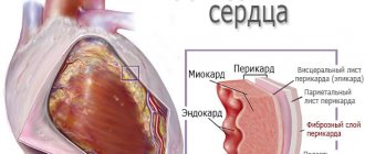
The development of portal hypertension with adrenal blockade of blood circulation is most often caused by restrictive pericarditis, narrowing of the hepatic veins or the inferior vena cava above its confluence with the hepatic veins (Chiari disease), narrowing of the inferior vena cava and/or hepatic veins as a result of a congenital septum, thrombus, tumor, hypertrophied caudate lobes of the liver, which ultimately leads to a disorder of blood and lymph circulation in the organs and tissues of the underlying sections.
The main methods of studying patients with suspected portal hypertension, in addition to the generally accepted physical ones, are angiographic, endoscopic and ultrasound in combination with Dopplerography (in color and normal modes). All of the above methods complement each other. Relative competition can be discussed between angiography and Dopplerography. However, given the harmlessness of the ultrasound method and the relative ease of its use, it must be recognized that it should be used first in every child with an enlarged spleen and other signs suggesting portal hypertension. And then, based on the results obtained, decide on the conduct of radiocontrast studies.
The most common form of portal hypertension in children compared to others is extrahepatic blockade of the portal circulation, the frequency of which in the differential diagnosis of portal hypertension reaches 50-88%. However, in adults this figure is within 10% [1, 2]. This difference is explained by the fact that the main cause of the development of hypertension is primary damage to the veins of the portal system, which in some cases is accompanied by fibrosis of the liver parenchyma.
An echographic examination of the abdominal organs in children with suspected extrahepatic form of portal hypertension allows us to correctly assume the diagnosis in almost 100% of cases. The liver is usually of normal or reduced size. The latter is due to a significant decrease in portal blood flow with the development of parenchymal fibrosis, observed in 60-70% of children. With congenital fibrosis, multiple small strong echo signals are recorded from the parenchyma, scattered throughout the entire section of the organ (Fig. 1). The portal vein is changed, and there may be an absence of lumen in its trunk or main branches (Fig. 2), the presence of multiple thin tortuous vessels with hyperechoic walls, collected together - angiomatous transformation (Fig. 3), the presence of a thrombus narrowing or closing the lumen of the vessel, or local narrowing of the lumen, which leads to the development of collaterals, visible as anechoic narrow, tortuous tubular structures running adjacent to the main trunk (Fig. 4). Such changes in the portal vein lead to a significant slowdown in blood flow. Color Doppler mapping facilitates a qualitative assessment of the state of the portal vein, allowing one to determine the direction of blood flow (hepatopetal - to the liver or hepatofugal - from the liver).
Rice. 1.
Echogram of the liver with congenital fibrosis.
Rice. 2.
Echogram of the portal vein without lumen.
Rice. 3.
Echogram of the portal vein with angiomatous transformation.
Rice. 4.
Echogram of the right branch of the portal vein with collaterals that developed due to thrombosis of the trunk.
Portal hypertension in its extrahepatic form gradually leads to a number of secondary changes. The spleen enlarges, small focal or diffuse small focal compaction of the parenchyma, compaction of the walls of small vessels are observed, some may be thrombosed (Fig. 5). The splenic vein is dilated, often tortuous in the hilum area. The speed of blood flow through it, as a rule, remains the same, but the rate of volumetric blood flow increases significantly. The splenic artery is slightly dilated, which leads to an increase in the resistance index and indicates an increase in peripheral resistance, indirectly confirming the ultrasound data on the state of the splenic parenchyma.
Rice. 5.
Echogram of the spleen, thrombosis of small branches of the splenic vein.
The lesser omentum in children is usually not visualized on sonographic examination, but in portal hypertension it often thickens and becomes visible as an echogenic tissue formation with indistinct edges located between the anterior surface of the pancreas and the posterior surface of the stomach, or, if the stomach is not visible, then the posterior surface liver (Fig. 6).
Rice. 6.
Echogram of a thickened lesser omentum. P - pancreas; ST - stomach.
Normally, the walls of the gallbladder are usually not visible, but they can thicken up to 2 mm. With portal hypertension, the gallbladder is usually deformed, with sharply thickened walls (Fig. 7). In some cases, varicose veins may be visible in the walls, which is especially well demonstrated when using color Doppler mapping in energy mode.
Rice. 7.
Echogram of the gallbladder with sharply thickened walls containing varicose vessels and a reduced lumen (CDC mode).
In severe forms of portal hypertension, venous collaterals develop, some of which may be visible: these are coronary, gastroesophageal, defined between the left lobe of the liver and the head, the body of the pancreas as anechoic round or tubular structures (Fig. 8). Esophageal varices can be identified by longitudinal scanning in the aortic area. Due to the different directions of blood flow due to the tortuosity of the vessels during CDK, its mixed nature is demonstrated. When examining the liver with fibrosis, a patent umbilical vein is noted, which sometimes has uneven walls and is directed from the region of the bifurcation of the portal vein to the surface of the liver in the cephalolateral direction (Fig. 9).
Rice. 8.
Echogram of the area of the head of the pancreas with vascular collaterals (arrow). L - liver; P - pancreas; VL - splenic vein; AO—aorta; IVC - inferior vena cava.
Rice. 9.
Echogram of the dilated umbilical vein. VU - umbilical vein; VP - portal vein.
In the hepatic form of portal hypertension, the main cause of which in children is cirrhosis of the liver, almost the same secondary changes are observed. The portal vein is significantly expanded, reaching a diameter of 20 mm, its walls are thickened. The speed of blood flow through the vein is reduced, but due to an increase in diameter, the volumetric blood flow also increases. An increase in this indicator also occurs in the splenic vein. Changes occurring in the portal system lead to a redistribution of blood volume - its increase in the vessels of the portal system and its decrease in other vital organs (brain, kidneys) [3]. With more severe circulatory disorders in the portal vein, fluid accumulates in the abdominal cavity, which, when its quantity is small, is primarily localized in the lateral canals and pelvis, and then in the perihepatic and perisplenic spaces.
The most effective way to treat extrahepatic portal hypertension and its main complication - recurrent acute bleeding from varicose veins of the esophagus and stomach - is the creation of portocaval anastomoses, which make it possible to create conditions for decompression of the portal system. The choice of vessels for creating a shunt is determined individually, taking into account the hemodynamic parameters and age of the patient. In recent years, preference has been given to splenorenal, mesocaval, side-to-side types. All data on blood flow in potentially used vessels can be obtained from Doppler examination. Observation of the nature of blood flow after the operation and comparison of the obtained data with the primary data makes it possible to talk about the effectiveness of the surgical treatment both immediately after it and in late follow-up. One of the complications after bypass surgery is the formation of a hematoma (4-5% of cases), which on an echogram appears as a round, echo-free formation with fairly clear boundaries. Dynamic research allows you to monitor the condition of the hematoma and regression of its size.
One of the treatments for intrahepatic portal hypertension is removal of the spleen. The main possible postoperative complications diagnosed by ultrasound include bleeding with the formation of a hematoma, the development of an abscess, and reactive changes in neighboring organs, most often the pancreas.
With Badd-Chiari syndrome, echograms reveal a narrowing or absence of the lumen of the inferior vena cava or one of the hepatic veins, in particular, the hypertrophied caudate lobe (Fig. 10). When the process becomes chronic, venous collaterals are formed, visualized by ultrasound, the vessel above the obliteration or narrowing is dilated, and the blood flow is significantly weakened.
Rice. 10.
Echogram of the inferior vena cava (longitudinal section). The lumen of the vein is narrowed by the hypertrophied caudate lobe.
Thus, ultrasound examination of the abdominal organs is the method of choice at the early outpatient and inpatient diagnostic stages of examining children with portal hypertension, includes a mandatory Doppler assessment of the state of blood flow, allows highly accurate confirmation and determination of the cause of portal hypertension, and a comparative analysis of data obtained at different periods of therapeutic and surgical treatment of the disease - to evaluate its effectiveness.
Literature
- Patsiora M.D. Surgery for portal hypertension. M.: Medicine. 1974. P. 232.
- Van Vroonhen T., Molenaar J. Distal splenorenal shunt for decompression of portal hypertension in children with cystic fibrosis// Surg.Ginecol.Obst. 1979. V.149. P.559-566.
- Abbas N.M. The state of central and peripheral hemodynamics in children with chronic liver diseases: Diss... cand. honey. Sci. - M., 1996. P.160.
Ultrasound scanner HS70
Accurate and confident diagnosis.
Multifunctional ultrasound system for conducting studies with expert diagnostic accuracy.
Principle of transplantation
Cirrhosis is considered to be the result of various inflammations. Pathology can develop quickly or slowly. At the stage of obvious functional disorders, portal hypertension begins. The disease is characterized by ascitic syndrome and bleeding from the vessels of the esophagus. Patients become disabled and often die due to complications. Conservative treatment methods at this stage do not give results.
Today, a large number of surgical methods are used. Most of them are palliative in nature. The possibility of a liver transplant allows you to get rid of serious disorders, a large number of different diseases and organ transformations. Today people have learned to consider the advantages of tissue and organ donation.
Modern views on the treatment of portal hypertension
Main objectives of treatment
- decreased pressure in the portal vein. For this purpose, nitrates are used (monotherapy is not recommended) - Isosorbide-5-mononitrate; non-selective beta blockers - Propranolol, Obzidan, Anaprilin, Timolol, Nadolol. The most effective use of a combination of two groups of drugs, but noticeable disadvantages are the negative effect on the kidneys;
- prevention and control of bleeding. The first point is to prescribe beta-blockers in the presence of small-diameter varicose nodes and their endoscopic doping (ligation with elastic rings). To stop bleeding, tamponade (compression from the inside) of the esophagus is used using a special Blackmore probe; endoscopic sclerotherapy - the introduction into the cavity of the esophagus of special substances that contribute to blockage of the lumen of the veins - Somatostatin, Octreotide.
- normalization of the blood coagulation system. It comes down to prescribing drugs containing vitamin K (Vikasol, etc.);
- correction of liver failure is carried out by prescribing hepatoprotective agents (Hepasol, Thiotriazolin, Berlition, Essentiale), protein replacement drugs and detoxification therapy (Albumin, Reopoliglyukin, Trisol);
- binding of toxic products of nitrogen metabolism. For this purpose, a drug is used that promotes the binding and removal of ammonia from the body - Hepa-merz.
Surgical treatment
If conservative therapy is ineffective, they proceed to surgical interventions. Currently used:
- portosystemic shunting - application of artificial collaterals to unload the liver;
- embolization of the splenic artery using a retroendovascular method - used to eliminate the phenomena of hypersplenism;
- splenectomy - comes down to the removal of the spleen if it is enlarged to such an extent that it compresses the abdominal organs;
- liver transplantation - allows to reduce the manifestations of portal hypertension, ascites, hepatic encephalopathy, repeated bleeding from varicose veins.